Case
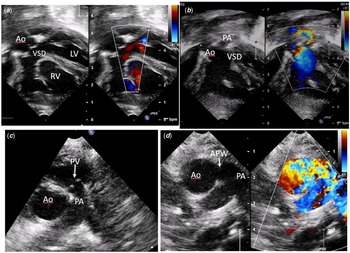
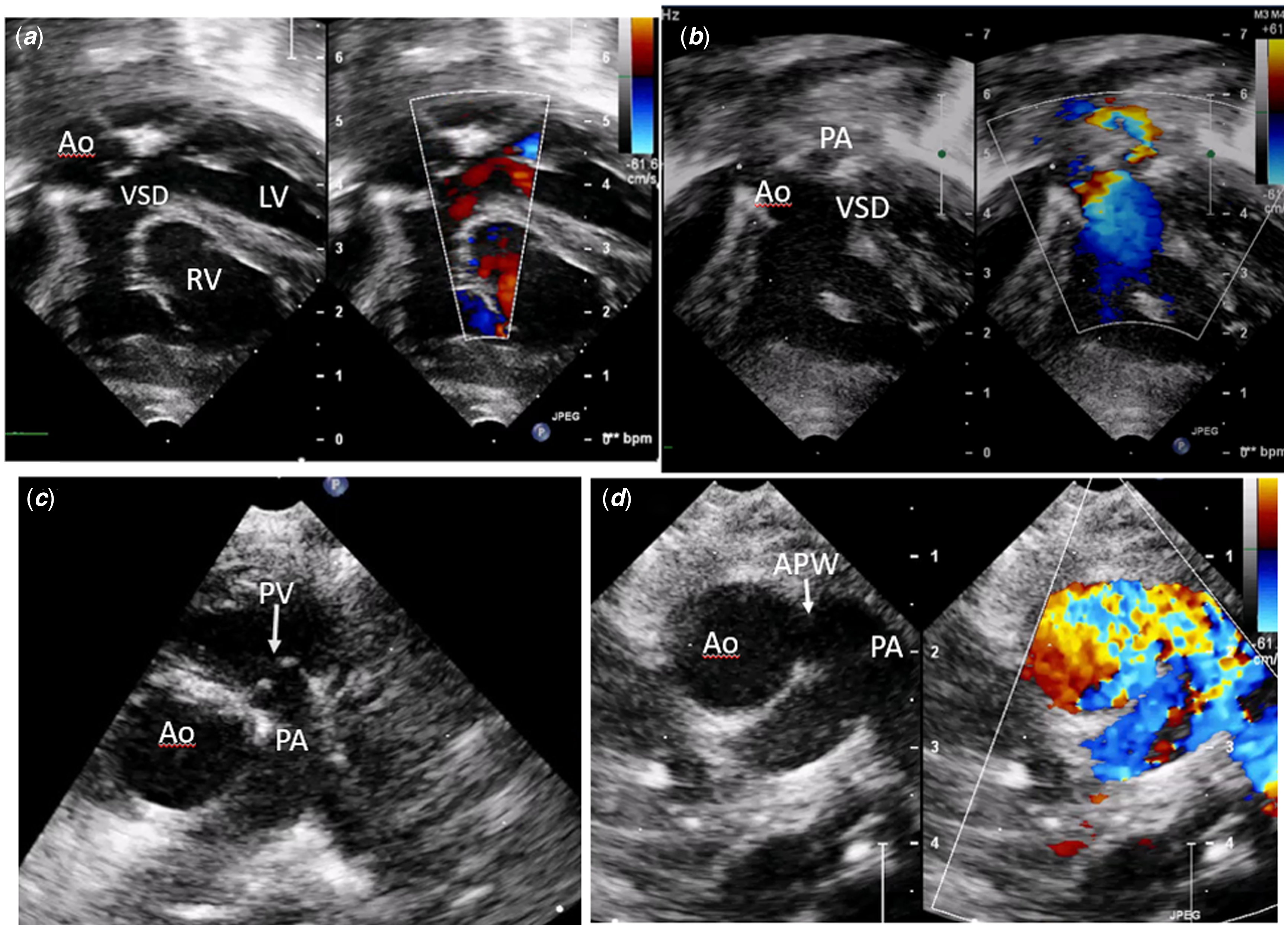
A 2.6-kg term newborn boy suspected antenatally to have common arterial trunk (Edward-Collet type I) presented on fifth postnatal day with respiratory distress needing ventilation. Echocardiography revealed large doubly committed ventricular septal defect with 60% aortic override (Fig 1a), hypoplastic pulmonary annulus (Z score −3), and pulmonary stenosis with gradient of 50 mmHg and no pulmonary regurgitation (Fig 1b and c). The arterial duct was absent. The branch pulmonary arteries were significantly dilated, like that seen in tetralogy of Fallot with absent pulmonary valve syndrome. Closer examination revealed a large type II aortopulmonary window (Fig 1d). He underwent aortopulmonary window repair and intracardiac repair with transannular patch and monocusp reconstruction of the pulmonary valve on postnatal day 25. He is doing well at 1-year follow-up.
( a ): Subcostal view showing large ventricular septal defect (VSD) with 50% aortic (Ao) override. ( b ): Subcostal right ventricular outflow tract view showing large doubly committed VSD with aorta (Ao) and pulmonary artery (PA) overriding the defect. ( c ): Parasternal short-axis view showing hypoplastic pulmonary valve (PV) and dilated right and left pulmonary arteries (RPA, LPA). ( d ): Parasternal short-axis view showing large aortopulmonary window (APW) from ascending aorta to right pulmonary artery. LV: left ventricle, RV right ventricle.
Discussion
Tetralogy of Fallot with aortopulmonary window causes heart failure secondary to large left to right shunt distal to the stenosed pulmonary valve. Reference Alborino, Guccione, Di Donato and Marino1 This can present in the newborn period if the defect is large, and the pulmonary vascular resistance falls rapidly. The diagnosis can easily be mistaken for a common arterial trunk as the pulmonary valve is commonly atretic or is very hypoplastic. The resemblance to the common arterial trunk is accentuated by the presence of doubly committed ventricular septal defect with absence of conal septum beneath the pulmonary valve. In our case, it was difficult to image the hypoplastic pulmonary annulus as it had a peculiar horizontal lie over a doubly committed ventricular septal defect with absent conal septum (Fig 1b).
Surgical management consisted of repairing the aortopulmonary window first through trans-window approach. This was followed by intracardiac repair with transannular patch. Our institutional policy is to perform monocusp reconstruction of the pulmonary valve after transannular patch, and it was particularly beneficial in this case as pulmonary artery pressure was higher than that for a usual tetralogy of Fallot and pulmonary regurgitation in this situation is more likely to cause post-operative right ventricular failure.
Less than 20 such cases have been reported in literature with association of pulmonary atresia being commoner than pulmonary stenosis. Poor prognosis has been noted in cases with delayed diagnosis. The case is of educational value as aortopulmonary window is rarely considered as a cause of heart failure in tetralogy of Fallot. The lesion can be easily missed on transthoracic echo, unless carefully looked for. Surgical repair yields excellent result and must be offered early to prevent pulmonary hypertensive changes. Reference Kynta, Rawat, Thingnam, Manoj and Sharma2
Supplementary material
To view supplementary material for this article, please visit https://doi.org/10.1017/S104795112200364X
Acknowledgements
None.
Author contribution
All authors contributed to the study conception and design. Material preparation, data collection, and analysis were performed by Saikiran Kakarla and Deepa Sasikumar. The first draft of the manuscript was written by Saikiran Kakarla; Deepa Sasikumar and Baiju S Dharan critically reviewed the manuscript. All authors read and approved the final manuscript.
Financial support
This research received no specific grant from any funding agency, commercial, or not-for-profit sectors.
Conflicts of interest
None.