Implications
Decubital shoulder ulcers are lesions on sows kept in production systems and to some extent comparable with human pressure ulcers. We review the pathogenesis, classification and consequences in terms of pain and animal welfare. Knowledge of welfare consequences of decubital shoulder ulcers is limited. On the basis of the tissue that is involved, we assume that the development and presence of decubital shoulder ulcers are a painful and prolonged condition. We conclude that the extent of the welfare problem cannot be fully determined until a valid ante-mortem classification system is available, and knowledge about the duration of the condition as well as possible consequences in terms of pain or discomfort have been established.
Introduction
In lactating sows, lesions on the shoulder regions can be observed in the weeks after farrowing – decubital shoulder ulcers induced by pressure from the flooring and to some extent comparable with human pressure ulcers. The lesions vary from superficial ulcers, in which redness of the skin is the only clinical sign, to deep ulcers involving subcutaneous layers or even bone tissue (Lund et al., Reference Lund, Aalbæk and Jensen2003; Vestergaard et al., Reference Vestergaard, Fruergaard, Nielsen and Madsen2005; Jensen, Reference Jensen2009). In production systems, the ulcers will normally heal after the lactation period but will often relapse when sows are returned to the farrowing pens for the next lactation (Davies et al., Reference Davies, Morrow, Miller and Deen1996). In a recent epidemiological survey based on 3831 Danish sows from 98 herds, Bonde (Reference Bonde2008) found decubital shoulder ulcers in 17% of lactating sows kept in conventional farrowing systems. In other European countries, smaller surveys based on herd visits have shown prevalence of 34% among sows in 60 Swedish herds (Ivarsson et al., Reference Ivarsson, Mattson, Lundeheim and Holmgren2009) and 10% among sows kept in- or outdoors in 86 herds in the United Kingdom (Kilbride et al., Reference KilBride, Gillman and Green2009), whereas meat inspection at four Norwegian slaughterhouses showed a prevalence of decubital ulcers of 10% (Baustad and Fredriksen, Reference Baustad and Fredriksen2006). A smaller North American investigation in one sow herd reported a decubital ulcer prevalence of 34% (Zurbrigg, Reference Zurbrigg2006), whereas Knauer et al. (Reference Knauer, Stalder, Karriker, Baas, Johnson, Serenius, Layman and McKean2007) found shoulder lesions on 18% of culled sows from two different US harvest plants. Today, decubital shoulder ulcers are considered a multi-factorial problem, connected to risk factors at both individual and herd levels, such as the body condition of the sows, their lying behaviour and the physical properties of the flooring (Cleveland-Nielsen et al., Reference Cleveland-Nielsen, Christensen and Ersbøll2004; Rolandsdottor et al., Reference Rolandsdottor, Westin and Algers2009) and may be inherited (Lundgren et al., Reference Lundgren, Zumbach and Lundeheim2009). As a consequence, no immediate solutions are available for modern pig production. Until now the majority of scientific knowledge about these lesions is epidemiological, and only little is known about the pathogenesis or the consequences in terms of animal welfare, basic knowledge of which is crucial for future scientific focus on the condition. The aim of this paper is to review the present knowledge about the pathogenesis, classification and possible pain associated with decubital shoulder ulcers in sows to relate this to animal welfare and point to areas within these fields in which new research is needed.
Description and definition of decubital shoulder ulcers
Scientifically, an ulcer of the skin is defined as a condition in which the epidermis and the underlying basal membrane (Laminae epithelialis) is damaged (McGavin and Zachary, Reference McGavin and Zachary2007). ‘Shoulder ulcers’ or ‘decubital shoulder ulcers’ are the terms most often used to describe visible skin lesions on the shoulder region(s) of sows kept in the production systems (Zurbrigg, Reference Zurbrigg2006). Decubital shoulder ulcers are, to some extent, comparable with human pressure ulcers that often appear over the underlying bony prominences, in which the amount of soft tissue (e.g. muscular and/or adipose tissue) between the skin and the bone is insufficient to distribute external pressure. In sows, decubital ulcers are most often seen near the tuber of the scapular spine (tuber spina scapulae), described as varying from a slight redness of the skin to deep ulcers measuring up to 10 cm in diameter (Vestergaard et al., Reference Vestergaard, Fruergaard, Nielsen and Madsen2005; Jensen, Reference Jensen2009) and present on one or both shoulders of an individual animal.
As the scientific literature on decubital shoulder ulcers in pigs is very limited, knowledge of human pressure ulcers is included in this review in order to elucidate the development of the ulcers, their classification and welfare consequences. This knowledge is considered of particular relevance because of the considerable histological and functional similarities between the skin of humans and pigs (Dyce et al., Reference Dyce, Sack and Wensing2002), and because pigs have been used as animal models in studies of human pressure ulcers (e.g. see Dinsdale, Reference Dinsdale1974; Kokate et al., Reference Kokate, Leland, Held, Hansen, Kveen, Johnson, Wilke, Sparrow and Iaizzo1995). In both species, the skin can be divided into the epidermis and the dermis, the latter covering the subcutis primarily consisting of loose connective tissue and fat (Maklebust and Sieggreen, Reference Maklebust and Sieggreen1996). Furthermore, due to the limited knowledge within this area, we have chosen to include reports and other non-peer reviewed material in the references. However, within this review, the use of references has been weighted according to their quality of documentation.
No concordant, scientific definition of decubital shoulder ulcers in pigs is available. Recently, the American National Pressure Ulcer Advisory Panel (NPUAP, 2007) has defined human pressure ulcers by their anatomical position and cause, as ‘a visible lesion of the skin and/or underlying tissue – often appearing over bony prominences – that is caused by pressure or pressure in combination with abrasion and/or friction’. Thus, in humans, pressure ulcers can be diagnosed without any lesions of the epidermis.
Pathogenesis: the emergence and development of the condition
Causal factors and underlying mechanisms for human pressure ulcers
Gottrup (Reference Gottrup2002) and Leigh and Bennett (Reference Leigh and Bennett1994) reviewed pressure ulcers in humans. The authors pointed out that under normal conditions, behavioural avoidance patterns induced by pain/discomfort due to long-term pressure, will be identified, and will ensure that the pressure will not exceed a pathological level. In cases in which a person is, for example, under the influence of drugs, unconscious or paralysed, the risk of pressure ulcers increases considerably.
It is widely recognized that one of the causal factors for pressure ulcers is tissue traumatizing pressure and that the force and the duration of pressure are important factors for the development of the characteristic tissue necrosis (Seiler and Stähelin, Reference Seiler and Stähelin1986; Vandeberg and Rudolph, Reference Vandeberg and Rudolph1995). Lowthian (Reference Lowthian2005) pointed out that not only the duration of one period of pressure but also the frequency of repeated periods of pressure are of importance, as the affected tissue will need sufficient restitution after each period of pressure in order to avoid tissue damage. Bouten et al. (Reference Bouten, Oomens, Baaijens and Bader2003) presented computer models simulating the consequences of mechanical stimulation on skin and underlying tissue and stated that pressure directed towards the skin surface is not representative of mechanical conditions in the underlying tissue. This applies, in particular, in cases of complex tissue structure and tissue geometry, for example, near bony prominences, implying that potentially harmful pressure cannot be described solely by means of the force and/or duration.
Shearing forces are believed to be of importance in relation to the initiation of human pressure ulcers (Gottrup, Reference Gottrup2002; Jørgensen, Reference Jørgensen2004). Maklebust and Sieggreen (Reference Maklebust and Sieggreen1996) defined shearing forces as mechanical forces that shear different types of tissue parallel to each other and are typically caused by non-perpendicular pressure. Lowthian (Reference Lowthian2005) mentioned consequences such as stretching, and, possibly also, rupture of the blood vessels, and stated that especially deep tissue is affected by shearing forces. One example is a human patient lying in bed with the head elevated, whereby bone tissue is sheared in one direction as a result of the body weight, whereas the skin surface is fixed by the bed linen and remains stationary. Consequently, the exposed tissue will be subjected to stretching as well as compression depending on the position of the tissue in relation to the direction of the force as well as bony prominences.
A related causal factor for the development of human pressure ulcers is believed to be friction (Dinsdale, Reference Dinsdale1974); however, there is mixed evidence for this, as some studies suggest that the major effect of friction is removal of devitalized epidermis (Witkowski and Parish, Reference Witkowski and Parish1982). As an example of friction contributing to the development of human pressure ulcers, Maklebust and Sieggreen (Reference Maklebust and Sieggreen1996) mentioned patients unable to lift themselves sufficiently from their bed when changing position.
Another causal factor for human pressure ulcers is believed to be increased temperature in tissue and the surrounding environment. This assumption is based on results from model studies using pigs (Kokate et al., Reference Kokate, Leland, Held, Hansen, Kveen, Johnson, Wilke, Sparrow and Iaizzo1995), in which the extent of tissue damage induced by 5 h of pressure (100 mmHg) depended on the temperature of the compression plate: thus, the fact that damage was more severe at 35°C or 45°C, than at 25°C. In sows, a non-pathological increase in body temperature can be observed around farrowing, peaking at 1 to 2 h after the birth of the last piglet (Littledike et al., Reference Littledike, Witzel and Riley1979). After farrowing, the body temperature decreases slightly, but remains increased during the entire lactation period (Havn et al., Reference Havn, Poulsen, Enøe and Nielsen2004; Damgaard et al., Reference Damgaard, Malmkvist, Pedersen, Jensen, Thodberg, Jørgensen and Juul-Madsen2009). Increase in body temperature has been suggested to be one of the releasing factors of decubital shoulder ulcers in sows. However, no data are available with regard to the effects of temperature of, for example, flooring or skin on the occurrence of decubital shoulder ulcers in sows.
The mechanisms behind the development of pressure ulcers in humans have not yet been fully elucidated. However, ischaemia (restriction in blood supply) is involved – either directly or indirectly depending on the type of exposed tissue. Depending on the proximity of, for example, bony or fatty tissue, the development of tissue destruction will differ. Bouten et al. (Reference Bouten, Oomens, Baaijens and Bader2003) described several possible ischaemic mechanisms, for example, localized ischaemia due to the occlusion of the blood vessels, which impeded the flow of interstitial fluid and lymphoid drainage resulting in the loss of nutritional supply and clustering of waste products in the tissue leading to increased cell death and necrosis. However, both Bouten et al. (Reference Bouten, Oomens, Baaijens and Bader2003) and Lowthian (Reference Lowthian2005) stated that these ischaemic mechanisms may not be equally relevant for different types of tissue, and may not fully explain the development of pressure ulcers. In relation to pressure ulcers near bony prominences, Lowthian (Reference Lowthian2005) suggested that pressure combined with shearing forces will result in stretching and pressure directed towards the surrounding tissue leading to capillary rupture in the area subjected to maximal pressure and thrombosis (obstruction of blood flow) that will subsequently lead to ischaemia.
Decubital shoulder ulcers in sows
In experimental studies using pig skin and the underlying tissue as a model for human pressure ulcers, and studying the consequences of pressure applied to the skin (however, not directed at the shoulder region), the force and duration of the pressure significantly affected the development of pressure ulcers. Daniel et al. (Reference Daniel, Priest and Wheatley1981) found that the destruction of the skin in anaesthetized but otherwise normal pigs required high pressure and relatively long duration (800 mmHg for 8 h). Other factors, such as friction or repeated periods of pressure, have been shown to have additive effects, reducing the threshold of the force and duration of the pressure (Dinsdale, Reference Dinsdale1974).
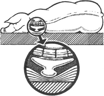
With regard to decubital shoulder ulcers, it is generally believed that persistent and constant compression of the blood vessels in the skin around the tuber of the scapular spine results in insufficient blood circulation, necrosis, and subsequently ulceration, and it is assumed that the development of ulcers primarily depends on the force and the duration of the pressure but is also affected by the robustness of the skin (e.g. texture). Recently, the importance of duration of pressure for the prevalence of decubital ulcers in sows has been emphasized in studies showing a lower frequency of decubital shoulder ulcers in sows fed eight v. three times daily (Sørensen, Reference Sørensen2009) and positive correlations between the maximum duration of lying bouts and the degree of decubital shoulder ulcers at weaning (Rolandsdottor et al., Reference Rolandsdottor, Westin and Algers2009). Reese et al. (Reference Reese, Straw and Waddell2005) estimated that the tuber of the scapular spine in a recumbent sow is one of the most weight-bearing body parts (Figure 1). However, this has not been demonstrated experimentally, and neither the cause and effect relationship between pressure and decubital shoulder ulcers nor the above-mentioned pathological development have been demonstrated scientifically. Thus, the pathogenesis of porcine decubital shoulder ulcers is subject to more uncertainty than human pressure ulcers.
Graphic drawing of the tuber of the scapular spine (tuber spina scapulae), a tongue-shaped bony prominence, in sow lying in lateral recumbency (printed from Vestergaard et al. (Reference Vestergaard, Fruergaard, Nielsen and Madsen2005) with permission).

Recently, Jensen (Reference Jensen2009) found subcutaneous lesions on the shoulders of approximately 50% of 95 slaughtered sows, in which the skin had appeared normal. In almost 80% of these cases, the lesions were situated caudally to the tuber of the scapular spine, suggesting that they did not only result from counter pressure from the floor. Correspondingly, Høgedal and Pedersen (Reference Høgedal and Pedersen2007) found deep tissue changes (e.g. reddish brown discolouration, oedemas) in shoulders from 15 slaughtered sows with prominent scapular spines and intact skin. Crated sows can interact quite forcefully with crate fixtures, for example, when getting up, and sows in farrowing crates can be observed to lean or rest their scapular spine against the horizontal bars. Jensen (Reference Jensen2009) suggested that some of the observed lesions under intact skin were inflicted shortly before the animals were killed, for example, during transport. Therefore, some shoulder lesions reported from slaughterhouses and pig production systems may not be decubital ulcers but include injuries induced by stimulation other than pressure from the flooring.
Rate of progression
We have not been able to find scientific data describing the rate of progression of decubital shoulder ulcers in sows or the exact time of appearance after farrowing. In humans, pressure ulcers caused by immobility during surgery have been reported to be visible after 2 days, and to start diminishing after 12 to 14 days (Vermillion, Reference Vermillion1990). In pigs, Daniel et al. (Reference Daniel, Priest and Wheatley1981) recognized experimentally induced pressure lesions 48 h after a tissue traumatizing pressure was applied to the great trochanter of the femoral bone, but the lesions were not clinically recognizable until after 7 days.
On the basis of cross-sectional examination of the incidence of shoulder lesions in an intensively kept American sow herd, Davies et al. (Reference Davies, Morrow, Miller and Deen1996) divided the shoulder lesions into six categories based on the presence of (i) normal skin; (ii) haemorrhage in the skin; (iii) callosity; (iv) ulcer; (v) ulcer with crusts; and (vi) scars. The results showed that the incidence of the various types of shoulder lesions depended on the interval from farrowing. Thus, the incidence of haemorrhage in the skin peaked around the time of farrowing; ulcers were observed approximately 2 weeks after farrowing, whereas ulcers with crusts peaked approximately 2 months after farrowing. The authors interpreted the temporal pattern of the various types of lesions as reflecting stages of the development and subsequent healing of the lesions (Davies et al., Reference Davies, Morrow, Miller and Deen1996). However, as these data were not collected by repeated observations on the same individuals, the temporal development of the lesions was not examined properly. In a follow-up study, Davies et al. (Reference Davies, Morrow, Rountree and Miller1997) used a prospective cohort design including several visits to the same herds from farrowing in the following 3 months. At each visit, shoulder lesions from a group of sows with normal shoulders at farrowing were examined. The majority of the 206 lesions observed was visible within the first 14 days after farrowing and all lesions were healed completely 10 weeks after farrowing. Consequently, when untreated, the individual sows may have had visible lesions for more than 1 month. In a Danish study in two production herds, Christensen et al. (Reference Christensen, Wachmann and Enøe2002) registered decubital shoulder ulcers in 427 sows during the first and third weeks of lactation, and found that 96% of the ulcers observed in the first week after farrowing were still there 2 weeks later, and that 40% of the superficial decubital shoulder ulcers (defined by the authors as lesions covered by skin) observed in the first week after farrowing had developed into more severe ulcers 2 weeks later. These results confirm that the majority of decubital shoulder ulcers on sows in production systems are present for at least 2 to 3 weeks and that some of the lesions develop into ulcers during this period.
Direction of progression
The progression of decubital shoulder ulcers is considered a process starting in the outer layer of the skin and developing into the deep tissue (top to bottom), in the most advanced cases involving the underlying bone (Lund, Reference Lund2003; Jensen, Reference Jensen2009). In human medicine, however, the existence of at least two pathogenesis scenarios for the development of pressure ulcers has recently been recognized. Besides the top-to-bottom development, these include a pressure-related injury of tissue below the intact skin, which may subsequently evolve into a severe ulcer (bottom to top; e.g. see Bouten et al., Reference Bouten, Oomens, Baaijens and Bader2003; Jørgensen, Reference Jørgensen2004; Black, Reference Black2005; NPUAP, 2007). According to Vermillion (Reference Vermillion1990) and Aronovitch (Reference Aronovitch1999), such ulcers may evolve from a single period of long-term immobilization, for example, as a consequence of prolonged surgery, and are known to develop into severe states more quickly than normal (Bliss, Reference Bliss1992). Bottom-to-top ulcers have also been described in animal models (as reviewed by Ankrom et al., Reference Ankrom, Bennett, Sprigle, Langemo, Black, Berlowitz and Lyder2005) using, for example, laboratory rats and pressure directed at the great trochanter of the femoral bone.
In pigs, there is evidence suggesting that pressure ulcers can develop from lesions under intact skin, but not in the shoulder region. On the basis of studies of critical pressure directed at the great trochanter of the femoral bone of healthy pigs, Daniel et al. (Reference Daniel, Priest and Wheatley1981) stated that, also in pigs, the muscular tissue is more sensitive to pressure than the skin, and that the initial pathological changes could be observed in muscular tissue. After increased force or duration of pressure, the changes could then be observed in the skin, and became clinically recognizable. Le et al. (Reference Le, Madsen and Barth1984) measured the pressure in the tissue above the great trochanter of the femoral bone in pigs and found that even with an external pressure below the capillary pressure (25 to 30 mmHg), the internal muscular pressure could be three to five times higher than the threshold of pressure ulcers. In addition, the tissue pressure was shown to increase inversely with distance to bony prominences (laterally as well in depth). Thus, the authors suggested that porcine pressure ulcers may also develop near bony prominences and grow outwards. Jensen (Reference Jensen2008) mentioned that bottom-to-top pressure ulcers have been observed over the great trochanter of the femoral bone in sows in production systems and that these lesions are pathologically comparable to bottom-to-top pressure ulcers in humans. However, all scientific documentation of bottom-to-top porcine pressure ulcers originates from studies of pigs as a model for humans, in whom the experimental pressure has been directed towards other body parts than the shoulder region and the pressure has not been caused by long periods of spontaneous lying behaviour. Furthermore, based on data from 516 shoulders of slaughtered sows with or without ulceration of the skin, Jensen (Reference Jensen2009) did not find muscle necrosis or osteitis (bone inflammation), which is considered a pathological indicator of bottom-to-top lesions in humans and other animals (Sugarman et al., Reference Sugarman, Hawes, Musher, Klima, Young and Pircher1983), and found no other evidence for bottom-to-top development in the examined shoulders.
Even though it cannot be precluded that some porcine shoulder lesions have developed from bottom to top, the available data as well the anatomical characteristics of the porcine shoulder region indicate that it is most likely that porcine decubital shoulder ulcers develop from top –to bottom. However, there is a need for more research in order to confirm this.
Classification of decubital shoulder ulcers in sows
Classification systems
Early studies on effects of housing systems on the occurrence of skin lesions in sows, focused on lesions on the whole body and not specifically the shoulder region (e.g. de Koning, Reference De Koning1985), and the classification systems used were divided into the following categories: (i) no lesions; (ii) moderate skin bruises; (iii) swellings and abrasion/bruises; (iv) severe callosity and many bruises/abrasion; and (v) open wound (Morris et al., Reference Morris, Hurnik, Friendship, Buhr, Evans and Allen1997), or (i) hair loss or callosity; (ii) discoloured skin, eventually in combination with subcutaneous swelling; (iii) damaged epidermis, no ulceration; (iv) hair loss and swelling (most often near the joints); (v) necrotic pressure ulcer or severe swellings with reddened skin, warmth and hyperalgesia; and (vi) severe wound and swelling (Boyle et al., Reference Boyle, Leonard, Lynch and Brophy1999). However, these classification systems are too imprecise to classify all decubital shoulder ulcers.
In both humans and animals, unambiguous classification of pressure ulcers is difficult. In human medicine, attempts have been made to achieve consensus with regard to a definition at the macroscopic level in order to be able to commence adequate and timely therapy and to be able to categorize pressure ulcers for clinical comparison and testing (e.g. Shea, Reference Shea1975; Yarkony et al., Reference Yarkony, Kirk, Carlson, Roth, Lovell, Heinemann, King, Lee and Betts1990; Haoli, Reference Haoli1998; Russell, Reference Russell2002; Black, Reference Black2005). Thus, in humans, the classification of pressure ulcers is exclusively used in live patients. Until recently, a four-level classification system, including only top-to-bottom injuries, has been used, but during the past few years, revision of the classification system has led to the inclusion of bottom-to-top injuries. In 2007, the American NPUAP introduced a revised classification system for pressure ulcers in humans (NPUAP, 2007). This system included two new levels – one in each end of the scale – and allowed the classification of lesions as bottom –to top in cases in which deep tissue damage was suspected but not visible during clinical examination.
At present, no clinical scientific classification system for decubital shoulder ulcers in sows has been established internationally – only a number of operational scales, of which each has been used in only a few studies (Davies et al., Reference Davies, Morrow, Miller and Deen1996; Christensen et al., Reference Christensen, Wachmann and Enøe2002; Jensen, Reference Jensen2002; Christensen, Reference Christensen2003; Thorup, Reference Thorup2006; Zurbrigg, Reference Zurbrigg2006; Kaiser et al., Reference Kaiser, Bach-Mose and Alban2007) and which are characterized by a lack of consensus with regard to, for example, the placing of certain criteria on the scales (e.g. existence of scar tissue) or the inclusion of measures of, for example, depth or diameter of the lesion.
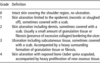
In his veterinary thesis, Lund (Reference Lund2003) argued for the use of a classification system for decubital shoulder ulcers in sows, and attempted to establish, and in part validate, such a classification system based on macro- and microscopic characteristics of sections of decubital shoulder ulcers in slaughtered sows. Access to a quick and reliable system would (i) make it possible to perform uniform recording of the extent of the problem and enable comparison of various studies and the development of the ulcers; and (ii) be the necessary basis for forensic evaluation of possible cases of mistreatment of animals. On the basis of the already established classification systems for humans, Lund (Reference Lund2003) found that the amount of granulation tissue, the extent of necrosis and the extent of infection in porcine decubital shoulder ulcers are not directly comparable with human pressure ulcers. Thus, the classification systems used in human medicine could not be adjusted to be used to classify decubital shoulder ulcers in sows. Although reddening of the skin is considered an early sign of a porcine decubital shoulder ulcer (Jensen, Reference Jensen2002; Nielsen and Vestergaard, Reference Nielsen and Vestergaard2003; Kaiser et al., Reference Kaiser, Bach-Mose and Alban2007), Lund (Reference Lund2003) decided only to include ulcerations – defined pathologically as a condition in which the epidermis and the underlying basal membrane is damaged (McGavin and Zachary, Reference McGavin and Zachary2007) – in his classification system. Therefore, lesions, which in human classification systems would have been defined as grade 1 (reddened but intact skin) and thus potentially be bottom-to-top pressure ulcers without damaged skin, were excluded. The post-mortem classification system suggested by Lund (Reference Lund2003), confirmed by Jensen (Reference Jensen2009) and described in Table 1 is presently the only available guideline with regard to the classification of decubital shoulder ulcers in slaughtered sows and is used by the Danish Veterinary and Food Administration (Anonymous, 2003 and 2007).
GrossFootnote a pathological characteristics of the scale for post-mortem classification of decubital shoulder ulcers in sows suggested by Lund (Reference Lund2003) and Jensen (Reference Jensen2009)

a See Jensen (Reference Jensen2009) for detailed description of histological characteristics.
Ante-mortem classification
It has been questioned whether a system designed for post-mortem classification is also applicable for live animals. Lund (Reference Lund2003) mentioned that the determination of the tissue types involved can be difficult based on visual inspection. Furthermore, based on macro- and microscopy of sections of 38 shoulders from slaughtered sows, Lund (Reference Lund2003) suggested that the carcass processing during slaughter may result in injuries making it difficult to identify the tissue structures involved, and that the existence of new bone formation could be overseen, whereby grade 4 ulcers incorrectly could be classified as grade 3. In a recent report from Danish Pig Production based on 809 shoulders from sows slaughtered at two Danish sow slaughterhouses in 2007 (in which only 1% was classified as grade 3 or 4), Vestergaard et al. (Reference Vestergaard, Kaiser, Petersen, Bækbo, Alban, Toft, Madsen and Friis2007) found a lack of accordance between the examination carried out ante mortem at the slaughterhouse and macroscopic carcass examination.
In the search for clinical signs of the presence of decubital shoulder ulcers corresponding to a grade 3 or 4 lesions, Strathe (Reference Strathe2007) carried out statistical analysis of data from clinical ante mortem as well as post-mortem examinations performed at a slaughterhouse. The data were collected at a Danish sow slaughterhouse during a period of 2 weeks in 2007 and included more than 700 shoulders of which only eight were classified as grade 3 or 4 lesions (based on post-mortem examination). The results showed that low-grade decubital shoulder ulcers may be overlooked at ante-mortem examination; that the seven sows with ulcers classified post mortem as grade 3 or 4 lesions all had at least one clinical symptom ante mortem; and that in order to be a valid indicator of severe shoulder lesions, the condition of the skin (e.g. the presence of crusts/scars) has to be combined with palpable findings such as (i) the adherence of the lesion to the underlying bone tissue; (ii) permanent swellings of more than 50 mm in diameter; or (iii) crusts and haemorrhage in the skin covering large permanent lesions. Jensen and Svendsen (Reference Jensen and Svendsen2006) mentioned that undermined shoulder lesions, which may not be visible, may break up, for example, during transport to the slaughterhouse, even though the lesions appear healed before transport.
As reviewed above, the concern about the lack of suitability of the post-mortem classification system for live sows appears to be well-founded. Lund’s (Reference Lund2003) classification system was designed for the examination of carcases and documentation of forensic material. Therefore, this classification system will not be optimal for the assessment of, for example, the prevalence of shoulder lesions in sows kept in herds, which is supported by the results of Strathe (Reference Strathe2007) and Vestergaard et al. (Reference Vestergaard, Kaiser, Petersen, Bækbo, Alban, Toft, Madsen and Friis2007). On live animals the examination should be based on visual examination as well as palpation rather than description of detailed pathological changes.
At present, a classification scale for ante-mortem determination of decubital shoulder ulcers in sows is under development in Denmark using the post-mortem scale suggested by Lund (Reference Lund2003) as a gold standard. Included herein – as one of several selected clinical measures – is the registration of the diameter of decubital shoulder ulcers. This measure does not take into consideration the types of tissue involved but focuses solely on the horizontal size of the lesion. However, the diameter has been used to score decubital shoulder ulcers in sows (Davies et al., Reference Davies, Morrow, Rountree and Miller1997; Zurbrigg, Reference Zurbrigg2006). In a small study including only lesions of grades 1 and 2, Kaiser et al. (Reference Kaiser, Bach-Mose and Alban2007) measured the diameter based on wound edges and showed that lesions of grade 2 were considerably larger than grade 1. Except for this study, there is no information on possible correlations between size measurements and other tools for classification of decubital shoulder ulcers in sows.
Dating of porcine decubital shoulder ulcers
In relation to animal protection it is of vital importance that the dating of an individual shoulder lesion can be done correctly. However, scientific documentation of the temporal progression of decubital shoulder ulcers in sows (e.g. the duration of each stage of development) is not yet available.
Reddening of the skin above the scapular spine was not included in Lund’s (Reference Lund2003) classification system, but in live sows it is often used as an early indicator of decubital shoulder ulcers (Nielsen and Vestergaard, Reference Nielsen and Vestergaard2003; Fruergaard, Reference Fruergaard2005). However, the duration of this condition has not been described.
In human forensic examinations, the appearance of granulation tissue and its transition into scar tissue are used to date lesions. Lund (Reference Lund2003) performed a histological comparison of decubital shoulder ulcers of the four stages (grades 1 to 4 in Table 1), and found that granulation tissue can be present in ulcers of grades 1 to 4, and that the thickness of the tissue may vary from 0 to 6 mm (grade 1) to 10 to 22 mm (grade 4). Later, this was confirmed by Jensen (Reference Jensen2009). The development of granulation tissue differs between animal species and has not been examined in sows. As a consequence, Lund (Reference Lund2003) used the knowledge from humans and other animal species to suggest that the lesions with considerable formation of granulation tissue must have been in progression for several days before the time of slaughter. In sows with decubital shoulder ulcers of grade 4, excessive bone formation and mechanical wear of bone tissue can be seen, indicating a long-term condition (Anonymous, 2003).
Healing of porcine decubital shoulder ulcers
Ulcer healing
The maintenance of skin integrity is important to prevent dehydration, bleeding and infections, and well-developed mechanisms for the re-establishment of skin integrity, in case lesions exist. Regeneration of skin is possible; however, skin damage may result in loss of tissue to such an extent that the process of regeneration implies embedding of connective tissue and formation of scars. In general, the healing of ulcers can be divided into three stages: (i) inflammation; (ii) proliferation including the formation of granulation tissue; and (iii) maturation/differentiation including regeneration of capillaries and increased strength of scar tissue (Gottrup, Reference Gottrup2002). Furthermore, healing can be described as primary, implying well-appositioned wound edges, sufficient blood supply and insignificant infection or necrotic debris (e.g. after surgery); or secondary, that is initiated at the wound bed and involving a number of factors that may delay or prevent primary healing (e.g. lack of supply of blood or oxygen or presence of bacteria). This type of healing is slow (weeks for ordinary human lesions) and often with the formation of scar tissue (Gottrup, Reference Gottrup2002).
Decubital shoulder ulcers
We have not been able to find longitudinal studies examining the temporal development of the healing of decubital shoulder ulcers in sows. Jensen and Svendsen (Reference Jensen and Svendsen2006) described that healing of decubital shoulder ulcers can be compromised by infection, and consequently, the slower secondary healing will take place. According to Jensen (Reference Jensen2008), the prominent type of healing for porcine decubital shoulder ulcers is secondary healing. Furthermore, the author suggested that apparently healed wounds may hide abscesses from the process of healing and, consequently, fistulas may later appear on the surface of the skin. Vestergaard et al. (Reference Vestergaard, Fruergaard, Nielsen and Madsen2005) mentioned that healing of decubital shoulder ulcers normally takes place within a few weeks, provided that the sows are weaned from their piglets and kept in hospital pens with sufficient bedding. This is in accordance with the assessment of the Danish Veterinary and Food Administration (Anonymous, 2003) and has also been suggested by Davies et al. (Reference Davies, Morrow, Miller and Deen1996) and Havn et al. (Reference Havn, Poulsen, Enøe and Nielsen2004). The latter examined decubital shoulder ulcers in 429 sows from a Danish herd and described that the ulcers apparently healed within a few weeks after weaning (Havn et al., Reference Havn, Poulsen, Enøe and Nielsen2004). However, data supporting this are not shown in the study. Thus, even though no scientific data are available, it is possible that most decubital shoulder ulcers present in production systems can heal by themselves – with or without therapeutic intervention – within a few weeks after weaning, thereby allowing a possible recovery. Importantly, the documentation of the welfare of these animals is insufficient, and therefore, it is currently not possible to draw conclusions as to whether such a period of recovery is acceptable in terms of animal welfare, and which requirements as regards housing etc. should be made during such a period.
As mentioned, there is no scientific documentation available of the timing of the healing process in decubital shoulder ulcers in sows or characterization of the tissue in the area around the healed ulcer. Thus, it is uncertain whether decubital shoulder ulcers may heal completely after correct therapeutic treatment, and how scar tissue can be characterized, for example, with regard to strength. However, it is evident that a history of previous decubital shoulder ulcers increases the risk of developing new pressure ulcers in subsequent lactations (Christensen et al. Reference Christensen, Wachmann and Enøe2002; Kaiser et al. Reference Kaiser, Mose and Alban2006; Thorup Reference Thorup2006). Therefore, it is recommended to pig producers that sows leaving the farrowing pen due to decubital shoulder ulcers are culled as they have a predisposition to develop more ulcers (Nielsen and Vestergaard, Reference Nielsen and Vestergaard2003).
Welfare consequences of decubital shoulder ulcers
The presence of shoulder ulcers is a welfare problem, indicating – like other skin injuries – that the production conditions prevent the animals from keeping their integrity intact (Broom, Reference Broom1988). More than two decades ago, the presence of skin lesions was described as a usable tool for measuring the welfare of animals in production systems (de Koning, Reference De Koning1985).
Pain and decubital shoulder ulcers in sows
We have not been able to find any scientific data which could be used to assess the level of pain in relation to the development, presence or healing of decubital shoulder ulcers in sows. In general, the established knowledge with regard to the expression of pain in pigs and the level of pain associated with frequently occurring diseases or pathological conditions in pig production is very limited. The existing knowledge is based primarily on studies of post-surgical pain, describing how pigs respond differently to acute and prolonged pain, in which the former typically triggers vocalization and escape responses. In contrast, signs of prolonged pain are described as more discrete, for example, inactivity, abnormal behaviour, postural changes or reluctance to change posture (Harvey-Clark et al., Reference Harvey-Clark, Gilespie and Riggs2000). The Danish Veterinary and Food Administration (Anonymous, 2007) has stated, however without any data, that sows with shoulder ulcers may show difficulties in getting up and that they express pain when touched. In a recent study of pain sensitivity in the shoulder region of healthy gilts, it was shown that short-term thermal nociceptive stimulation induced rubbing of the shoulder region against fixtures in the barn (Herskin et al., Reference Herskin, Ladewig and Arendt-Nielsen2009), suggesting that this behaviour might be included in future studies of pain sensitivity in sows with decubital shoulder ulcers.
In humans, pressure ulcers have been described as painful, especially lesions corresponding to grade 1 on the post-mortem porcine classification system (Bermark et al., Reference Bermark, Zimmerdal and Müller2003). According to Bliss (Reference Bliss1992), the pain related to human pressure ulcers is particularly pronounced in cases with bottom-to-top development. However, patients suffering from pressure ulcers are often non-verbal or with a reduced ability to communicate, and even in humans, the knowledge about pain related to pressure ulcers is rather limited.
The presence of infection will, in general, increase pain related to any lesion (e.g. review by Dray, Reference Dray1995; Gregory, Reference Gregory1998; Julius and Basbaum, Reference Julius and Basbaum2001) leading to spontaneous pain and hyperalgesia in the lesion as well as in the surrounding tissue. This is caused by increased activation and sensitivity of nociceptors in skin and muscles as well as increased sensitivity in afferent nerve cells induced by inflammatory mediators. Furthermore, the recurrence of cutaneous or muscular pain will gradually increase the pain experienced due to processes in nerve cells in the dorsal horn of the spinal cord (Arendt-Nielsen, Reference Arendt-Nielsen2003). Thus, over a period of time, the presence of tissue damage and infection may imply that the pain threshold will decrease or even that previously painless stimulation, for example, touching or exposure to changes in temperature, is now perceived as painful (allodynia). However, at present no knowledge exists with regard to these neural and inflammatory mechanisms in porcine decubital shoulder ulcers.
On the basis of the tissue structures involved in porcine decubital shoulder ulcers, the Danish Veterinary and Food Administration has stated that ulcer development implies considerable pain and suffering (Anonymous, 2003 and 2007). On the basis of macroscopic and histological studies of decubital shoulder ulcers as well as the literature, Lund (Reference Lund2003) suggested that a serious ischaemic condition including necrosis and possibly also infection and involvement of underlying structures including the periodeum will cause considerable pain, which can be expected to be prolonged. If so, the presence of decubital shoulder ulcers in sows without pain-relieving therapy is a significant welfare problem.
In a recommendation to pig producers, Vestergaard et al. (Reference Vestergaard, Fruergaard, Nielsen and Madsen2005) mentioned the possibility of providing pain relief to sows with decubital shoulder ulcers, and suggested that analgesic treatment may enhance healing and improve the welfare of the animals. However, no data with regard to the effects of pain relief given to sows with decubital shoulder ulcers are available.
It should be noted that often pain is not only related to existing tissue damage, as the pain system is plastic, implying a risk of the pain becoming chronic (experienced after the lesion has healed), leading to irreversible damage followed by permanent pain and hyperalgesia and/or allodynia (Jensen et al., Reference Jensen, Dahl, Arendt-Nielsen and Bach2003). One example is that damage to peripheral afferent nerves may lead to nerve sprouting and a thickening of nerve ends (neuromas). Generally, these sprouts are extremely sensitive, show spontaneous activity, as well as increased sensitivity to touch, pressure and temperature (Gottrup, Reference Gottrup2002; Jensen and Sindrup, Reference Jensen and Sindrup2003). Abnormal activity in neuromas will affect the central nervous system, in which sensitivity will also increase. Jensen and Sindrup (Reference Jensen and Sindrup2003) described the process as domino-like and with considerable consequences with respect to, for example, increased sensitivity and recruitment of otherwise inactive nerve cells. In human medicine, the consequences of peripheral nerve damage are described as the presence of permanent and/or attacks of pain (Gottrup, Reference Gottrup2002) characterized as burning sensations, increasing on repeated stimulation (summation) and continuing after stimulation (Jensen and Sindrup, Reference Jensen and Sindrup2003). During healing, sprouting of nerves may also take place in pressure ulcers. Until now, however, potential neuromas in healed shoulder ulcers of sows have not received scientific attention, but it is possible that sows with partly/fully healed injuries may suffer from long-term pain as a result of changes in the nervous system.
Sequelae
Several reports have mentioned an increased risk of sequelae (pathological condition resulting from a prior disease) after decubital shoulder ulcers; however, often there is no evidence of this. Deep decubital shoulder ulcers are described as a possible port of entry for bacteria to the circulation, which may lead to septicaemia and abscesses (Nielsen and Vestergaard, Reference Nielsen and Vestergaard2003; Vestergaard et al., Reference Vestergaard, Fruergaard, Nielsen and Madsen2005). Hassing and Nielsen (Reference Hassing and Nielsen2000) mentioned that decubital shoulder ulcers are often infected because of the high risk of contamination from the environment. Furthermore, according to Cleveland-Nielsen et al. (Reference Cleveland-Nielsen, Christensen and Ersbøll2004) correlations between decubital shoulder ulcers and remarks from the slaughterhouses with regard to infected skin lesions and abscesses have been found. However, based on macroscopic examination of 139 shoulder lesions, Davies et al. (Reference Davies, Morrow, Miller and Deen1996) stated that obvious signs of infection such as pus or a foul smell seldom occurred. Contrarily, after culture of samples from shoulder ulcers and adjacent lymph nodes in 36 slaughtered sows, Lund (Reference Lund2003) found that 85% of the shoulder lesions were infected and approximately half of the lymph nodes as well, and that Arcanobacterium pyogenes was the primary cause of infection. On the basis of these findings, the Danish Veterinary and Food Administration (Anonymous, 2003) emphasized the risk of infection.
Taken together, the existence of decubital shoulder ulcers in sows is a welfare problem, the extent of which cannot be fully determined until a valid ante-mortem classification system is available and knowledge about the duration of the condition (including the various stages), as well as the possible consequences in terms of pain or discomfort have been established.
Conclusions
This paper reviews the existing knowledge about decubital shoulder ulcers in sows, focusing on the pathogenesis, classification and consequences in terms of pain and animal welfare. Decubital shoulder ulcers are lesions on the shoulders of sows, reported to have a relatively high prevalence, and to some extent comparable with human pressure ulcers. In sows, the ulcers are caused by pressure inflicted by the flooring leading to oxygen deficiency in the skin and the underlying tissue.
The temporal development of decubital shoulder ulcers in sows is not clear. The rather few available descriptions suggest that the ulcers initiate as reddening of the skin in the area above the tuber of the scapular spine in the days around farrowing, that they develop into open ulcers over the next days or weeks and remain clinically recognizable for weeks, until healing after the sow is weaned. It is, therefore, presumably a rather prolonged condition.
There has been some debate about the pathogenesis of the ulcers and their tendency of progression, including whether the damage starts in the outer layer of skin or in the underlying tissue. However, the present evidence suggests that the ulcerations start on the surface as a result of obstructed blood flow and gradually progress inwards to the tuber spina scapulae, but there is a need for more systematic knowledge within this field, including knowledge of ulcer progression, the releasing factors and involved mechanisms.
In relation to, for example, animal protection, decisions on adequate and timely therapy, assessment of preventive initiatives and authority checks, it is important to have access to usable and reliable systems for the classification of decubital shoulder ulcers. Currently, only one classification system, primarily designed for the classification of carcasses, is available. However, this system is not optimal for the classification of decubital shoulder ulcers in live sows.
The knowledge of the welfare consequences of decubital shoulder ulcers is limited. In general, the presence of skin lesions such as ulcers is a welfare problem reflecting that the production conditions prohibit the normal ability of the animals to adapt. Therefore, steps should be taken to prevent the development of this condition. The extent of the welfare problem related to decubital shoulder ulcers in sows, however, cannot be determined until evidence of the prevalence of shoulder ulcers in herds and the duration of the condition (including the various stages) has been established, knowledge of which requires access to a classification system suited for use on live animals.
On the basis of the knowledge of the tissue structures involved, we assume that the development and presence of decubital shoulder ulcers in sows are painful. However, scientific evidence of this has not yet been established. If so, the welfare of the affected sows is significantly reduced and we believe that there is a need for scientific effort to clarify the possible consequences of decubital shoulder ulcers in terms of pain and to identify proper therapy.
Acknowledgements
The authors thank Ellen Fritze for technical support, Professor H. E. Jensen for helpful advice and senior scientist Birte L. Nielsen for suggestions to an earlier version of this manuscript.