Sudden cardiac death in athletes is a tragic event; 12-lead electrocardiography is an established tool for the evaluation of athletes, providing important diagnostic and prognostic information on a variety of cardiovascular diseases that are associated with an increased risk of sudden cardiac death during sports.Reference Corrad, Pelliccia and Heidbuchel 1 , Reference Uberoi, Stein and Perez 2
Pathological T-wave inversion is rarely observed in electrocardiograms of healthy athletes, but is common in patients with certain cardiac diseases. T-wave inversion is present in several sudden cardiac death-related disorders in athletes, including hypertrophic cardiomyopathy, dilated cardiomyopathy, arrhythmogenic right ventricular cardiomyopathy, and myocarditis.Reference Corrad, Pelliccia and Heidbuchel 1 – Reference Pelliccia, Di Paolo and Quattrini 4 Although benign T-wave inversion is sometimes observed in asymptomatic African/Afro-Caribbean athletes,Reference Papadakis, Carre and Kervio 5 no such accounts exist in healthy Japanese athletes.Reference Kervio, Pelliccia and Nagashima 6
We report the case of a Japanese adolescent soccer player found to have T-wave inversion in precordial leads in resting electrocardiography. A transient recovery during a short detraining period helped in diagnosing athlete’s heart.
Case report
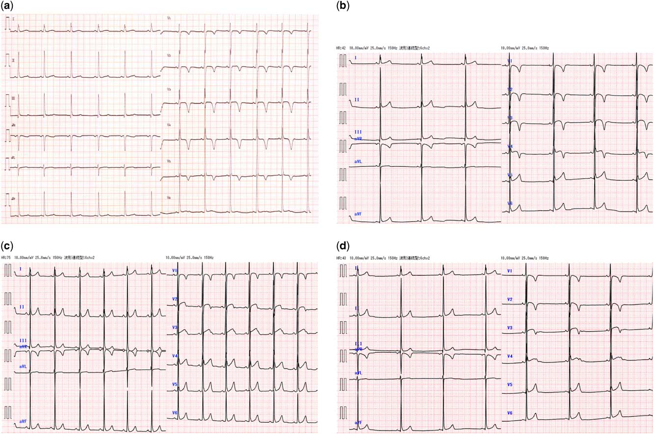
A 17-year-old male soccer player who had been training regularly for several hours a day since the age of 6 years was referred to our outpatient clinic because of non-specific thoracic pain while jogging before practice. T-wave inversion in leads V1–V5 had been noted in an earlier junior high school-based electrocardiographic screening (Fig 1a), although further examinations were normal. He did not notice any chest pain during soccer games, nor did he have a family history of arrhythmia, cardiomyopathy, heart failure, or sudden cardiac death.
( a ) Electrocardiogram at an earlier junior high school-based electrocardiographic screening examination. ( b ) Electrocardiogram at outpatient clinic presentation. ( c ) Electrocardiogram after 2 weeks of detraining. ( d ) Electrocardiogram after 2 weeks of resuming exercise training.

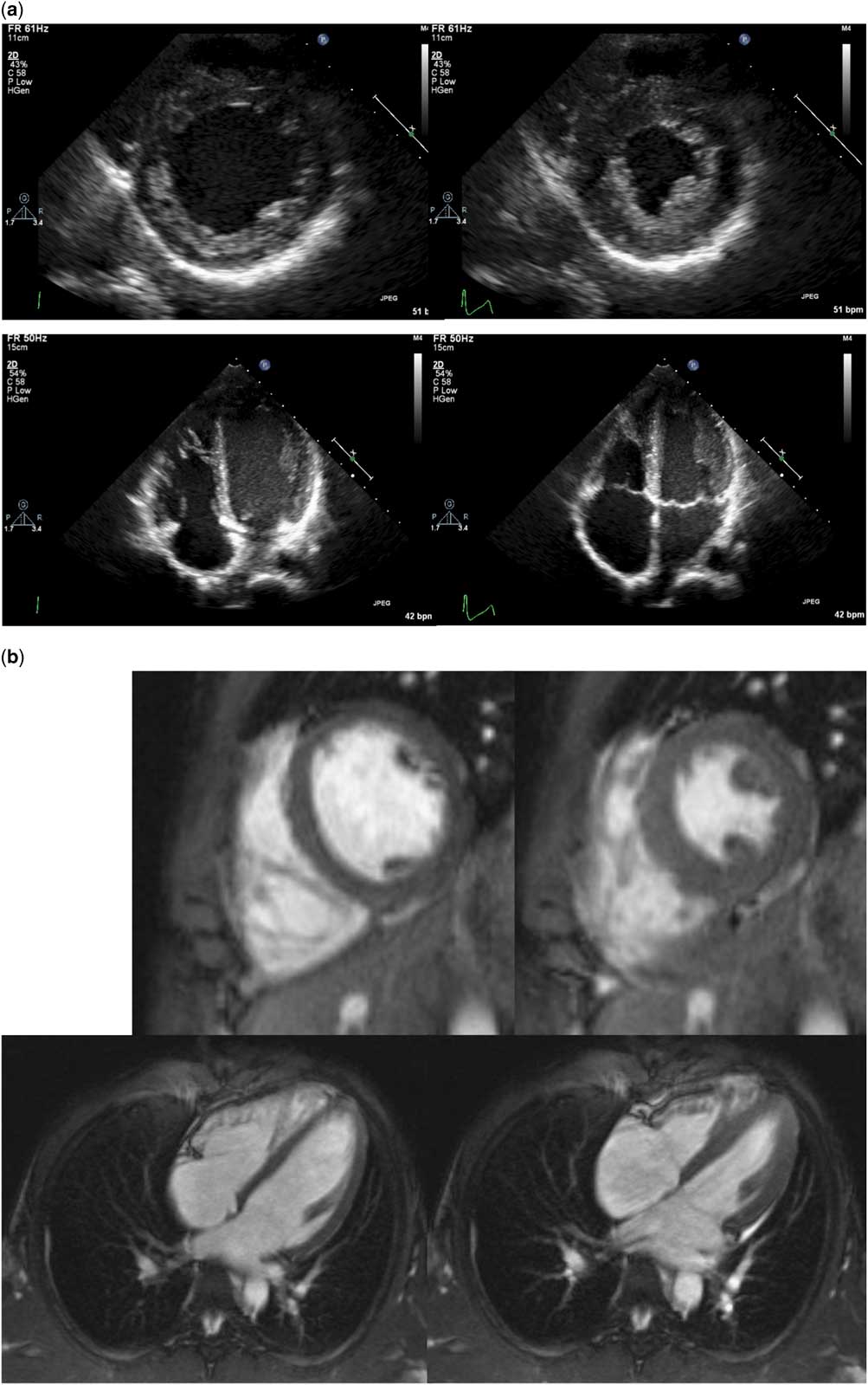
On presentation, the patient was asymptomatic, with normal cardiovascular physical findings and blood pressure. His electrocardiogram revealed sinus bradycardia of 42 beats per minute, isolated QRS voltage criteria for left ventricular hypertrophy, early re-polarisation, and giant negative T-waves in precordial leads (Fig 1b), all of which were strongly suggestive of cardiomyopathy. Laboratory data, including blood cell counts, electrolytes, C-reactive protein, creatine kinase MB isoenzyme, troponin T, and brain natriuretic peptide, showed no abnormalities. Echocardiography revealed normal left ventricular wall thickness with normal systolic and diastolic functions as follows: left ventricular end-diastolic diameter 45.3 mm, left ventricular posterior wall diameter 6.6 mm, left ventricular end-diastolic volume 137 ml, ejection fraction 76%, ratio of early to late diastolic inflow velocity 2.0, and tissue Doppler annular early diastolic velocity 15.3 cm/second (Fig 2a). Right ventricle volume and systolic function were normal. Right ventricular outflow tract dimension was normal value (diameter of short axis: 24.5 mm and long axis: 23.6 mm). Exercise stress test and Holter electrocardiogram disclosed neither significant ST segment depression nor any signs of ventricular arrhythmia. Late potential by signal average electrocardiography was negative. There were no coronary abnormalities as detected by coronary CT. Gadolinium-enhanced cardiac MRI revealed slightly dilated ventricular chambers (left ventricular end-diastolic volume: 134.6 ml [115% of normal value], right ventricular end-diastolic volume: 135.0 ml [112% of normal value]) without late gadolinium enhancement, suggesting myocardial fibrosis (Fig 2b).
Echocardiography ( a ) and cardiac MRI ( b ) findings at discovery of T-wave inversion by electrocardiography. ( a ) Short-axis view in the diastolic phase (upper left) and systolic phase (upper right) and apical four-chamber view in the diastolic phase (lower left) and systolic phase (lower right). ( b ) Short-axis view in the diastolic phase (upper left) and systolic phase (upper right) and apical horizontal long-axis view in the diastolic phase (lower left) and systolic phase (lower right).

Soon after presentation, the patient temporarily stopped athletic training owing to leg pain. After only 2 weeks of detraining, his resting heart rate increased and T-wave inversion in leads V2–V4 disappeared (Fig 1c). Stress test electrocardiogram at this time did not show ST segment change or arrhythmia. The praecordial T-wave inversion re-appeared 2 weeks after resuming his training (Fig 1d); thus, his electrocardiographic abnormality was considered to be athlete’s heart because of T-wave inversion normalisation and re-surfacing over a short detraining period.
Discussion
Post-pubertal persistence of T-wave inversion beyond V1 is present in 2–4% of athletes, whereas T-wave inversion in two leads is rarely observed in electrocardiograms of healthy athletes.Reference Uberoi, Stein and Perez 2 , Reference Schnell, Riding and O’Hanlon 3 T-wave inversion should be considered pathological in all cases as it is associated with cardiac pathology in 45% of athletes. Despite echocardiography identifying disorders in half of these cases, cardiac MRI is strongly advised in young athletes with T-wave inversion and normal echocardiography;Reference Schnell, Riding and O’Hanlon 3 those with T-wave inversion or other uncommon electrocardiographic findings have a higher incidence of subclinical cardiac disease than matched, control athletes with normal electrocardiographic findings and require long-term monitoring of clinical outcome.Reference Pelliccia, Di Paolo and Quattrini 4
T-wave inversion in leads V1–V4 appears to represent an ethnic variant of athlete’s heart. African/Caribbean athletes exhibited a greater prevalence of T-wave inversion confined to anterior leads compared with Caucasian athletes.Reference Papadakis, Carre and Kervio 5 Another study of soccer players revealed that no Japanese athletes possessed T-wave inversion.Reference Kervio, Pelliccia and Nagashima 6 As T-wave inversion in anterior leads is very uncommon in Japanese athletes, it is essential to clinically differentiate normal physiological re-modelling from heart disease, especially arrhythmogenic right ventricular cardiomyopathy. Electrocardiographic T-wave inversion and balanced symmetrical biventricular dilation likely represent benign manifestations of intensive training in the majority of asymptomatic athletes without a relevant family history. Other markers of physiological re-modelling include early re-polarisation and voltage criteria for right or left ventricular hypertrophy.Reference Zaidi, Sheikh and Jongman 7
Endurance exercise triggers structural re-modelling and acute dysfunction of the right ventricle, but not the left ventricle, as the right ventricle is placed under greater haemodynamic stress.Reference La Gerche, Burns and Mooney 8 , Reference Arbab-Zadeh, Perhonen and Howden 9 Brosnan et alReference Brosnan, Classen, Heibduchel, Prior and La Gerche 10 reported that praecordial T-wave inversion in endurance athletes was associated with displacement of the right ventricle towards the left axilla rather than right ventricular dilatation or hypertrophy. In the present case, however, no data were available on the volumetric change or spatial relationship of the left ventricule and right ventricule at the time of disappearance of T-wave inversion by echocardiography or MRI.
There is a case report in which giant negative T-waves in the praecordial leads of a young soccer player became normalised after a long detraining period.Reference Vessella, Cardillo, Bianco, Palmieri and Zeppilli 11 The present case also contained only minimal cardiac abnormalities of slight dilatation of both ventricles. Electrocardiographic findings indicated cardiac structural re-modelling or acute haemodynamic stress triggered by intensive endurance training for soccer, as T-wave inversion improved during a short abstinence period of 2 weeks and re-appeared soon after re-starting exercise. We therefore considered that the patient’s T-wave inversion in the anterior leads reflected normal structural re-modelling of the right ventricle.
Recent studies, however, have focussed on the overlap between structural re-modelling of the right ventricle and arrhythmogenic right ventricular cardiomyopathy.Reference Zaidi, Sheikh and Jongman 7 Low amplitudes in praecordial leads, unbalanced right ventricular dilation, right ventricular wall motion abnormality, a burden of ventricular extrasystoles, abnormal response of blood pressure to exercise testing, and abnormal findings of signal-averaged electrocardiogram indicate pathological re-modelling.Reference Zaidi, Sheikh and Jongman 7 These studies suggest the importance of meticulous, life-long follow-up even in apparently asymptomatic athletes with T-wave inversion in anterior leads. Accordingly, careful surveillance of this patient will be continued indefinitely.
In conclusion, T-wave inversion normalisation after a short period of detraining may be considered a diagnostic significance for athlete’s heart, although it cannot completely rule out the presence of underlying disease such as arrhythmogenic right ventricular cardiomyopathy.
Acknowledgements
The authors sincerely thank the patient and his parents for providing all of clinical and laboratory information and samples.
Financial Support
This research received no specific grant from any funding agency or from commercial or not-for-profit sectors.
Conflicts of Interest
None.