Introduction
The autonomic nervous system (ANS) is a component of the peripheral nervous system whose principle function is to maintain homeostasis. Langley first conceived the term in the early 20th century, describing the absence of sensory nerve cell bodies in autonomic ganglia.Reference Langley1 It has motor and sensory pathways that convey and relay neurons to every organ in the body. Autonomic dysfunction produces a complex array of clinical problems including cardiovascular dysregulation resulting in inappropriate blood pressure and heart rate responses to physiological stressors, erectile dysfunction, and thermoregulatory and sweating problems producing an inability to adapt to environmental thermal extremes.Reference Myllyla, Tulppo, Bolis, Licinio and Govoni2–Reference Collins4
Search methods
Search methods involved Medline using the key words: autonomic dysfunction, autonomic failure, geriatrics and older people. Subsection searches used the words autonomic dysfunction, diabetes mellitus, neuronal cellular changes, alpha synuclein, toll-like receptors, paraneoplastic syndrome, autonomic function tests, orthostatic hypotension, neurocardiogenic syncope, carotid sinus hypersensitivity, erectile dysfunction, gastrointestinal dysfunction and thermoregulation.
Anatomy and physiology
The ANS consists of sensory neurons arising from both the primary viscera and the cranial sensory ganglia from nerves VII, IX and X, which then project to second-order neurons in the nucleus solitarius of the medulla oblongata. These monitor and regulate carbon dioxide arterial tension, blood glucose and pH.Reference Widicombe and Sterling5–Reference Kopp and DiBona7 They have afferents to the chemoreceptor trigger zone stimulating vomiting from noxious substances. Motor neurons comprise the sympathetic, parasympathetic and enteric nervous systems. Sympathetic ganglia lie within the prevertebral and para-aortic chains close to the spinal cord,Reference Bulygin8 and neuronal stimulation produces a ‘fight or flight’ response, diverting blood flow away from the gastrointestinal tract and skin towards skeletal muscle and lung.Reference Bulygin8 This increases oxygen exchange, cardiac chronotropy and inotropy and subsequently cardiac output, and results in pupillary dilatation.Reference Woodcock, Du, Reichelt and Graham9 The parasympathetic nervous system dilates the splanchnic circulation, aiding digestion.Reference Kvietys and Granger10 It causes bronchoconstriction in the lungs when oxygen demands falls, produces pupillary constriction in the eye during accommodation, stimulates salivary gland secretion in the mouth, and controls erectile function in the genitalia.Reference Lewis, Short and Lewis11–Reference Rampin13
Autonomic failure and ageing
Several studies have demonstrated deteriorating autonomic function with increasing age. Collins et al. examined three physiological tests of autonomic function in 29 young adults and 64 healthy older patients (age range 66–86 years).Reference Collins, Exton-Smith, James and Oliver14 These included heart rate response to standing, changes in hand blood flow in response to differing thermal conditions, and the cardiovascular effects of lower body negative pressure. Heart rate response to standing increased by 20 beats/min in younger patients compared with only 10–15 beats/min in the older group. Interestingly, younger patients with diabetic neuropathy showed a similar response to the older age group.Reference Oliver15 The effects of changes in body temperature including shivering and sweating on hand blood flow were measured using volume plethysmography. One-third of older volunteers had higher than expected blood flow in colder temperatures, showing a diminished ability to vasoconstrict. Patients with abnormal blood flow responses had higher rates of autonomic dysfunction elsewhere. Forty-three per cent of those with abnormal vasomotor responses to cooling and 10% of those with normal responses were found to have a postural drop in systolic blood pressure of greater than 20 mmHg. Lower body negative pressure produces an increase in heart rate, fall in blood pressure, falls in central and peripheral blood flow and a decrease in cardiac output. Ten young adults and ten healthy older patients underwent lower body negative pressure (–10 to –50 mmHg) testing. Blood pressure fell earlier in the older group, but heart rate increased significantly in the younger group. Younger subjects showed a much greater reduction in peripheral blood flow as measured by a photo electric pulsimeter measured at the thumb, suggesting a greater ability to redirect blood flow in response to negative pressure. These tests demonstrate reduced cardiovascular responses to physiological stressors in older people and are partially replicated in younger patients with diabetic peripheral neuropathy, suggesting similar mechanisms of loss of autonomic control in both groups.
Autonomic failure and diabetes mellitus
The prevalence of autonomic related conditions such as type 2 diabetes mellitus and neurogenic orthostatic hypotension increases with age. It has been estimated that 18–20% of people over 65 have type 2 diabetes in the USA, and 20–25% have impaired glucose tolerance, a condition associated with a twofold increase in macrovascular complications.16, Reference Abrass, Schwartz and Fitzgerald17 Distal sensorimotor neuropathy commonly presents as an autonomic neuropathy. Cardiac involvement produces an increased resting heart rate when the vagus nerve is affected. If sympathetic and parasympathetic fibres are involved, the heart rate becomes less able to increase in response to physiological demands.Reference Ewing, Campbell and Clarke18 Orthostatic hypotension occurs due to arteriolar efferent sympathetic denervation, producing reduced vasoconstriction in the major blood vessels on standing.Reference Low, Walsh, Huang and McLeod19 Autonomic dysfunction can cause sudden death in diabetes via a number of postulated mechanisms. These include absent or altered perception of myocardial ischaemia, inadequate haemodynamic responses to physiological stressors, cardiac arrhythmias due to QT interval lengthening, and alterations in regional myocardial function following changes in innervationsReference Langer, Freeman, Josse, Steiner and Armstrong20–Reference Stevens, Raffel, Allman, Dayanikli, Ficaro, Sandford, Wieland, Pfeifer and Schwaiger23 predisposing to ischaemia and arrhythmia.
Autonomic cellular changes and ageing
Autonomic ganglia do not change in number with ageing. However, ageing is associated with dendritic alterations, changes in synapse function and neuroaxonal dystrophy (NAD). NAD produces dramatic axonal swellings and neurofilamentous and tubulovesicular deposits as a result of impaired axonal regeneration. Normal synaptic turnover is exaggerated, producing excess swelling and sprout formation.
The relationship between ageing and peripheral nervous system ganglia is largely unknown, although several hypotheses have been proposed. These include oxidative stress in nerve terminals resulting in lipid peroxidation, alterations in membrane structure and function, and mitochondrial dysfunction. Neurotrophic substances become deficient, depleting end organ innervation. Exogenous neurotrophic growth factor was found to increase sympathetic innervation in older patients, suggesting that growth factor deficiency may be detrimental to neuronal function. Conversely, neurotrophic excess causes dendritic and neuronal swelling and axonal sprouts contributing to neuroaxonal dystrophy, which have been found in the senile plaques of Alzheimer's disease.Reference Gavazzi24, Reference Cotman and Gomez-Pinilla25 Excessive neurofilament glycosylation and phosphorylation occurs in ageing and diabetes, and results in their accumulation in axonal terminals.Reference Vlassara, Bucula and Striker26 These cellular processes diminish synaptic transmission, reduce end organ innervation, and the overall functionality of the autonomic nervous system.
Alpha synucleinopathies and autonomic dysfunction
Alpha synuclein (AS) is an inclusion protein found in patients with several neurodegenerative conditions including Lewy body dementia, Parkinson's disease and multiple system atrophy.Reference Arima, Uéda, Sunohara, Hirai, Izumiyama, Tonozuka-Uehara and Kawai27, Reference Trojanowski28 Alpha synuclein is deposited in neurons and glial cells in both the central nervous system and the spinal cord, and in key areas involved in the control of autonomic function. Basal ganglia AS deposition in the thalamus has been demonstrated in patients with Parkinson's diseaseReference Rüb, Del Tredici, Schultz, Ghebremedhin, de Vos, Jansen Steur and Braak29 and failure of AS protein degradation occurs in oligodendrocytes in multiple system atrophy.Reference Rüb, Del Tredici, Schultz, Ghebremedhin, de Vos, Jansen Steur and Braak29 In 102 cases of multiple system atrophy, Yoshida et al. found glial and neuronal cytoplasmic inclusions and positive alpha synuclein immunostaining in the pontine nuclei, putamen, substantia nigra, locus coeruleus, inferior olivary nucleus and the intermediolateral column of the thoracic spinal cord.Reference Yoshida30 The intermediolateral column is a key area of autonomic control which is damaged in multiple system atrophy.Reference Daniel, Matthias and Bannister31
Can early AS protein deposition predict later onset of neurodegenerative disease? Surgical specimens from 100 patients (age range 44–84 years) undergoing resection of an abdominopelvic organ were examined by anti-AS immunoassaying.Reference Minguez-Castellanos, Chamorro, Escamilla-Sevilla, Ortega-Moreno, Rebollo, Gomez-Rio, Concha and Munoz32 Nine per cent of patients had positive immunostaining for antibodies against AS. At 16 months, none of the subjects had clinical features of Parkinson's disease, dementia or autonomic failure. However, when asked to perform a Valsalva manoeuvre, the AS-positive group showed a longer blood pressure recovery time. Cardiac metaiodobenzylguanide uptake was reduced in the AS group and striatal muscle Reference Danzl and Pozos123I-ioflupane was low in one AS-positive subject. At 30 months, these lower cardiac and striatal uptake values reflect similar findings in patients with higher UKPDS III scores. This suggests that AS aggregates in the peripheral autonomic nervous system may pre-date the onset of a number of neurodegenerative disorders.
The autonomic nervous system, response to infection and toll-like receptors
Dendritic cells and macrophages process antigens and act as antigen-presenting cells in response to systemic infection. The innate immune response is activated, involving the interaction between microbe molecules and surface receptors on antigen-presenting cells. These molecules are collectively known as pathogen-associated molecular patterns (PAMP) and the set of receptors with which they interact are known as pattern recognition receptors (PRR). PRRs can be subdivided into signalling and endocytic receptors and secreted proteins. The main signalling receptor is the toll-like receptor (TLR) of which there are thought to be 10–15 different types in most mammalian species.Reference Rittirsch, Flierl and Ward33 In septic shock, damaged and dying tissues release intracellular proteins or mediators known as ‘alarmins’, which stimulate TLR as a crucial part of the initial stages of the inflammatory response. The TLR-4 receptor responds to the lipopolysaccharide component of the cell wall of gram-negative bacteria, forming a complex with CD14 and MD2.Reference Medzhitov and Janeway34 There is considerable interaction between TLR-4 and the complement system. The complement anaphylatoxin C5a, which is released early in sepsis, has been found to negatively regulate TLR4-mediated responses correlating with lower levels of complement activation products.Reference Hawlisch, Belkaid, Baelder, Hildeman, Gerard and Köhl35 Lymphoid tissue is richly innervated by the autonomic nervous system, and cytokines produced peripherally can stimulate the induction of brain cytokines in macrophages and microglial cells through central toll receptors. These brain-produced cytokines act on cortical tissue to produce the central factors associated with infection, namely fever (IL-6), neuroendocrine activation and sickness behaviour. Munford and Tracey have suggested that severe sepsis is a neuroendocrine disorder.Reference Munford and Tracey36
The parasympathetic nervous system plays a key neuromodulatory role in the systemic response to infection. Activation of α7 acetylcholine nicotinic receptors by vagal nerve or direct agonist stimulation decreases intracellular cytokine synthesis in macrophages, dampening down the inflammatory response to sepsis. Inflammation is thus under neuronal control by the autonomic nervous system, which can reflexively modulate the inflammatory response by inhibiting the production of pro-inflammatory cytokines.
Catecholamines are released early by the sympathetic nervous system in response to systemic sepsis. Leucocytes are also an abundant source, and themselves have catecholamine receptors suggesting autocrine and paracrine effects. α- and β-receptor stimulation results in alterations in lymphocyte trafficking, changes in vascular perfusion and cell proliferation and apoptosis. Pro-inflammatory cytokine release by macrophages and neutrophils is also tightly regulated by α-adrenergic receptors.
Toll-like receptors have been found in murine intestine and human ileum as well as in dorsal root ganglia with TLR-3, -7 and -4 the predominant receptor subtypes.Reference Barajon, Serrao, Arnaboldi, Opizzi, Ripamonti, Balsari and Rumio37 This suggests a local enteric neural network, which may activate in response to local bacterial and viral invasion through closely regulated cytokine production utilizing both the autonomic nervous system and toll-like receptor mechanisms.
Autonomic dysfunction has been demonstrated to contribute to multiple organ failure in septic shock. Loss of cholinergic inflammatory inhibition, reduced catecholamine sensitivity in providing cardiovascular stability and reduced macrophage and Europhile-mediated cytokine release could be potential explanations. Heart rate variability as a marker of autonomic dysfunction has been shown to be an independent predictor of 28- and 60-day mortality in critically ill patients with multiple organ dysfunction.Reference Schmidt, Hoyer, Hennen, Heinroth, Rauchhaus, Prondzinsky, Hottenrott, Buerke, Müller-Werdan and Werdan38
Autonomic receptor downregulation in response to inflammation
There is increasing evidence to support autonomic receptor downregulation in response to inflammation. Young healthy volunteers were injected with a bolus of lipopolysaccharide endotoxin, and muscle sympathetic nerve activity, heart rate and blood pressure were observed and compared with placebo. Muscle sympathetic nerve burst activity was suppressed, whilst heart rate increased and blood pressure was unchanged following endotoxin injection. This suggests a blunting of postsynaptic receptor and uncoupling of baroreceptor activity in humans in septic shock.Reference Sayk, Vietheer, Schaaf, Wellhoener, Weitz, Lehnert and Dodt39 Rat models in which colitis was pharmacologically induced resulted in diminished enteric smooth muscle activity related to β-adrenoreceptor downregulation and may well explain the diarrhoea associated with inflammatory bowel disease.Reference Zhao, Bossone, Piñeiro-Carrero and Shea-Donohue40
Autonomic dysfunction and paraneoplastic syndromes
Guichard and Vignon first used the term ‘paraneoplastic’ in 1949 in a patient with metastatic uterine cancer.Reference Guichard and Vignon41 The commonest paraneoplastic syndromes associated with autonomic dysfunction include small cell lung cancer,Reference Anderson, Rosenblum and Graus42 tumours of the breast and ovary, and lymphoma.Reference Ficker and Hammack43 Dysautonomias with a well-recognized paraneoplastic association include paraneoplastic autoimmune autonomic gangliopathy (AAG), paraneoplastic sensory neuropathy, paraneoplastic encephalomyeloneuropathy, and the Lambert-Eaton myaesthenic syndrome. Common clinical features include orthostatic hypotension, impaired gastrointestinal motility, lack of heart rate changes with respiration, bladder dysfunction, impotence, abnormal sweating, and dry eyes and mouth. Autonomic symptoms may arise prior to the diagnosis of malignancy, and during and after treatment, with orthostatic hypotension, constipation and pseudo-obstruction being the commonest presentations.Reference Oskarsson, Quan and Wiley44 The clinical course is subacute but usually rapidly progressive, with many patients immobile within a few weeks.Reference Lorusso, Hart, Ferrari, Ngonga, Gasparetto and Ricevuti45
Autoimmune antibodies produced by the tumour can attack parts of the autonomic nervous system with lymphocytic infiltrates a histological finding in anti-Hu encephalomyeloneuropathy and in autonomic postganglionic and myenteric neurons with antineuronal antibodies. Antibodies associated with paraneoplastic autonomic dysfunction include Hu, CV2, Yo and voltage-gated calcium channels.
Investigations include identification of the tumour and its anatomical location through imaging such as CT, MRI and PET scanning. Tissue diagnosis can be made through biopsy and cellular washings for cytology. CSF analysis may also detect malignant cells, and often shows an inflammatory pleocytosis and raised total protein. Blood tests include serum profiles for antineuronal autoantibodies.
Treatment of the underlying cancer occasionally improves autonomic dysfunction, but in the majority of cases there is little response. Therapeutic options include immunotherapy via intravenous immunoglobulin, steroids, plasma exchange and cyclophosphamide, although results have generally been disappointing. Early aggressive immunotherapy may benefit some patients with anti-Hu paraneoplastic sensory neuropathy.Reference Oh, Dropcho and Claussen46
Clinical investigation
Ewing and Clarke developed five tests to assess diabetic autonomic neuropathy, which can be performed with simple equipment over 20 min.Reference Ewing and Clarke47 These include a heart rate response to a Valsalva manoeuvre, heart rate variability during deep breathing, blood pressure response to sustained handgrip, immediate heart rate response to standing, and blood pressure response to standing.
During the Valsalva maneoeuvre, expiration against a closed glottis usually produces a fall in blood pressure and rise in heart rate. Following release, the blood pressure usually rises producing an increase above the resting value and the heart rate slows. In patients with autonomic failure, blood pressure falls slowly during expiration and returns to normal but with no overshoot in blood pressure or change in heart rate.
Deep breathing causes heart rate variability in normal subjects and this is dependent on an intact parasympathetic nerve supply. Patients with autonomic failure have a reduction in and sometimes a complete absence of heart rate variation. Subjects sit quietly taking 6 breaths per minute and the maximum and minimum R–R intervals during each breathing cycle are measured. One study suggested that this is the most reliable test in patients with diabetes compared with heart rate on standing, the Valsalva manoeuvre and metronomic breathing tests.Reference Eckberg48
Standing produces heart rate increases peaking at the 15th beat, followed by slowing which peaks at the 30th beat.Reference Ewing, Campbell, Murray, Neilson and Clarke49 This effect can be abolished in normal subjects by intravenous atropine but not propranalol, suggesting mediation via the vagus nerve.Reference Ewing50 Diabetic patients with autonomic neuropathy show only a gradual or no increase in heart rate after standing.Reference Risk, Bril, Broadbridge and Cohen51 Blood pressure falls on standing, and in patients with severe sympathetic neuropathy, standing blood pressure remains lower than that when sitting.
Sustained handgrip produces a rise in blood pressure related to a heart rate-dependent increase in cardiac output with no change in systemic vascular resistance.Reference Ewing, Irving, Kerr, Wildsmith and Clarke52 Blood pressure rises in autonomic failure are attenuated.
Vasoconstrictor tests include isometric exercise where a sustained handgrip is held for several minutes,Reference Ebert, Stowe, Barney, Kalbfleisch and Smith53 cold immersion of the hand in ice cold water and mental arithmetic, which stimulate sympathetic outflow producing a rise in blood pressure and heart rate. In autonomic failure, these cardiovascular responses to vasoconstrictor stimuli are blunted.Reference Callister, Suwarno and Seals54
Sympathetic cholinergic response
1. Thermoregulatory sweat test. Body temperature is raised by using heat blankets or hot water vessels, and the patient's skin is covered in quinizarine or Ponceau red, which turns purple on exposure to moisture. Autonomic failure resulting in reduced sweating at higher ambient temperatures attenuates this response.Reference Birklein, Spitzer and Riedl55
2. Sudomotor axon reflex test. The test has three parts and measures resting skin temperature, resting sweat output, and stimulated sweat output. Measurements are typically taken on arms, legs or both. A small plastic cup is placed on the skin and the temperature and amounts of sweat under the skin are measured. To stimulate sweat pilocarpine is delivered intradermally to a sweat gland, but the patient will only feel warmth. Changes in temperature and sweat output are recorded and analysis detects local reflex dysfunction.Reference Low, Caskey, Tuck, Fealey and Dyck56
3. Sympathetic skin response. Electrical activity on the hand or foot increases with increasing sweat gland activity and this is recorded in response to a variety of stimuli including sound, touch and nerve stimulation, e.g. median nerve. Cholinergic dysfunction (e.g. primary autonomic failure) produces no response, but those with adrenergic failure (e.g. multisystem atrophy) may see some response.
Clinical consequences of autonomic failure
Orthostatic hypotension
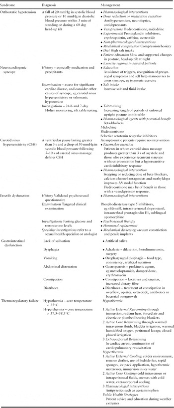
Orthostatic hypotension is defined as a fall of 20 mmHg in systolic blood pressure or 10 mmHg in diastolic blood pressure within 3 min of standing, or during a 60 deg head-up tilt.Reference Schatz, Bannister and Freeman57 It affects 30% of those >75 years, and up to 50% of the frail elderly living in nursing homes.Reference Harris, Lipsitz, Kleinman and Cornoni-Huntley58 500–1000 ml of blood normally pool in the lower limbs on standing, and this results in reduced cardiac filling sensed by the baroreceptors located in the aortic arch and carotid arteries.Reference Lipsitz59 This produces a reflex activation of the sympathetic nervous system, increasing heart rate through β-receptor stimulation and peripheral vasoconstriction to maintain blood pressure and cardiac output. As humans age, in addition to loss of α1-mediated vasoconstriction and decreased baroreceptor sensitivity, parasympathetic activity decreases resulting in a withdrawal of vagal tone.Reference James and Potter60 Reduced levels of renin, angiotension, and aldosterone, and an elevation of natriuretic peptide prevent salt and water conservation in the volume-depleted older patient. Increased vascular resistance results in impaired diastolic cardiac filling, stiffened ventricles, a reduction in stroke volume and a decrease in preload. Symptoms may include dizziness, light-headedness, blackouts, nausea, paracervical, lower back and chest pain. Disturbed speech, visual changes, falls, confusion and impaired cognition are often seen.Reference Low, Opfer-Gehhrking and McPhee61 Patients are at significant risk of falls, fractures, transient ischaemic attack, and myocardial infarction.Reference Ooi62–Reference Luukinen, Koski, Laippala and Airaksinen65 Recurrent orthostatic hypotension and hypoperfusion have been associated with the development of vascular dementia.Reference Román66
Initial evaluation should include a full history and examination. A clear history is essential in determining an accurate account of symptoms and their temporal relationship to a change in posture. Where there is loss of consciousness, establishing a witness account can distinguish syncope from other causes such as seizures. A review of current medications may reveal a culprit such as a potent antihypertensive drug. A thorough examination identifies not only the presence of postural hypotension, but may also detect peripheral neuropathy due to diabetes, peripheral vascular disease and evidence of ischaemic heart disease. The blood pressure and heart rate should be measured once the patient has been supine for 5 min, then after 1 min and 3 min of standing.
Investigations should include a 12-lead electrocardiogram, which may show evidence of sinus tachycardia, reduced R–R variability, a prolonged QTc interval and changes consistent with pre-existing ischaemic heart disease, all of which potentially signify underlying autonomic dysfunction. Blood tests including a full blood count, urea, electrolytes and fasting blood glucose should be performed, as should liver function tests and a bone profile to potentially detect alcoholic or malignancy-related neuropathy. Neuroimaging should be considered if an underlying neurological abnormality or cerebrovascular disease is suspected. Further clinical investigation may include the Ewing and Clarke protocol to evaluate cardiovascular autonomic function in more detail.Reference Ewing and Clarke47
Management consists of pharmacological and non-pharmacological interventions, and patient education. Non-pharmacological interventions include compression stockings, adequate fluid intake, slow and supported changes in posture, and a liberal salt intake to achieve a 24 h urinary volume of 1.5–2 litres, which may attenuate orthostatic hypotension in sympathetic dysfunction. Head-up tilt at night, and reducing the quantity of meals and alcohol may be helpful in susceptible patients. In older patients prone to muscular deconditioning, exercise regimes involving aerobic exercise have led to a significant reduction in reported symptoms.Reference van Lieshout, ten Harkel and Wieling67, Reference Petrella, Cunningham and Smith68 Pharmacological interventions include mineralocorticoid therapy with fludrocortisone,Reference Decaux70 and vasopressors such as midodrine, an α-agonist with selective α-activity.Reference Freeman69–Reference McClellan, Wiseman and Wilde72 Reducing or stopping relevant medications, such as antihypertensives, antidepressants and neuroleptics is a simple measure that can have a dramatic effect on orthostatic hypotension. Paradoxically, some orthostatic changes have been found to improve with time following commencing antihypertensive therapy, and these drugs should be used at the lowest dose possible and titrated upwards but preventing large changes in blood pressure following postural change.Reference Masuo, Mikami, Ogihara and Tuck73 Other potential agents of benefit include prostaglandin inhibitors such as indomethacin, erythropoietin in those with coexistent anaemia, caffeine, and octreotide.Reference Hoeldtke and Streeten74–Reference Woo, Or, Teoh and Nicholls76
Supine hypertension and postural hypotension
Supine hypertension can occur in up to 50% of patients with orthostatic hypotension.Reference Shannon, Jordan, Costa, Robertson and Biaggioni77 Autonomic failure results in lability of blood pressure as demonstrated by profound hypotension on standing in the heat and on exercise, but a marked pressor effect on water drinking.Reference Robertson, Wade and Robertson78, Reference Jordan, Shannon, Black, Ali, Farley, Costa, Diedrich, Robertson, Biaggioni and Robertson79 Proposed mechanisms for supine hypertension include baroreflex dysfunction, high blood volume with low blood pressure due to plasma redistribution in primary autonomic failure, and hypersensitive postsynaptic adrenoreceptors with an increase in resting sympathetic tone.Reference Biaggioni and Robertson80–Reference Naschitz, Slobodin, Elias and Rosner82 Treatment options include prevention by avoiding vasopressor agents in the early evening and sleeping with elevation of the head of the bed. Vasodilators such as glyceryltrinitrate can help when supine blood pressures remain elevated. Pyridostigmine may improve postural hypotension without causing a rise in lying blood pressure.Reference Singer, Sandroni, Opfer-Gehrking, Suarez, Klein, Hines, O'Brien, Slezak and Low83
Cerebral autoregulation changes with ageing and autonomic dysfunction
Cerebral autoregulation is the maintenance of stable perfusion to the brain despite variations in systemic blood pressure. In healthy patients without autonomic dysfunction, mean arterial pressure ranges of 50–150 mmHg produce an insignificant change in cerebral perfusion with increasing systemic pressure producing cerebral arteriolar dilatation and lower pressures resulting in vasoconstriction.Reference Paulson, Strandgaard and Edvinsson84 Postulated control mechanisms include a myogenic theory in which vascular smooth muscle tone is altered in response to changes in mean arterial pressure, a metabolic mechanism in which vasoactive substances regulate cerebral blood flow to maintain autoregulation, and neurogenic control where perivascular nerve fibres influence cerebral arteriolar constriction and dilatation.
In autonomic failure and ageing, cerebral autoregulation appears to be preserved despite loss of baroreceptor sensitivity. Dynamic cerebral blood flow as measured by transcranial doppler imaging and beat-to-beat arterial blood pressure measured at the radial artery suggested little effect on cerebral autoregulation with increasing age.Reference Yam, Lang, Lagopoulos, Yip, Griffith, Mudaliar and Dorsch85 There was no reduction in cerebral blood flow despite a fall in middle cerebral artery velocity measured by transcranial doppler in patients with pure autonomic failure and multiple system atrophy undergoing tilt-table testing.Reference Brooks, Redmond, Matthias, Bannister and Symon86 In orthostatic hypotension, the autoregulation range is expanded at both upper and lower limits so that cerebral perfusion remains relatively constant when the patient is supine and in response to standing, and may explain why many patients are asymptomatic.Reference Novak, Novak, Spies and Low87
Neurocardiogenic syncope
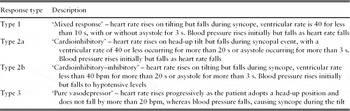
Neurocardiogenic syncope is characterized by a sudden failure of the autonomic nervous system to maintain blood pressure and heart rate leading to insufficient cerebral perfusion pressure. It has an unknown trigger, but the afferent arc synapses with the brainstem cardioinhibitory and vasodepressor centres causing vasodepressive hypotension and heart rate slowing or asystole resulting in loss of consciousness. It can therefore be subdivided into a vasodepressor, cardioinhibitory response with and without asystole, or a mixed response.Reference Sutton, Petersen and Brignole88 Three phases may exist, including a prodrome, loss of consciousness and post-syncope episode.Reference Wayne89 Prodromal symptoms include palpitations, nausea, sweating, light-headedness, blurred vision, paraesthesiae and pallor.Reference Zaqqa and Massumi90 An identifiable precipitant is common, and in older people is mostly attributable to vasodilator medication or prolonged standing. Normally, prolonged standing leads to the pooling of blood in the lower extremities, and the subsequent arterial hypotension is sensed in the carotid bodies. Afferent fibres trigger a reflex increase in heart rate and contractility. Pressure receptors located in the infero-posterior wall of the left ventricle may respond to a variety of stimuli including arterial hypotension, stretch, chemical substances and drugs that produce an increase in parasympathetic and decrease in sympathetic activity.Reference Wieling, van Lieshout and Low91 The resultant heart rate slowing, vasodilatation and hypotension is known as the Bezold-Jarisch reflex and is a possible physiological explanation for neurocardiogenic syncope.Reference Samoil and Grubb92 Loss of consciousness is usually brief, lasting 30 s to 5 min, and some patients develop myoclonic or tonic-clonic jerks mimicking epileptic attacks. Recovery is rapid, although a small proportion of patients may experience distressing symptoms for a number of hours.
Investigations include 24-h and 7-day Holter monitoring for those with an abnormal ECG, postural blood pressure measurements, and tilt-table testing. Indications for tilt testing include following a single episode for those in a high risk setting, syncope associated with exercise, and for further evaluation of recurrent syncope in patients in whom a cause has yet to be identified.93 Contraindications include severe ventricular outflow obstruction, severe mitral stenosis, syncope in the setting of critical proximal coronary artery disease, and cerebrovascular disease. Tilt testing in selected older patients can produce a reasonable yield.Reference Benditt, Ferguson, Grubb, Kapoor, Kugler, Lerman, Maloney, Raviele, Ross, Sutton, Wolk and Wood94 In 176 patients aged over 65 referred for tilting due to unexplained syncope, 49 patients over 65 and 10 patients over 80 had a positive tilt test.Reference Bloomfield, Maurer and Bigger95
Outcomes following head-up tilt

Treatment of neurocardiogenic syncope involves avoidance of precipitants. A vasodepressor response can be attenuated by isometric manoeuvres such as leg crossing or intense handgrip, patient education, and manoeuvres to avert syncope when presyncopal symptoms arise.Reference Krediet, van Dijk, Linzer, van Lieshout and Wieling96, Reference Brignole, Croci, Menozzi, Solano, Donateo, Oddone, Puggioni and Lolli97 Dietary measures such as increased salt intake and oral supplements may be helpful.Reference El-Sayed and Hainsworth98, Reference Younoszai, Franklin, Chan, Cassidy and Allen99 Beta-blockers suppress autonomic activity and blood pressure variability, and although there is much anecdotal and short term data to suggest a benefit, there is little longer term controlled data providing an evidence base to support their use.Reference Ventura, Maas, Zeidler, Schoder, Nienaber, Schuchert and Meinertz100 Other agents of potential benefit include mineralocorticoids that promote salt and water retention, midodrine, and in refractory cases selective serotonin inhibitors such as paroxetine.Reference Da Costa, McIntosh and Kenny101–Reference Di Girolamo, Di Iorio, Sabatini, Leonzio, Barbone and Barsotti103 Patients with a predominant cardioinhibitory or mixed response should be considered for dual chamber permanent pacemaker insertion.Reference Connolly, Sheldon, Roberts and Gent104
Carotid sinus hypersensitivity
Carotid sinus hypersensitivity is defined as a ventricular pause lasting longer than 3 s and a drop of 50 mmHg in systolic blood pressure following 5–10 s of carotid sinus massage.Reference McIntosh, Lawson and Kenny105 It was present in 39% of 272 asymptomatic older subjects, and an equally high prevalence has been suggested in patients with Alzheimer's and Lewy body dementia.Reference Kerr, Pearce, Brayne, Davis and Kenny106 In a consecutive series of older patients with carotid sinus hypersensitivity who required pacemaker insertion, over 30% had presented to hospital with unexplained falls prior to pacing. Significant injuries occurred within this group, with 25% having fallen and sustained a fractured neck of femur prior to presentation.Reference Brignole and Menozzi107 Interestingly, amongst those with carotid hypersentivity, there is no increased risk of death compared with unexplained syncope in a population matched for age and sex.
Three subtypes of carotid sinus syndrome exist,Reference McIntosh, Lawson and Kenny105 and include:
(1) cardioinhibitory, or when carotid sinus massage produces 3 s or more of asystole with symptom reproduction;
(2) a vasodepressor response, or where carotid sinus massage produces a drop in systolic blood pressure of 50 mmHg or more with symptoms;
(3) a mixed response where the criteria for both vasodepressor and cardioinhibitory responses are met.
Carotid sinus massage should be considered in elderly patients who present with syncope, presyncope or unexplained falls and in whom history, examination and investigation have not identified an underlying cause.93 Contraindications include myocardial infarction, transient ischaemic attack or stroke, and a history of ventricular fibrillation or ventricular tachycardia. The right carotid should be massaged first for 5–10 s, followed by the left carotid after 1–2 min if no response. There should be continuous cardiac and blood pressure monitoring, and it should be performed in both supine and upright positions. It is positive if it induces syncope during or straight after massage, produces asystole of >3 s, or a fall in systolic blood pressure of 50 mmHg or more. Neurological complications of carotid sinus massage vary between 0.07 and 0.14%.Reference Davies and Kenny108
Treatment is not necessary in asymptomatic patients. Carotid sinus massage producing more than 3 s of asystole is a mandatory criterion for pacemaker insertion, whereas recurrent syncope without provocative events but a hypersensitive cardioinhibitory response is a strong indication for pacing.109 Drugs such as beta-blockers, calcium channel blockers and methyldopa can affect atrioventricular node function, and stopping them may improve carotid hypersensitivity in up to 50%. Management of the vasodepressor response is more challenging. Fludrocortisone can help but causes many unwanted adverse effects in older patients.Reference Hussain, McIntosh, Lawson and Kenny110
Erectile dysfunction
Erectile dysfunction is defined as an inability to achieve and sustain an erection sufficient to permit satisfactory sexual intercourse.111 Some 5–20% of men have moderate to severe dysfunction. The greatest risk factor is age. The Massachusetts Male Ageing Study showed that the incidence of erectile dysfunction increases with every decade of life.Reference Johannes, Araujo, Feldman, Derby, Kleinman and McKinlay112 These were 12.4 per 1000 man years aged 40–49, 29.8 between 50 and 59 years and 46.4 between 60 and 69 years.
Diabetes mellitus, smoking, hypertension and hyperlipidaemia are strongly associated, and there is a correlation between coronary artery and cerebrovascular disease when erectile dysfunction is present.Reference Morley, Korenman and Kaiser113
Validated psychosexual questionnaires as well as detailed clinical examination focusing on penile structure, the presence of prostatic disease, signs of hypogonadism and cardiovascular and neurological status are key in making the diagnosis.Reference Wespes, Amar, Hatzichristou, Hatzimouratidis, Montorsi, Pryor and Vardi114 Routine investigations should include a fasting glucose and testosterone level.
Management includes psychosexual therapy for those with a psychological cause, hormonal replacement for those deficient in testosterone, treatment of prostatic disease, mechanical devices such as vacuum constriction and penile implants, and pharmacological agents such as sildenafil, tadafil and vardenafil. Second- and third-line therapeutic options include aloprostadil which may be administered by intracavernous injection, prostaglandin E1 which can be given intraurethrally, and sublingual apomorphine.
Sildenafil,Reference Goldstein, Lue, Padma-Nathan, Rosen, Steers and Wicker115 a phosphodiesterase type 5 inhibitor which is a smooth muscle dilator improving penile blood flow, and other drugs from the same class have significantly improved sexual function and outcomes in older men.
Gastrointestinal dysfunction
Dysphagia may result from reduced salivary gland function and also lower oesophageal dysfunction related to autonomic innervations of the lower two-thirds of the oesophagus. Gastric motility is reduced with age, as is the perception of gastric distension, and this is related to progressive autonomic nerve dysfunction. These features are found in diabetic gastroparesis in which there is abdominal distension and vomiting. Large intestinal motility also diminishes, and constipation is a common clinical problem. There are a reduced number of myenteric plexus neurones and reduced large bowel transit time is found in both primary autonomic failure and Parkinson's disease, in which there is a reduction in myenteric plexus neurones with Lewy body accumulations.Reference Singaram, Ashraf and Gaumnitz116 Diarrhoea can result from overflow in which constipation and faecal impaction result in loose motions and from changes in bowel flora and motility in diabetes mellitus.
Thermoregulation
Attenuated responses to thermal stress have been well documented in older patients. Nearly fifteen thousand people lost their lives in France in 2003 due to exceptionally high temperatures, the vast majority of whom were in the older age group.Reference Belmin, Auffray, Berbezier, Boirin, Mercier, de Reviers and Golmard117 Autonomic failure can produce sudomotor dysfunction, resulting in hypo- or anhidrosis, gustatory sweating, hypo- and hyperthermia and heat stress.Reference Cheshire and Freeman118
Hyperthermia occurs when thermoregulatory cooling mechanisms are overwhelmed by excessive metabolic production of heat, excessive environmental heat, or inadequate heat dissipation.Reference Simon119 Heat loss during periods of high temperatures occurs through diversion of cardiac output with increases in skin blood flow and a reduction in splanchnic and renal blood flow, and sweat evaporation.Reference Wilson, Sauder, Kearney, Kuipers, Leuenberger, Monahan and Ray120 These are the principle mechanisms through which homeostatic cooling occurs. Kenney et al. found lower cardiac outputs and heart rates in older men exercising in warm environments compared with younger subjects.Reference Kenney121 Skin blood flow at a given temperature during passive and active heat stress was considerably less in older patients, suggesting less ability to lose heat through cutaneous vasodilatation than in younger patients.Reference Kenney, Morgan and Farquar122 This lack of vasodilatory response can be explained partially by an imbalance between noradrenaline and nitric oxide, producing an increase in vascular smooth muscle tone. Cardiac output diversion from the core to the periphery and heat dissipation is therefore more difficult to achieve in older patients during periods of heat stress.
Major syndromes encountered in autonomic failure
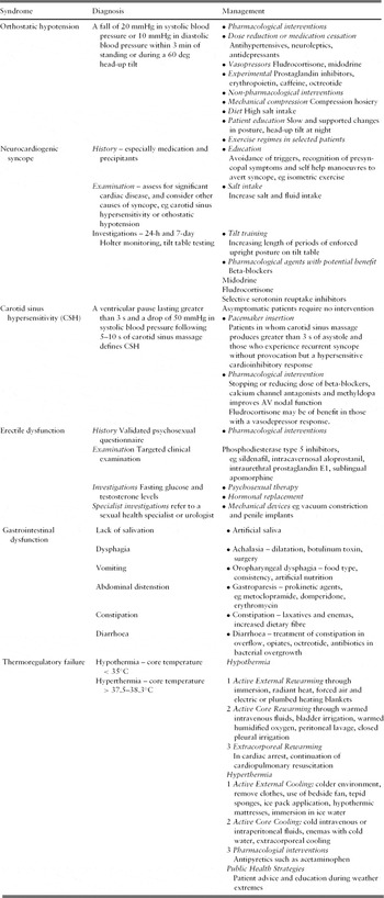
Hypothermia is defined as an unintentional decline in core temperature below 35°C where co-ordinated physiological responses to retain heat by minimizing conduction, convection, radiation, evaporation and respiration begin to fail.Reference Danzl and Pozos123 In the USA in 2001, hypothermia was thought to have caused up to 600 deaths, whereas between 1982 and 1997 in England and Wales, there was an average of 491 deaths per year.124, Reference Chantler and Kelly125 When exposed to cold temperatures, thermoreceptors in the skin, spinal cord and brain transmit impulses to the hypothalamus which produce cutaneous vasoconstriction, heat generation by shivering of skeletal muscle and other processes such as increased metabolism in brown adipose tissue. Cold perception is critical in preventing hypothermia, and thermoreceptor sensitivity is significantly reduced in older patients.Reference Collins, Exton-Smith and Dore126 Shivering is reduced due to reduced skeletal muscle bulk, and onset of shivering is delayed compared with younger subjects.Reference Kenney and Buskirk127 Reduced skeletal muscle enzyme activity in aerobic metabolism has also been postulated as a mechanism for reduced shivering. Cutaneous blood vessels show a reduced ability to vasoconstrict coupled with an overall reduction in skin blood flow, producing cold stress in the older patient. Collins et al. measured changes in hand blood flow response to the cold in 43 older males over a period of 8 years. The proportion in whom there was no cold-induced reduction in blood flow was 14% at baseline, increasing to 32% after 4 years, with a higher percentage still at 8 years.Reference Collins, Dore, Exton-Smith, Fox, MacDonald and Woodward128 Blood flow has been shown to decrease in both young and old subjects in response to the cold, but it increases again to pre-cooling levels in the later group after 1 min. This suggests that older subjects are unable to maintain sustained vasoconstriction.Reference Richardson, Tyra and McCray129
Thermoregulatory dysfunction increases with age, but factors such as physical fitness, socioeconomic status, co-existent disease states and prior environmental acclimatization play an important role. Tight environmental temperature control with adequate heating in the winter months and cooling methods in the summer, as well as adequate social and financial support are vital in preventing thermal stress in the older patient.
Conclusions
The ageing autonomic nervous system produces complex physiological changes that manifest in a number of clinical syndromes, some of which can be very difficult to treat. Cardiovascular disease and diabetes mellitus directly damage autonomic nerve fibres and are particularly prevalent in older people. Orthostatic hypotension due to reduced baroreceptor sensitivity and reduced vasoconstriction is a significant cause of falls, morbidity and mortality in the elderly. Early identification of risk factors in middle age, careful diagnosis, treatment of vascular disease and diabetes, and potential future interventions to treat neurodegenerative processes may delay or prevent the onset and reduce the severity of autonomic-related disorders. In established autonomic failure, better patient information, education and understanding to avoid changes in posture, heat extremes and situational circumstances precipitating events such as neurocardiogenic syncope are key to successful long-term management. Clinical judgement is required to achieve a fine balance in supporting aberrant autonomic function in response to physiological stress without creating further medical complications.
Conflicts of interest
The authors declared no conflicts of interest.