It was in 1897 that EisenmengerReference Eisenmenger 1 described and illustrated a heart with a large ventricular septal defect. The defect was characterised by antero-cephalad malalignment of the muscular outlet septum, with obvious biventricular connection of the aortic root, but with normal expansion of the subpulmonary infundibulum, and dilation of the pulmonary trunk. Subsequent to further observations by Baumgartner and Abbott,Reference Baumgartner and Abbott 2 it was AbbottReference Abbott 3 herself who emphasised that the lesion led progressively in the postnatal period to pulmonary hypertension, with progressive right ventricular hypertrophy, and dilation of the pulmonary trunk. As far as we can establish, it was also AbbottReference Abbott 3 who suggested that the entity be called the Eisenmenger complex. WoodReference Wood 4 subsequently pointed out that other lesions promoting post-tricuspid shunting in the postnatal period could lead to pulmonary hypertension, and he named this cascade of events the Eisenmenger reaction. The defect itself has a characteristic phenotype, and has subsequently been studied by several additional groups of investigators.Reference Van Mierop and Wiglesworth 5 – Reference Chen, Xu, Wang, Shen, Zhang and Hong 9 Its obvious similarity to tetralogy of Fallot in terms of the deviation of the muscular outlet septum has led to ongoing discussions regarding the associations between the two lesions.Reference Oppenheimer-Dekker, Gittenberger-de Groot, Bartelings, Wenink, Moene and van der Harten 8 , Reference Gasul, Dillon, Vrla and Hait 10 , Reference Fukuda, Suzuki and Ito 11 More recently, discussions have been centred on the significance of the rightward rotation of the aortic root, engendering debates relating to the value of “dextroposition” when describing hearts characterised by overriding of the aortic root.Reference Bozok, Kestelli and Ilhan 12 , Reference Anderson 13 In this regard, one of us had suggested that the similarity between the Eisenmenger defect and tetralogy of Fallot reflected the biventricular origin of the overriding aorta, but that the differences relate to the degree of expansion of the subpulmonary infundibulum.Reference Restivo, Unolt, Putotto and Marino 14 Another of us, however, had disputed the significance of this finding when considering the arrangements in which both arterial trunks arise predominantly from the right ventricle.Reference Anderson 15 Therefore, to adjudicate these previous differences, and hopefully to elucidate the nosological significance of its phenotypic features, we have now carried out a detailed analysis of the subpulmonary infundibular morphology of the hearts catalogued as having the Eisenmenger defect in the archive of the University of Rome. We have compared these findings with those from a comparable number of specimens with intact ventricular structures, and others with perimembranous ventricular septal defects in the absence of overriding of the aortic root.
Materials and methods
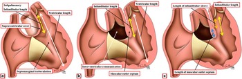
We examined in detail 11 hearts that had previously been catalogued in the cardiac pathology collection of the Museum of Pathologic Anatomy, Policlinico Umberto I, University of Rome “La Sapienza”, as exhibiting the Eisenmenger variant of ventricular septal defect (Table 1). The specimens, most of which consisted of heart–lung blocks, had already been opened by following the flow of blood, thus permitting examination of all four cardiac chambers at the time of autopsy. The specimens had been fixed in 10% formaldehyde. The age of death of the patients ranged from 2 months to 3 years. In three cases, where only the heart specimen was available, the clinico-pathological reports revealed that all patients had had a normal left-sided aortic arch. Whenever necessary, for photographic reasons, the margins of cut areas of the specimens were reconstituted without stitching. All hearts had, as their essential phenotypic feature, overriding of the aortic root, with antero-cephalad malalignment of the muscular outlet septum. As a consequence of the malalignment, the outlet septum was located exclusively within the right ventricle, rather than occupying an interventricular position. The subpulmonary infundibulum was unobstructed in all the specimens. Having selected the hearts on this basis, we then assessed the extent of overriding of the aortic root, establishing the relationships between the left and non-coronary leaflets of the aortic valve relative to the aortic, or anterior, leaflet of the mitral valve, and comparing these findings with those found in the hearts with intact ventricular septal structures. This permitted us to grade the hearts as showing mild, intermediate, or severe degrees of aortic rightward rotation. We then established the length of the subpulmonary infundibulum as assessed from its ventral aspect, taking all measurements using a calibrated ruler. By comparing the values of the length of the subpulmonary infundibulum with the overall length of the right ventricle (Fig 1a and b), each heart was able to serve as its own control (Table 2). In the hearts having Eisenmenger defects, we determined the length of the subpulmonary infundibulum by measuring from the proximal hingepoint of the left pulmonary valvar leaflet to the leading edge of the muscular outlet septum (Fig 1b). We then compared these values with similar measurements made in 11 hearts from the archive with intact ventricular septal structures (Table 3), matching as far as possible the age range to those of the patients with the Eisenmenger defects. In the normal hearts, as it is not possible to recognise the leading edge of the muscular outlet septum when the ventricular septum itself is intact, we measured the length of the subpulmonary infundibulum by measuring the distance from the hinge of the pulmonary valve leaflet to the bifurcation of the limbs of the septomarginal trabeculation (Fig 1a). It is at this point, when the ventricular septum is intact, that the outlet component of the supraventricular crest inserts between the limbs of the septomarginal trabeculation. When feasible, we also assessed the length of the muscular outlet septum, which was possible only in the hearts with deficient ventricular septation. Thus, we measured the distance between the leading edge of the muscular outlet septum and the attachments of the aortic valvar leaflets on its dorsal aspect (Fig 1c). We then compared this value to the overall length of the subpulmonary muscular infundibulum. Accurate assessment of this value, however, was possible in only eight of the hearts (Table 1). Comparable measurements were not feasible in the hearts with intact ventricular septal structures, as it is not possible, in this setting, to assess the length of the muscular outlet septum.
The cartoon illustrates the measurements taken to establish the length of the subpulmonary infundibulum relative to the length of the right ventricle in the normal heart ( a ) compared with the Eisenmenger defects ( b ). These morphometric data are reported in Tables 2 and 3. In eight of the Eisenmenger defects, we also established the length of the muscular outlet septum, as discrete from the free-standing infundibular sleeve, by measuring the distance from its free edge to the attachment of the aortic valvar leaflets on its dorsal aspect ( c ). These morphometric data are included in Table 1. We have labelled ( c ) as a separate entity so as not to unduly crowd the cartoon.

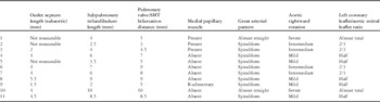
Summary of the relevant features of the hearts examined with Eisenmenger defects, including the measurements made, when possible, of the length of the subpulmonary infundibulum.

SMT=septomarginal trabeculation
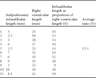
The lengths of the subpulmonary infundibulum relative to the lengths of the right ventricle in the hearts with Eisenmenger defects.

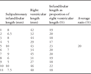
The lengths of the subpulmonary infundibulum relative to the lengths of the right ventricle in the hearts with intact septal structures.

We did, nonetheless, compare the findings in the hearts with Eisenmenger defects with another nine hearts from the archive with perimembranous ventricular septal defects opening centrally within the right ventricle, but in the absence of any rightward rotation or overriding of the aortic root. With regard to our use of “centrally” when describing the location of the selected perimembranous defects, themselves defined on the basis of fibrous continuity postero-inferiorly between the leaflets of the aortic and tricuspid valves, we took the stance that the membranous septum itself, when the ventricular septum is intact, occupies the central component of the cardiac base, filling the base of the gap – when assessed from the left ventricle – between the non-adjacent and right coronary leaflets of the aortic valve. The area occupied by the membranous septum, therefore, is flanked postero-inferiorly by the inlet component of the right ventricle, guarded by the tricuspid valve, and antero-superiorly by the right ventricular outflow tract, specifically by the supraventricular crest inserting between the limbs of the septomarginal trabeculation. The hearts with perimembranous defects chosen for comparison, therefore, all showed septal deficiencies opening centrally to the base of the right ventricle through the space between the non-adjacent and right coronary leaflets of the aortic valve, with the supraventricular crest being normally inserted between the limbs of the septomarginal trabeculation, and with no extension of the defect itself towards the inlet of the right ventricle.
Results
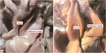
Our overall findings in the hearts with Eisenmenger defects are summarised in Table 1. In nine of the hearts with Eisenmenger defects, the intrapericardial arterial trunks spiralled as they extended to the margins of the pericardial cavity (Fig 2a). In two of the hearts, in contrast, specifically those with the greatest rightward rotation of the aortic root, the arterial trunks extended to the margins of the pericardial cavity in an almost parallel manner (Fig 2b).
The images compare the spiralling arrangement of the arterial trunks found in the majority of our hearts ( a ) with the almost parallel arrangement seen in two of our specimens ( b ).

The subpulmonary infundibulum
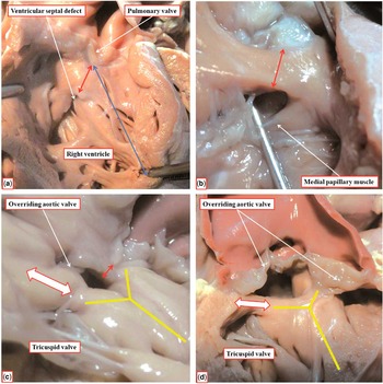
In only three of the hearts with Eisenmenger defects was the subpulmonary infundibulum comparable in length with those with intact ventricular septation (cases 8, 10, and 11 in Table 1). In these three hearts, furthermore, the leading edge of the subpulmonary infundibulum – formed by the malaligned muscular outlet septum – was at the level of the bifurcation of the limbs of the septomarginal trabeculation. The length of the subpulmonary infundibulum in these three hearts, assessed as the distance between the proximal hingepoint of the left pulmonary valve leaflet and the cranial basal bifurcation of the septomarginal trabeculation, was comparable with the average length of the subpulmonary infundibulum in hearts with intact ventricular septal structures (Fig 3a; Tables 1–3). The leading edge of the subpulmonary infundibulum did not reach the bifurcation of the septomarginal trabeculation in the remaining eight hearts (Fig 3b). In these eight hearts, the length of the subpulmonary infundibulum was reduced, being particularly short in case 9 (Fig 3c).
The images show the varying length of the subpulmonary infundibulum (double-headed red arrows), which was always capacious. The double-headed blue arrow in panel ( a ) shows the overall length of the right ventricle. In this heart, the subpulmonary infundibulum, as in the normal hearts, accounted for 20% of the right ventricle. As revealed by our measurements, the subpulmonary infundibulum was deficient in the heart illustrated in panel ( b ), with the leading edge of the outlet septum not extending to the cranial bifurcation of the septomarginal trabeculation, and with the subpulmonary infundibulum accounting for only 16% of the right ventricular length. The subpulmonary infundibulum was appreciably shorter in the heart illustrated in panel ( c ) (double-headed red arrow), accounting for only 9% of the right ventricular length. Note in panel ( c ) the discontinuity between the leaflets of the aortic and tricuspid valves (double-headed white arrow), with the yellow Y showing the limbs of the septomarginal trabeculation. Panel ( d ) shows the overriding of the aortic root in the same heart as shown in panel ( c ), but with the outlet septum reflected in the antero-cephalad direction. The double-headed white arrow again shows the ventriculo-infundibular fold interposed between the leaflets of the aortic and tricuspid valves. The fold is fused with the caudal limb of the septomarginal trabeculation (yellow Y).

In all the hearts, nonetheless, along with those having intact ventricular septal structures, the subpulmonary infundibulum was capacious, showing no evidence of obstruction.
In only eight of the Eisenmenger defects was it possible to assess the component of the subpulmonary infundibulum made up by the muscular outlet septum, this being achieved by determining the level of the attachment of the leaflets of the aortic valve on its dorsal aspect (Fig 1c; Table 1). In these eight hearts, there was marked variability in its extent, relative to the length of the free-standing subpulmonary infundibular sleeve. In most of the hearts, the outlet septum accounted for around half of the overall length of the subpulmonary infundibulum; however, in case 9, with a particularly short subpulmonary infundibulum, three-quarters of its length was made up of the outlet septum.
Rotation of the aortic root
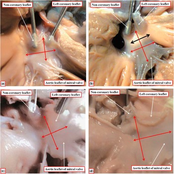
In hearts with intact ventricular septal structures, which we take as representing the lack of any aortic rightward rotation, the area of fibrous continuity between the valvar leaflets was made up approximately two-thirds by the non-coronary, or non-adjacent, leaflet of the aortic valve and about one-third by the left coronary leaflet (Fig 4a). In the hearts with Eisenmenger defects, we found five cases where the left coronary leaflet accounted for half of the zone of fibrous continuity, deeming these to have mild aortic rotation (Fig 4b). In four hearts, the left coronary leaflet made up two-thirds of the area of fibrous continuity, this being the consequence of moderate rotation (Fig 4c). In two of the hearts, the anterior leaflet of the mitral valve was related only with the left coronary aortic leaflet, being the result of severe rightward rotation (Fig 4d). Both the hearts with severe aortic rotation also had almost parallel arterial trunks, as shown in Figure 2b. In one of the hearts, the mid portion of the ventriculo-infundibular fold interposed in the roof of the left ventricle between the hinges of the aortic and mitral valves (Fig 4b). This heart also exhibited discontinuity between the leaflets of the aortic and tricuspid valves (Fig 3c). This specimen, therefore, exhibited a completely muscular subaortic infundibulum in addition to the muscular subpulmonary infundibulum.
The images show the variation in the relationships between the leaflets of the aortic valve and the aortic leaflet of the mitral valve. In hearts with intact septal structures ( a ), two-thirds of the non-coronary, or non-adjacent, leaflet of the aortic valve are in fibrous continuity with the aortic leaflet of the mitral valve (crossed double-headed red arrows). When there is minimal rightward rotation of the aortic root ( b ), about half of the non-coronary leaflet is in fibrous continuity with the aortic leaflet of the mitral valve leaflet. In cases with moderate rotation, only one-third of the non-coronary leaflet is in fibrous continuity with the aortic leaflet of the mitral valve, with the left coronary leaflet occupying two-thirds of the mitral leaflet itself ( c ). With severe rightward aortic rotation ( d ), the aortic leaflet of the mitral valve is almost exclusively in continuity with the left coronary aortic valvar leaflet. Note that, in panel ( b ), the muscular ventriculo-infundibular fold interposes between the leaflets of the aortic and mitral valves, producing a completely muscular subaortic infundibulum (double-headed black arrow). In this heart, therefore, it is the spatial relationship of the aortic and mitral valvar leaflets that has been compared, rather than the area of fibrous continuity.

Overriding of the aortic root
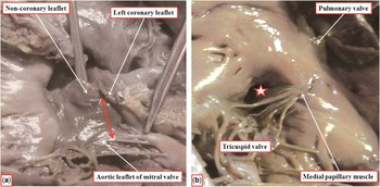
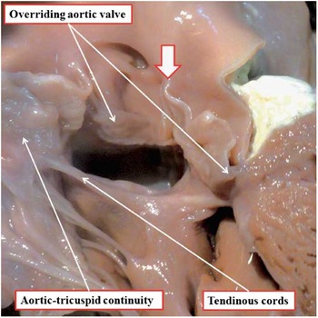
In all the hearts from our archive having perimembranous defects opening centrally to the base of the right ventricle, and with alignment between the muscular outlet septum and the apical muscular ventricular septum – in other words, in the absence of any rightward rotation or overriding of the aortic root – the area of fibrous continuity between the aortic and mitral valves was formed as in the hearts with intact ventricular septal structures – namely, with two-thirds of the mitral valvar leaflet in continuity with the non-coronary, or non-adjacent, leaflet of the aortic valve (compare Figs 4a and 5a). As emphasised above, when judged from the aspect of the right ventricle, the perimembranous defect in the hearts with alignment of the ventricular muscular septal components opened posterior to the supraventricular crest, which was normally inserted between the limbs of the septomarginal trabeculation, or septal band (Fig 5b). In these hearts, the entirety of the circumference of the aortic valve was supported above the cavity of the left ventricle. In the five hearts with Eisenmenger defects and mild rightward rotation, up to one-third of the aortic valvar orifice was supported by right ventricular structures. In the four cases with intermediate overriding, almost half of the aortic root was overriding the cavity of the right ventricle, but the ventriculo-arterial connections remained concordant. In the two hearts with severe dextrorotation, and with almost parallel arterial trunks, almost two-thirds of the aortic root was supported by the right ventricle (Fig 6), with the larger part of the non-coronary aortic valvar leaflet being attached within the right ventricle. The two hearts with severe rightward rotation, therefore, had effective double-outlet ventriculo-arterial connection from the right ventricle.
The images show the left ( a ) and right ( b ) views of a perimembranous defect (star) opening centrally into the right ventricle, and entering the ventricle behind the postero-caudal limb of the septomarginal trabeculation. As can be seen in panel ( a ), the non-coronary aortic leaflet takes up the greater part (two-thirds) of continuity with the aortic leaflet of the mitral valve.

In this specimen, the greater part of the aortic root is supported above the morphologically right ventricle. The heart, therefore, has double-outlet ventriculo-arterial connection, despite the presence of fibrous continuity between the leaflets of the aortic, tricuspid, and mitral valves in the postero-inferior margin of the interventricular communication. Note that there is no medial papillary muscle in this heart, with the septal leaflet of the tricuspid valve attached by tendinous cords along with the inferior margin of the septal defect. The white arrow with red borders shows the location of the attachment of the zone of apposition between the coronary leaflets of the aortic valve at the sinotubular junction. This point, usually described as the valvar commissure, is shifted leftwards in keeping with the marked rightward rotation of the aortic root.

The medial papillary muscle
In hearts with intact ventricular septal structures, the medial papillary muscle, or the muscle of Lancisi, takes its origin from the postero-caudal limb of the septomarginal trabeculation, although there is marked variability in its specific morphology.Reference Restivo, Smith, Wilkinson and Anderson 16 In seven of the 11 hearts with Eisenmenger defects, we failed to identify a muscle in this location. In these seven hearts, the zone of apposition between the antero-superior and septal leaflets of the tricuspid valve was supported either by minor papillary muscles or by direct tendinous cords. These arose either from the postero-caudal limb of the septomarginal trabeculation or along the inferior margin of the defect (Fig 6). In the remaining four hearts, the muscle was found at its expected site (Fig 3c), but was rudimentary in one. The subpulmonary infundibulum was longest in four of the seven cases where it was not possible to identify the medial papillary muscle (Table 1).
The ventricular septal defect
When the borders of the hearts diagnosed as having Eisenmenger defects were assessed from their right ventricular aspect, all except one were perimembranous, with their postero-inferior margins being formed by an area of fibrous continuity between the leaflets of the aortic, tricuspid, and mitral valves (Figs 6 and 7). In the outstanding heart, which also had aortic-to-mitral valvar discontinuity, and thus a completely muscular subaortic infundibulum, the right ventricular margin of the channel between the ventricles had exclusively muscular borders. This was because the rightward margin of the ventriculo-infundibular fold was fused postero-inferiorly with the caudal right posterior limb of the septomarginal trabeculation or septal band (Fig 3d).
The image shows the fibrous continuity between the leaflets of the aortic and tricuspid valves, which is the phenotypic feature of a perimembranous defect. Note the length of the muscular outlet septum between the attachments of the aortic valve on its dorsal aspect and its leading edge (double-headed red arrow). Note also the location of the commissure between the two coronary aortic valvar leaflets (white arrow with red borders). The location of this commissure can be taken as a surrogate of the extent of aortic rightward rotation, which in this case is mild compared with the severe example shown in Figure 6.

Discussion
The Eisenmenger malformation is characterised by dextro-anterior and cephalad deviation of the outlet septum. In this position, it is exclusively within the right ventricle, rather than being an interventricular structure. This feature, of course, is also found in the setting of tetralogy of Fallot.Reference Anderson, Spicer, Giroud and Mohun 17 The Eisenmenger defect, nonetheless, is distinguished from tetralogy of Fallot because the subpulmonary infundibulum, although being of variable length as in tetralogy,Reference Becker, Connor and Anderson 18 is capacious, showing no evidence of obstruction. Although of variable length in tetralogy of Fallot, the subpulmonary infundibulum is longer on average when compared with the length of the normal subpulmonary infundibulum.Reference Becker, Connor and Anderson 18 In contrast, in our hearts with the Eisenmenger defect, the average length of the subpulmonary infundibulum was shorter compared with control hearts. In our normal hearts, we found that the subpulmonary infundibulum, on average, accounts for 20% of the overall length of the right ventricles (Table 3). In the hearts with Eisenmenger defects (Table 2), the subpulmonary infundibulums, on average, made up only 15.4% of the right ventricular length. In three of the hearts, nonetheless, in which the outlet septum extended proximally to the level of the bifurcation of the septomarginal trabeculation, the subpulmonary infundibulums were of normal length. The phenotypic feature of tetralogy of Fallot is the stenosis of the subpulmonary infundibulum, with a “squeeze” at its mouth between the malaligned muscular outlet septum, or its fibrous remnant, and the septoparietal trabeculations, which become hypertrophied in postnatal life.Reference Anderson, Spicer, Giroud and Mohun 17 This was not found in any of our specimens.
The capacious nature of the subpulmonary infundibulum in the Eisenmenger defect has been well emphasised in previous studies,Reference Van Mierop and Wiglesworth 5 , Reference Goor, Lillehei and Edwards 7 , Reference Oppenheimer-Dekker, Gittenberger-de Groot, Bartelings, Wenink, Moene and van der Harten 8 as has its reduced length when compared with normal values.Reference Goor, Lillehei and Edwards 7 In one of these studies, however, the muscular outlet septum was also always said to be hypoplastic.Reference Van Mierop and Wiglesworth 5 We found that, in those hearts in which we were able to make precise measurements, the component of the subpulmonary infundibulum made up by the muscular outlet septum was variable in its length. It was always appreciably longer, nonetheless, when compared with hearts with intact ventricular septal structures. This is because hardly any part of the subpulmonary infundibular musculature occupies a septal location when the ventricular septum is intact.Reference Anderson, Spicer, Giroud and Mohun 17
Our investigation, although at variance with the findings of Van Mierop and WiglesworthReference Van Mierop and Wiglesworth 5 with regard to the length of the muscular outlet septum, does lend some support to their findings regarding the presence or absence of the medial papillary muscle of Lancisi. They pointed out that the muscle was rarely recognisable as normal any time the components of the supraventricular crest were divorced from each other. They also suggested that the muscle was lacking when the supraventricular crest was deficient. This was not the case in our specimens. The muscle could be identified in three of our hearts with short subpulmonary infundibulums, yet was absent in the three hearts with the longest subpulmonary infundibulums.
In all but one of our cases with the Eisenmenger defects, defined on the basis of aortic overriding, but with unobstructed subpulmonary infundibulums, the ventricular septal defects were perimembranous. There were bilaterally complete muscular infundibulums in the outstanding hearts, with the subaortic infundibulum overriding the crest of the apical muscular ventricular septum. A similar arrangement, with bilateral muscular infundibulums, has been found occasionally in the setting of tetralogy of Fallot,Reference Dickinson, Wilkinson, Smith, Hamilton and Anderson 19 when the malalignment defect can again be either perimembranous or have exclusively muscular right ventricular borders.Reference Anderson, Spicer, Giroud and Mohun 17 This shows that, completely to categorise holes between the ventricles, it is necessary not only to take note of how they open between the ventricles, and to note the presence or absence of septal malalignment, but also to account for the precise anatomic nature of their borders as viewed from the right ventricle. Our observations also show that, when both arterial trunks are arising predominantly from the right ventricle, the roof of the geometric interventricular communication can be made up of fibrous continuity between the leaflets of the aortic and mitral valves. It is now becoming increasingly well recognised that the absence of a subaortic infundibulum does not preclude the diagnosis of double-outlet right ventricle;Reference Anderson, Spicer, Giroud and Mohun 17 however, the presence of bilateral infundibulums is one element of variability in the make-up of congenitally malformed hearts. Indeed, even normal hearts can, on rare occasions, show persistence of a subaortic muscular infundibulum.Reference Rosenquist, Clark, Sweeney and McAllister 20 Moreover, in the heart in our series with bilateral infundibulums, the aortic root was supported predominantly within the morphologically left ventricle, showing only mild rightward rotation of the aortic root.
In this respect, we assessed the extent of rightward rotation of the aortic root by comparing the proportions of the leaflets of the aortic valve in continuity with the aortic, or anterior, leaflet of the mitral valve. Others have assessed the relationships between the zone of apposition between two coronary leaflets of the aortic valve and the zone of apposition between the adjacent leaflets of the pulmonary valve, showing that this does not change according to the extent of aortic override.Reference Goor, Lillehei and Edwards 7 , Reference Isaaz, Cloez, Marcon, Worms and Pernot 21 Our findings suggest that, as the dorsal component of the aortic root rotates rightward, its overall axis is displaced leftward, correlating in this way with the observations regarding the interrelations of the zones of apposition between the adjacent valvar leaflets (Fig 7). In other words, the more the aortic root is rotated to the right, the more its longitudinal axis, coincident with the zone of apposition, usually described as the commissure, between the two coronary aortic valvar leaflets, is shifted to the left, as shown in Figure 6.
The key feature we chose to serve as the defining phenotype of the Eisenmenger defect was the malalignment of the muscular outlet septum relative to the apical muscular ventricular septum. The muscular outlet septum is divorced from the apical muscular septum in all hearts with perimembranous defects, defined on the basis of fibrous continuity between the leaflets of the aortic and tricuspid valves, but in most cases it retains its alignment with the apical muscular septum. In these hearts with divorced, but aligned, ventricular septal structures, the defect itself then opens either centrally to the base of the right ventricle or extends so as to open additionally to the right ventricular inlet. In all the hearts we examined with such aligned septal structures, the relationship between the leaflets of the aortic valve along the area of fibrous continuity with the mitral valve was as found in the hearts with intact ventricular septal structures. In these hearts, therefore, the geometric interventricular communication was effectively the same curved surface as the ventricular septal defect, the latter defined on the basis of the area of putative ventricular septation. This is not the case, however, when there is malalignment between the apical and outlet muscular septal components, as in the Eisenmenger defects.
This is because, when the aortic root is overriding, the muscular outlet septum is exclusively attached within the right ventricle. The curved surface that forms the right ventricular border of the channel providing interventricular shunting, therefore, will also be contained exclusively within the right ventricle. It is the borders of this curved surface that form the locus around which the surgeon will place a patch to reconstitute septal integrity. The curved surface, however, is no longer congruent with the geometric interventricular communication. This area is the cranial continuation of the long axis of the ventricular septum. These considerations become more significant when the larger part of the aortic root, along with the subpulmonary infundibulum, is supported by the right ventricle. In this latter setting, the ventriculo-arterial connection is one of double-outlet. During surgical connection, it is then essential that the geometric interventricular communication, which is the exit from the left ventricle, be distinguished from the curved surface representing the area of putative ventricular septation. In the ideal world, it is the latter surface, rather than the geometric interventricular communication, which should be described as the “ventricular septal defect”.
It is noteworthy that those studying cases in the clinical arena have reported progression of right ventricular hypertrophy in patients with Eisenmenger defects, this process then leading to narrowing of the subpulmonary infundibulum.Reference Chen, Xu, Wang, Shen, Zhang and Hong 9 , Reference Fukuda, Suzuki and Ito 11 When studying autopsy specimens, we see only the findings at the time of death. Of the 11 cases examined in our series, however, only the heart from the oldest patient, who was aged 3 years, showed any degree of hypertrophy of the right ventricle. In this heart, we observed prominent muscle bands, including the septoparietal trabeculations, but still detected no evidence of narrowing of the subpulmonary infundibulum. We cannot use our current findings, therefore, to determine with any degree of certainty whether the Eisenmenger malformation should be considered, in some circumstances, to be an intermediate lesion in the progression towards the so-called “pink” tetralogy of Fallot.Reference Altshuler 6 , Reference Gasul, Dillon, Vrla and Hait 10 Our findings demonstrate, at least in the archive of the University of Rome, the fundamental differences between the phenotypes of the Eisenmenger defects and tetralogy of Fallot, not only with regard to the absence of any stenosis and “squeeze” at the mouth of the subpulmonary infundibulum but also with regard to its variation in length when compared with the overall length of the right ventricle. Our major finding, nonetheless, is that even in the setting of the Eisenmenger defect there is marked variability in the specific phenotypic features of the individual hearts.
Acknowledgements
Authors’ contributions: A.R. conceived the study, made all the anatomic observations and measurements, and wrote the initial draft of the manuscript. P.G. provided access to the specimens, checked the anatomic observations, and approved the drafting of the manuscript. C.d.G also contributed in this direction. R.C. was of particular help in the production of the photographic material. R.H.A produced the final version of the manuscript subsequent to extensive discussions with his co-authors.