INTRODUCTION
The intertidal zone of the North Pacific rim hosts multiple species of sea anemone (Cnidaria: Anthozoa: Actiniaria) that brood their offspring. In these species, the offspring are retained within or upon the body through development to a fully formed polyp. This definition excludes offspring that are released as larvae or embryos. The genus Epiactis Verrill, Reference Verrill1869a is notable among actiniarians for its relatively high proportion of such species and for the diversity of strategies by which its members brood: members of E. arctica Verrill, Reference Verrill1869b and E. marsupialis Carlgren, Reference Carlgren1921 brood offspring within individual pits that form by invagination of the column, whereas the offspring of members of E. prolifera Verrill, Reference Verrill1869a and E. lisbethae Fautin & Chia, Reference Fautin and Chia1986 are fully exposed upon the surface of the parent. In other species, such as E. fernaldi Fautin & Chia, Reference Fautin and Chia1986, offspring are retained within the gastrovascular cavity of the adult and released as juveniles via the mouth of the parent. The genus is widely distributed globally, but most species-rich in the Pacific (seven of 18 species). In the North Pacific, external brooding is reported in E. prolifera (see Verrill, Reference Verrill1869a), E. lisbethae (see Fautin & Chia, Reference Fautin and Chia1986), and E. japonica (see Uchida, Reference Uchida1934); internal brooding has been reported in E. fernaldi (see Fautin & Chia, Reference Fautin and Chia1986) and E. ritteri (see Hand & Dunn, Reference Hand and Dunn1974). The specific identity of brooding individuals in the last species is contested (Sanamyan & Sanamyan, Reference Sanamyan and Sanamyan1998), reflecting different interpretations of features not described in the original description, such as adherence of debris to the column.
Epiactis ritteri Torrey, Reference Torrey1902 was described from specimens collected from Popof Island, Alaska. These individuals were not brooding externally at the time of collection. The description did not include any mention of internal brooding or provide any details about brooding behaviour or structures, except in noting the absence of external brooding as a point of contrast with the externally brooding species E. prolifera. Reproduction is a seasonal phenomenon in many species (Shick, Reference Shick1991), however, and presence or absence of offspring may be circumstantial. None of the subsequent publications mentioning E. ritteri from Alaska (Carlgren, Reference Carlgren1934, Reference Carlgren1947, Reference Carlgren1950) discussed individuals that brooded offspring internally or externally.
Dunn (Reference Dunn1972) identified internally brooding anemones from Bodega Bay, California as belonging to Epiactis ritteri. This was the first report of the species from outside Alaska and the first account of brooding for E. ritteri. The redescription of E. ritteri by Hand & Dunn (Reference Hand and Dunn1974) based on internally brooding specimens from California and Oregon is consistent with previous work (Torrey, Reference Torrey1902; Carlgren, Reference Carlgren1934) with respect to features like marginal sphincter morphology, mesenterial arrangement and concentration of holotrichous isorhizae (holotrichs) in the basal column. Hand & Dunn (Reference Hand and Dunn1974) also provided important details not mentioned in earlier work, most notably the adherence of foreign material to the column and the batteries of holotrichs in the distal column. Sanamyan & Sanamyan (Reference Sanamyan and Sanamyan1998) contested the identification by Dunn (Reference Dunn1972) and by Hand & Dunn (Reference Hand and Dunn1974), arguing that the details noted by Hand & Dunn (Reference Hand and Dunn1974) but not described by previous workers were indicative of this misidentification, and suggesting that the southern specimens studied by Hand & Dunn (Reference Hand and Dunn1974) actually represent a new species.
The identity of Epiactis ritteri was also challenged on the basis of its similarity with E. japonica, an externally brooding species from Japan. Uchida (Reference Uchida1934, Reference Uchida1938), Uchida & Iwata (Reference Uchida and Iwata1954) and Carlgren (Reference Carlgren1940, Reference Carlgren1947, Reference Carlgren1950, Reference Carlgren1952) hypothesized that the Alaskan individuals of E. ritteri belonged to E. japonica. Sanamyan & Sanamyan (Reference Sanamyan and Sanamyan1998) concurred, considering E. japonica and E. ritteri synonymous based on externally brooding specimens from the Commander Islands, Kamchatka Peninsula, Kurile Islands and Sea of Japan. Further complicating this situation, Uchida (Reference Uchida1934, Reference Uchida1938) and Uchida & Iwata (Reference Uchida and Iwata1954) used the name E. prolifera, which belongs to an Eastern Pacific species, to refer to what was subsequently identified as E. japonica, raising the possibility that E. ritteri is a junior synonym of E. prolifera. Synonymy between E. prolifera and E. ritteri was invariably rejected by Carlgren (e.g. Reference Carlgren1940, Reference Carlgren1947, Reference Carlgren1950) and eventually dropped by Uchida (see Dunn, Reference Dunn1972).
The taxonomic history of Epiactis ritteri is further complicated by Carlgren's (Reference Carlgren1934) establishment of the genus Cnidopus Calrgren for that species. The genus Epiactis is characterized by a smooth column (Verrill, Reference Verrill1869a; Stephenson, Reference Stephenson1922; Carlgren, Reference Carlgren1921, Reference Carlgren1949; Hand, Reference Hand1955) that may be modified temporarily: grooves or pits may form during external brooding in some species, but these are not present in animals without attached offspring. In the Alaskan specimens of E. ritteri that Carlgren (Reference Carlgren1934) examined, there is a band of holotrich-packed protuberances surrounding the base of the animal. This band of holotrich-dense protuberances, present even when an animal is not externally brooding, is the diagnostic feature of Cnidopus (see Carlgren, Reference Carlgren1934). Holotrichs in the column is one of the features that differs between the specimens of E. ritteri from Alaska and those from California and Oregon: Carlgren (Reference Carlgren1934, Reference Carlgren1945) described the column of the Alaskan specimens as being free of holotrichs except in these protuberances, whereas Hand & Dunn (Reference Hand and Dunn1974) reported batteries of holotrichs throughout the column of animals from California and Oregon.
Different authors have attributed more or less significance to the occurrence of holotrichs and to the basal protuberances in the column, and have thus considered Cnidopus a valid genus or a junior synonym of Epiactis. Fautin & Chia (Reference Fautin and Chia1986) considered these two features to be independent, citing the presence of holotrichs elsewhere on the column of their E. ritteri specimens and in species lacking basal protuberances such as E. prolifera. Fautin & Chia (Reference Fautin and Chia1986) asserted that neither was of generic significance and synonymized Epiactis and Cnidopus. Sanamyan & Sanamyan (Reference Sanamyan and Sanamyan1998) concurred with Carlgren (Reference Carlgren1934, Reference Carlgren1945) in considering Cnidopus a valid genus. Like Carlgren (Reference Carlgren1934, Reference Carlgren1945), Sanamyan & Sanamyan (Reference Sanamyan and Sanamyan1998) found holotrichs associated only with the basal protuberances and therefore considered the protuberances as a single complex structure analogous to the acrorhagi that characterize e.g. Anthopleura Duchassaing de Fonbressin & Michelotti, Reference Duchassaing de Fonbressin and Michelotti1860, or Actinia (Linnaeus, 1767), and thus sufficient for generic distinction (Sanamyan & Sanamyan, Reference Sanamyan and Sanamyan1998).
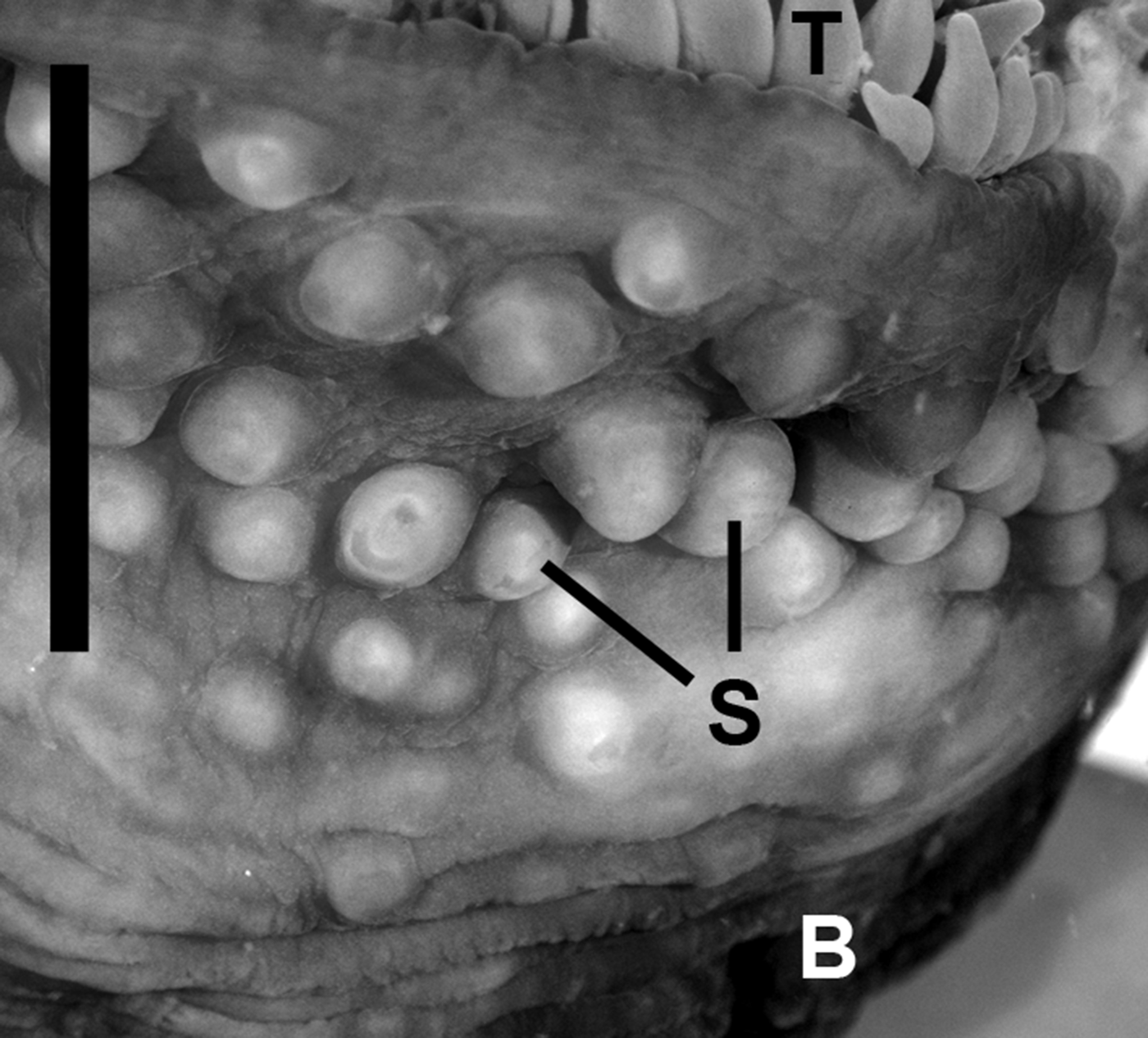
The taxonomic debate about the importance of these features has resulted in both ‘Cnidopus’ and ‘Epiactis’ being used in reference to the North American species: Zamponi & Excoffon (Reference Zamponi and Excoffon1988, Reference Zamponi and Excoffon1995) use C. ritteri whereas Edmands (Reference Edmands1995, Reference Edmands1996) uses E. ritteri; Song (Reference Song1992) and Daly et al. (Reference Daly, Lipscomb and Allard2002) use E. japonica whereas Ishimura & Nishihira (Reference Ishimura and Nishihira2002), Kostina et al. (Reference Kostina, Tsurpalo and Frolova2006), Yanagi & Daly (Reference Yanagi and Daly2004) and Uchida & Soyama (Reference Uchida and Soyama2001) use C. japonicus. England (Reference England and Morton1992) suggested establishing a new genus for the Japanese species to account for a modification of the column commonly found in it, namely the ‘spherules’ occupying the middle region of the column (Figure 1). Fautin et al. (Reference Fautin, Zelenchuk and Raveendran2007) listed the genus Cnidopus as ‘valid’, but at the time of writing, it is considered ‘not valid’ in Fautin's (Reference Fautin2013) online database. Here we follow the most recent genus-level revision (Fautin & Chia, Reference Fautin and Chia1986) and use Epiactis for E. ritteri and E. japonica, but acknowledge that the synonymy of these genera requires evaluation.
Preserved Epiactis japonica. B, band of basal vesicles; S, mid-column spherules; T, tentacles.

The confusion and debate about the attributes of Epiactis ritteri cannot easily be resolved because the critical species lack designated type specimens and vouchers. Neither Torrey (Reference Torrey1902), nor Carlgren (Reference Carlgren1934), nor Hand & Dunn (Reference Hand and Dunn1974) identified individual specimens upon which their respective findings were based. Therefore, it is impossible to consult the original material to adjudicate conflicting taxonomic hypotheses. We attempt to mitigate this obstacle in two ways: by making new collections from type localities and other locations across the range in which these species ostensibly occur, and by borrowing specimens which have been determined to species by the authors of these studies. Furthermore, we timed sampling in Alaska to coincide with the brooding season of the Japanese species to investigate Uchida's (Reference Uchida1934) concern that Torrey's (Reference Torrey1902) omission of brooding in E. ritteri was simply due to seasonality of reproduction. As a result, we are able to identify characteristics by which the species can be individuated. We redescribe the internally brooding species E. ritteri sensu Hand & Dunn (Reference Hand and Dunn1974) as E. handi sp. nov. We redescribe E. ritteri to eliminate confusion about the attributes of that species, document its mode of brooding, provide additional localities, and discuss attributes that may serve to maintain species boundaries between E. ritteri and E. japonica.
MATERIALS AND METHODS
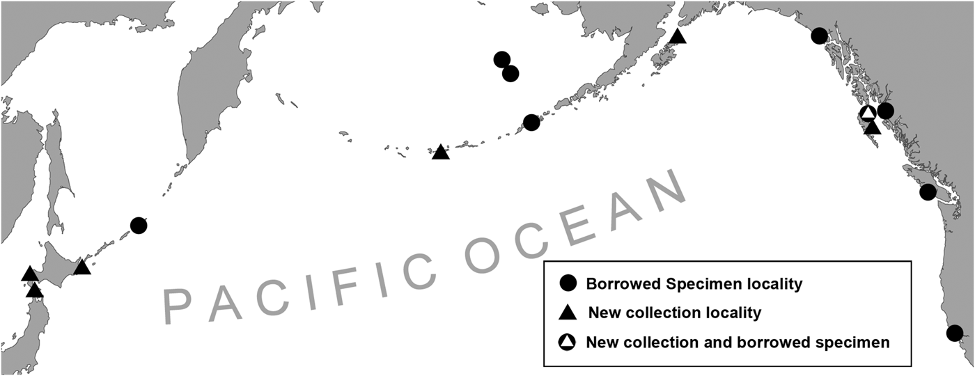
Individuals of species of Epiactis were observed in the field and collected from their substrate at low tide with tools such as forceps and a spatula. Collection sites span the North Pacific (Figure 2). Specimens were preserved in 10% buffered formalin seawater solution (4% formaldehyde) or 95% ethanol. Newly collected material has been deposited at the American Museum of Natural History (AMNH).
Sampling sites and borrowed specimen localities in the North Pacific Ocean. (Some clustered sites omitted for clarity.)
Histological sections were prepared by dehydrating tissue in an alcohol series, clearing it in xylene, and embedding it in Paraplast. Sections were cut at 12–18 μm, mounted on glass slides, and stained with a modified Heidenhain Azan (Presnell & Schreibman, Reference Presnell and Schreibman1997). General anatomical observations were made by eye and under dissecting microscope.
Slide preparations of cnidae were made from squashed tissue samples (less than 1 mm2 tissue/body region/animal) and observed and measured at 1000× under DIC. Length and width were measured using a digital video measurement system. At least 40 capsules of each type were measured for the more abundant types in each tissue; fewer were measured when the type was less abundant. Measured cnidae were selected using a search pattern described by Williams (Reference Williams1996). Cnida terminology follows Mariscal (Reference Mariscal, Muscatine and Lenhoff1974). Measurement range, mean, and standard deviation are reported and intended for qualitative comparison among species rather than for statistical evaluation (see Williams, Reference Williams1996, Reference Williams1998, Reference Williams2000).
RESULTS
SYSTEMATICS
Order ACTINIARIA Hertwig, Reference Hertwig1882
Suborder Enthemonae Rodríguez et al. Reference Rodríguez, Barbeitos, Brugler, Crowley, Grajales, Gusmão, Häussermann, Reft and Daly2014
Superfamily Actinioidea Rodríguez et al. Reference Rodríguez, Barbeitos, Brugler, Crowley, Grajales, Gusmão, Häussermann, Reft and Daly2014
Family ACTINIIDAE Rafinesque, Reference Rafinesque1815
Genus Epiactis Verrill, Reference Verrill1869a
Epiactis handi sp. nov.
(Figures 3–6)
Epiactis handi in situ: (A) partially contracted specimen on rock with adherent debris and bluish basal ring; (B) oral disc of expanded individual; (C) variously coloured individuals attached to rocks slightly below sand surface. D, adherent debris; M, mouth; O, oral disk; T, tentacle.
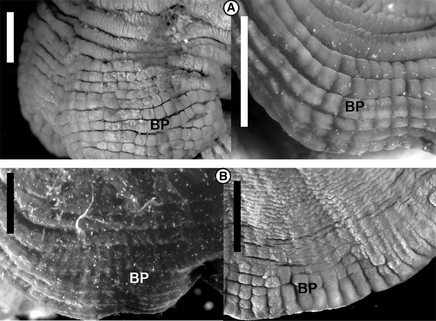
Features of the proximal column in E. ritteri and E. handi. (A) two specimens of E. ritteri showing the continuous band of regular, squarish protuberances characteristic of the species; (B) two specimens of E. handi showing discontinuous, irregular sculpturing at the base of the column. BP, basal protuberances.
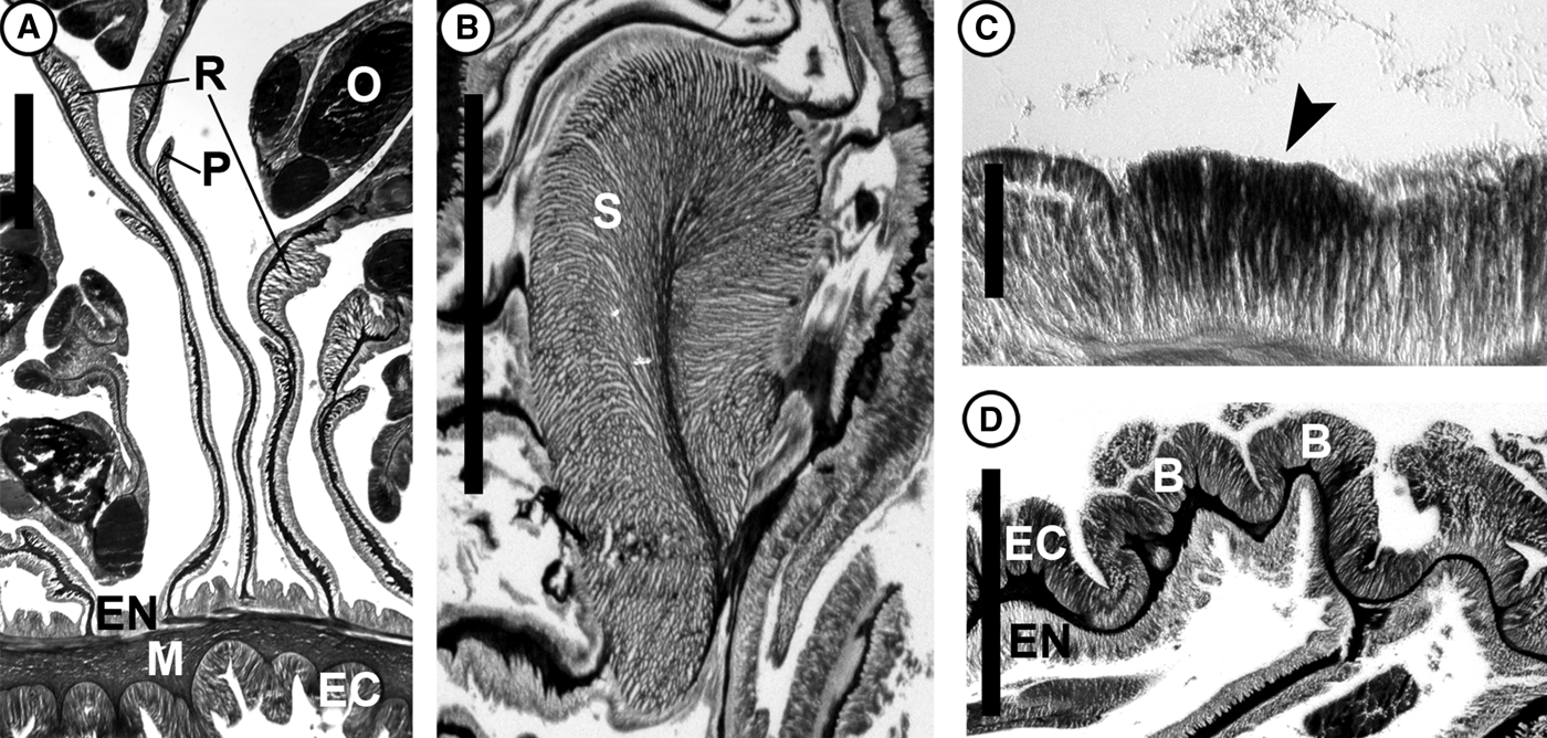
Histological sections of E. handi: (A) cross-section through column and mesenteries; (B) longitudinal section through sphincter; (C) specialized region of ectoderm (arrow); (D) irregular protuberances near limbus. B, basal protuberance; EC, ectoderm; EN, endoderm; M, mesoglea; O, oocyte; P, parietobasilar muscle; R, retractor muscle; S, sphincter. Scale bars: A, 0.5 mm; B, 2 mm; C, 100 μm; D, 1 mm.
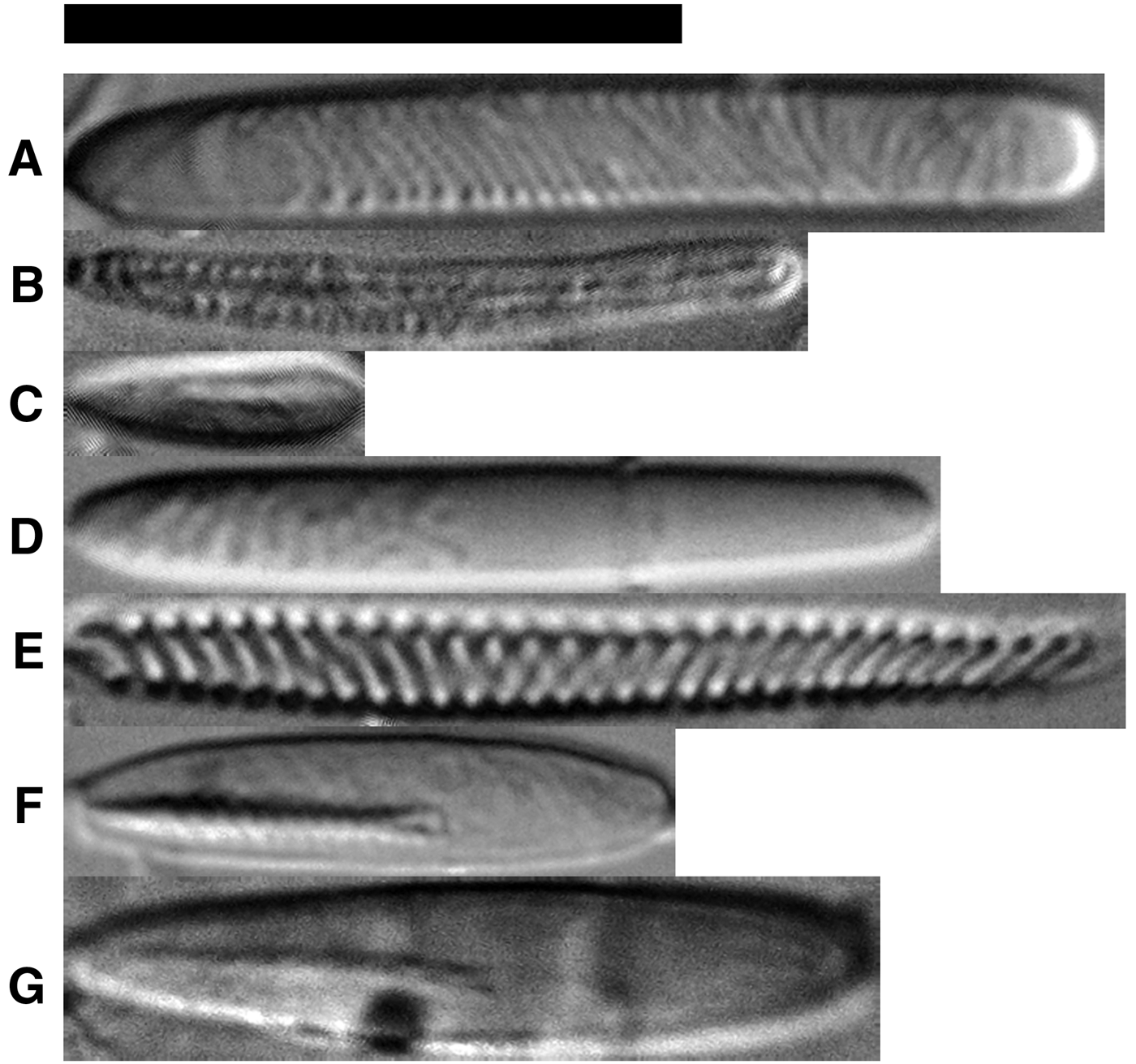
Cnidae of E. handi, E. ritteri, and E. japonica. Letters refer to types in Table 1. Scale bar: 20 μm.
Synonymy
Cnidopus ritteri (Dunn, Reference Dunn1972: 139, 147–153, 156, 166, 173. –Hand & Dunn, Reference Hand and Dunn1974: 187–192. –Fautin & Chia, Reference Fautin and Chia1986: 1665, 1673. –Zamponi & Excoffon, Reference Zamponi and Excoffon1988: 43, 45–48, Reference Zamponi and Excoffon1995: 5. –Zamponi, Reference Zamponi1989: 1–43, Reference Zamponi1993: 13, 15, Reference Zamponi2000: 48, 49.)
Epiactis ritteri (Fautin & Chia, Reference Fautin and Chia1986: 1665, 1670, 1673. –Fautin, et al., Reference Fautin, Siebert, Kozloff and E.N.1987: 76–77. –Zamponi & Excoffon, Reference Zamponi and Excoffon1988: 48. –Edmands & Fautin, Reference Edmands and Fautin1991: 59. –Edmands, Reference Edmands1995: 723–731, Reference Edmands1996: 228–235. –Edmands & Potts, Reference Edmands and Potts1997: 485–489, 491–495.)
Pro parte Epiactis ritteri (Kostina, Reference Kostina1988: 16, 18.)
Non Cnidopus ritteri (Carlgren, Reference Carlgren1934: 350, 351, Reference Carlgren1945: 10, Reference Carlgren1949: 62. –Uchida & Iwata, Reference Uchida and Iwata1954: 224.)
Non Epiactis ritteri (Torrey, Reference Torrey1902: 393–394. –Stephenson, Reference Stephenson1922: 274. –Carlgren, Reference Carlgren1934: 351, Reference Carlgren1947: 92, Reference Carlgren1950: 138, Reference Carlgren1952: 387. –Uchida, Reference Uchida1934: 18, 23, 29, 30. –Fautin et al., Reference Fautin, Zelenchuk and Raveendran2007: 198.)
Non Epiactis riterii [sic] (Uchida & Iwata, Reference Uchida and Iwata1954: 224.)
TYPE SPECIMEN
One specimen in formalin. North of Copper Bay, Moresby Island, British Columbia, Canada; 53.2°, –131.8°; intertidal; [AMNH5293]; coll. P. Larson/M. Daly, 17 October 2013.
OTHER MATERIAL
Three specimens in formalin and 10 microscope slides. West side of Yakan Point, Graham Island, British Columbia, Canada; intertidal; [California Academy of Science (CAS) 61560]; coll. D.G. Fautin/R.W. Buddemeier, 19 August 1986.
Five specimens in formalin and 13 microscope slides. Tow Hill, Graham Island, British Columbia, Canada; intertidal; [CAS 61565]; coll. D.G. Fautin/R.W. Buddemeier, 19 August 1986.
Four specimens in formalin. Tow Hill, Graham Island, British Columbia, Canada; 54.1°, –131.8°; intertidal; [AMNH5291]; coll. P. Larson/M. Daly, 20 October 2013.
Two specimens in formalin. Skidegate, Graham Island, British Columbia, Canada; 53.3°, –132.0°; intertidal; [AMNH5292]; coll. P. Larson/M. Daly, 24 October 2013.
Five specimens in formalin and nine microscope slides. Gray Bay, East coast of Moresby Island, British Columbia, Canada; intertidal; [CAS 61562]; coll. D.G. Fautin/R.W. Buddemeier, 21 August 1986.
One specimen in formalin. North of Copper Bay, Moresby Island, British Columbia, Canada; 53.2°, –131.8°; intertidal; coll. P. Larson/M. Daly, 17 October 2013.
Three specimens in formalin and 10 microscope slides. Newcombe Harbour entrance, McCutcheon Point, Pitt Island, British Columbia, Canada; 53.7°, –130.1°; intertidal; [CAS 61564]; coll. D.G. Fautin, 12 August 1986.
Three specimens in formalin and 10 microscope slides. North Rocks of West Beach, Calvert Island, British Columbia, Canada; 51.65°, –128.15°; intertidal; [CAS 61567]; coll. D.G. Fautin, 4 August 1986.
Seven specimens in formalin and 13 microscope slides. Kooryet Bay, Principe Channel, Banks Island, British Columbia, Canada, 53.33°, –129.87°; intertidal; [CAS 61561]; coll. D.G. Fautin, 11 August 1986.
Thirteen specimens in formalin and 10 histological microscope slides. Wadham's Landing, British Columbia, Canada; 51.48°, –127.53°; intertidal; [CAS 61580]; coll. D. Fautin, 2 August 1986.
Nineteen specimens in formalin and 26 microscope slides. Off Astoria, Oregon, USA; depth: 73 m; [Smithsonian Institution National Museum of Natural History (USNM) 51607]; coll. L. Marriage, 6 December 1947.
One specimen in formalin. NW of Bodega Marine Laboratory, Sonoma Co., California, USA; intertidal; [CAS 103187]; coll. S. Edmands. No date given.
Four specimens in formalin. West of Bodega Marine Laboratory, Bodega Head, Sonoma County, California, USA; intertidal; [CAS 10747]; coll. D.F. Dunn, 1 December 1979.
DIAGNOSIS
Gonochoric Epiactis; females brood internally. Column adherent with sand, bits of shell, or other debris. Ectoderm of column with specialized regions of densely packed, non-glandular cells. Limbus longitudinally furrowed, with abundant holotrichous isorhizae.
Description
COLOUR
Column variable: maroon, salmon, brown, olive green, teal. Limbus usually with lighter longitudinal stripes (solid or interrupted), sometimes with bluish ring overlaying background colour (Figure 3A). Column often finely flecked with spots slightly darker than background colour. Oral disk may differ in colour from column and tentacles, frequently purple; monochrome (Figure 3B), radially striped, or with chevrons at base of tentacles (Figure 3C). Tentacles coloured as column, unornamented except by chevrons at base in some specimens.
COLUMN
Widest at base, to 80 mm in diameter, smooth distally, longitudinally wrinkled proximally. In preserved specimens, annular wrinkles near limbus may intersect with longitudinal wrinkles, producing irregular protuberances of various size and shape: squarish bulges, oblong ridges or slight mounds. These bulges may be less pronounced, variable in shape, or absent across the circumference of a single specimen (Figures 4B, 5D). Distal column smooth, lacks verrucae or vesicles, but ectoderm and ectodermal surface of mesoglea finely corrugate in histological section (Figure 5A). Holotrichs concentrated in clusters on and between basal protuberances, may be present in batteries distally on column (see Hand & Dunn, Reference Hand and Dunn1974). Regions of tightly packed, non-glandular cells (Figure 5C) which lack cnidae and resemble suckers sensu Stephenson (Reference Stephenson1928) present throughout ectoderm of column, possibly responsible for adherence of foreign materials (Figure 3A).
ORAL DISK AND TENTACLES
Mouth slightly raised. Oral disk bare medially; tentacles restricted to periphery. Radial muscles of oral disk ectodermal, circular muscles endodermal. Tentacles hexamerously arranged, slender, conical, terminally perforate, slightly blunt at tips (Figure 3B, C). Circular muscles of tentacle endodermal, longitudinal muscles ectodermal.
INTERNAL ANATOMY
Two siphonoglyphs extend aborally beyond actinopharnyx. Up to five cycles of mesenteries: first and second cycles perfect but sterile, third through fifth imperfect but fertile. Gonochoric. Oral and marginal stomata.
Retractor muscle restricted to diffuse. Parietobasilar muscle strong, with thin flap extending freely from mesentery (Figure 5A). Marginal sphincter endodermal, strong, pinnate, lateral lobe extends proximally (Figure 5B).
CNIDOM
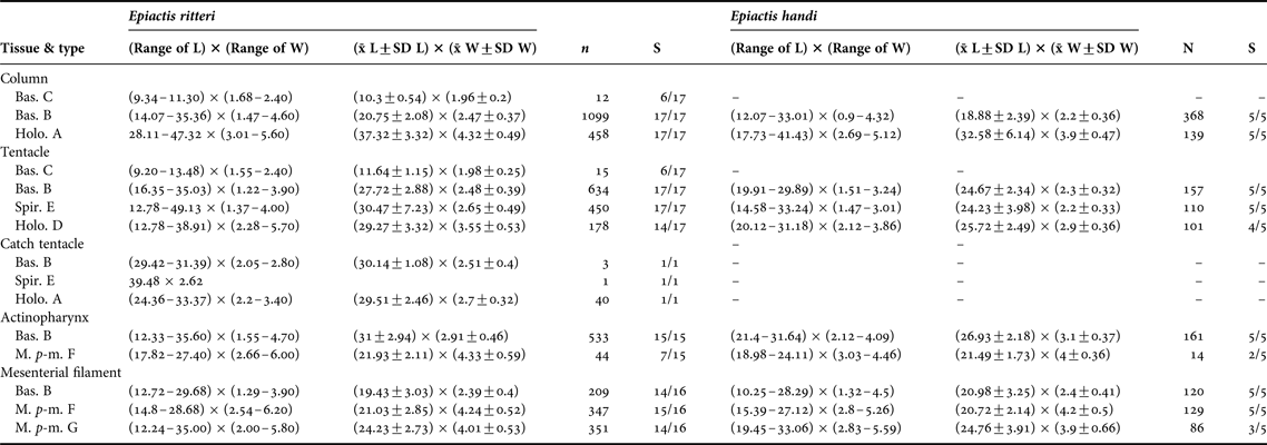
Basitrichs, holotrichs, microbasic p-mastigophores, spirocysts (Figure 6). See Table 1 for size and distribution.
Cnida types found in E. ritteri, E. prolifera and E. handi. Bas., basitrich; Spir., spirocyst; Holo., holotrich; M. p-m., microbasic p-mastigophore; L, length; W, width;  , mean; N, total number of capsules measured; S, proportion of examined specimens with cnida type; SD, standard deviation; Letters correspond with types depicted in Figure 6.
LIFE HISTORY
Internally broods relatively few offspring, but to large size (e.g. one preserved adult in USNM lot 51607 with 37 mm pedal disk diameter contained one offspring; it had 10 mm pedal disk diameter and at least 44 tentacles). Asexual reproduction via budding reported in two individuals from British Columbia, Canada (Zamponi & Excoffon, Reference Zamponi and Excoffon1988); none of our specimens have scars or other indications of this process.
HABITAT, GEOGRAPHIC AND BATHYMETRIC DISTRIBUTION
Eastern Pacific Ocean: Central California to Graham Island, Canada. Occurs from mid intertidal to at least 73 m; attaches to various substrates (e.g. cobbles and boulders in sandy intertidal; mollusc shells, wood, skate egg cases in deeper waters). May be fully exposed upon bare rock at low tide or partially covered with base attached to rocks beneath sand surface. Often most abundant actiniarian in cobble habitats in Haida Gwaii (Queen Charlotte Islands), British Columbia, where sympatric species typically include Urticina coriacea (Cuvier, Reference Cuvier1798), U. clandestina Sanamyan et al., Reference Sanamyan, Sanamyan and McDaniel2013 and occasionally include Anthopleura artemisia (Pickering in Dana, Reference Dana1846) and A. elegantissima (Brandt, Reference Brandt1835). Less abundant and more cryptic towards southern end of range (see Hand & Dunn, Reference Hand and Dunn1974).
REMARKS
Hand & Dunn (Reference Hand and Dunn1974) reported holotrich batteries distally in the column in addition to those on and among basal protuberances. Hand & Dunn (Reference Hand and Dunn1974) suggested the possibility of sequential hermaphrodity based on a single relatively large male specimen, but present material includes large males and females.
ETYMOLOGY
Epiactis handi honours Dr Cadet Hand, former director of the Bodega Marine Laboratory, and senior author of the first report of this species (as E. ritteri, see Hand & Dunn, Reference Hand and Dunn1974).
Epiactis ritteri Torrey, Reference Torrey1902
(Figures 4 & 7–9)
Epiactis ritteri in situ: Three colour morphs in various habits and brooding state: (A) fully emersed, partially contracted individual at low tide with several fully exposed offspring, maroon type; (B) submerged individual bearing zygotes or embryos in a groove which has begun closing (circled area), brown type; (C) submerged individual without offspring, orange type. B, basal band of vesicles; J, juvenile; T, tentacles; Z, zygotes or embryos. Scale bars: 2 cm.
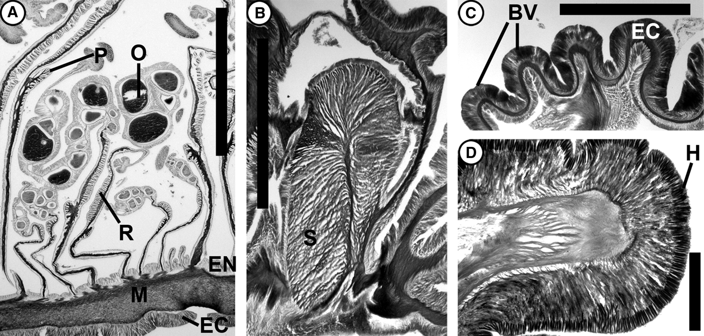
Histological sections of E. ritteri: (A) cross section through column of female; (B) longitudinal section through sphincter; (C) longitudinal section through basal vesicles; (D) basal vesicle detail with stained holotrichs. BV, basal vesicle; EC, ectoderm; EN, endoderm H, holotrichs; M, mesoglea; O, oocyte; P, parietobasilar muscle; R, retractor muscle; S, sphincter. Scale bars: 2 mm.
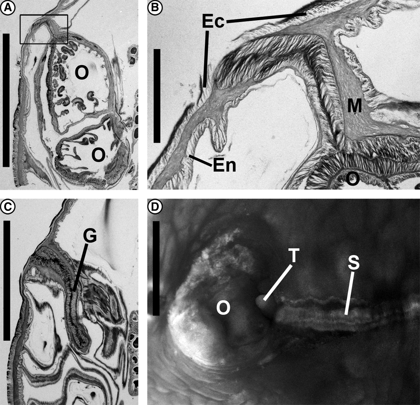
Epiactis ritteri Torrey, Reference Torrey1902: (A) longitudinal section through column of adult showing cross-section of brood groove containing two offspring; (B) detail of brood groove seal boxed area in (A); (C) collapsed groove in space between offspring; (D) external view of brood groove where seam has torn open to reveal an offspring. Ec, ectoderm; En, endoderm; M, mesoglea; G, groove; O, offspring; S, brood groove seam; T, tentacle (of offspring). Scale bars: A, C, D, 4 mm; B, 0.5 mm.
Synonymy
Cnidopus ritteri (Carlgren, Reference Carlgren1934: 350, 351, Reference Carlgren1945 : 10, Reference Carlgren1949 : 62. –Uchida & Iwata, Reference Uchida and Iwata1954 : 224.)
Epiactis ritteri (Torrey, Reference Torrey1902 : 393–394. –Stephenson, Reference Stephenson1922 : 274. –Carlgren, Reference Carlgren1934 : 351, Reference Carlgren1947 : 92, Reference Carlgren1950 : 138, Reference Carlgren1952 : 387. –Uchida, Reference Uchida1934 : 18, 23, 29, 30. –Kostina, Reference Kostina1988 : 16, 18. –Fautin et al., Reference Fautin, Zelenchuk and Raveendran2007 : 198.)
Epiactis riterii [sic] (Uchida & Iwata, Reference Uchida and Iwata1954 : 224.)
Pro parte Epiactis ritteri (Kostina, Reference Kostina1988 : 16, 18.)
Non Cnidopus ritteri (Dunn, Reference Dunn1972 : 139, 147–153, 156, 166, 173. –Hand & Dunn, Reference Hand and Dunn1974 : 187–192. –Fautin & Chia, Reference Fautin and Chia1986 : 1665, 1673. –Zamponi & Excoffon, Reference Zamponi and Excoffon1988 : 43, 45–48, Reference Zamponi and Excoffon1995: 5. –Zamponi, Reference Zamponi1989 : 1–43, Reference Zamponi1993 : 13, 15, Reference Zamponi2000 : 48, 49.)
Non Epiactis ritteri (Fautin & Chia, Reference Fautin and Chia1986: 1665, 1670, 1673. –Fautin et al., Reference Fautin, Siebert, Kozloff and E.N.1987 : 76–77. –Zamponi & Excoffon, Reference Zamponi and Excoffon1988 : 48. –Edmands & Fautin, Reference Edmands and Fautin1991 : 59. –Edmands, Reference Edmands1995 : 723–731, Reference Edmands1996 : 228–235. –Edmands & Potts, Reference Edmands and Potts1997 : 485–489, 491–495.)
MATERIAL EXAMINED
One specimen in 75% ethanol and 13 microscope slides. Sumisher[sic] Island, Kuril Islands, Sea of Okhotsk, Russia); [CAS 31235]; coll. unknown, on or prior to 14 May 1982.
Two specimens in formalin and 35 microscope slides. Palisade Rocks, Adak Island, Alaska, USA; 51.9°, –176.6°; intertidal; [AMNH5294]; coll. P. Larson, 13 April 2012.
Three specimens in formalin and 54 microscope slides. Breakwater, Adak Island, Alaska, USA; 51.9°, –176.6°; intertidal; [AMNH5295]; coll. P. Larson, 14 April 2012.
Two specimens in 75% ethanol. Dutch Harbor, Unalaska Island, Alaska, USA; [CAS 3437]; coll. unknown, 26 May (Year not given).
Two specimens in 70% ethanol and 15 microscope slides (St. George Island, Alaska, USA; intertidal); [USNM 53298]; coll. G D. Hanna, 13 June 1914.
Three specimens in 70% ethanol St. George Island, Alaska, USA; [USNM 43065]; coll. G D. Hanna, 20 August 1914.
Five specimens (plus brooded offspring) in 70% ethanol and 15 microscope slides (St. Paul Island, Alaska, USA); [USNM 32969]; coll. W. L. Hahn, 7 March 1911.
Four specimens in formalin and 41 microscope slides; Mill Bay, Kodiak Island, Alaska, USA; 57.8°, –152.3°intertidal; [AMNH5296]; coll. P. Larson, 19 April 2012.
Three specimens in formalin and 40 microscope slides; Mill Bay, Kodiak Island, Alaska, USA; 57.8°, –152.3°intertidal; [AMNH5297]; coll. P. Larson, 19 April 2012.
Two specimens in 70% ethanol and 15 histological microscope slides. St. George Island, Alaska, USA; intertidal; [USNM 53298]; coll. G D. Hanna, 13 June 1914.
Three specimens in 70% ethanol. St. George Island, Alaska, USA; [USNM 43065]; coll. G D. Hanna, 20 August 1914.
Five specimens (plus brooded offspring) in 70% ethanol and 15 histological microscope slides. (St. Paul Island, Alaska, USA); [USNM 32969]; coll. W.L. Hahn, 7 March 1911.
Three specimens in formalin and four histological microscope slides. Torch Bay, Gulf of Alaska, Alaska, USA; [CAS 52757]; coll. M. Dethier, 1978.
One specimen in formalin and four histological microscope slides. Torch Bay, Gulf of Alaska, Alaska, USA; [CAS 52756]; coll. M. Dethier, 1978.
DIAGNOSIS
Gonochoric Epiactis; females brood offspring externally. Column cylindrical and smooth except for broadly flared, flattened limbus ornamented with bands of square-based protuberances densely packed with holotrichous isorhizae.
Description
COLOUR
Bright orange, maroon, brown, or slightly reddish-purple (Figure 7). Adjacent individuals commonly differ in colour. Monochromatic, excepting one individual in Kodiak with white chevrons at base of tentacles. Inflated tentacles may appear lighter than column, due to slight translucence of thinned tissue.
COLUMN
Moderately sized, 18–53 mm basal diameter preserved, wider in life. Limbus much wider than rest of column, flared out and flattened significantly in live animal (Figure 7). Smooth in histological section, except for a proximal band of regularly arrayed non-adhesive square vesicles (Figures 7B, C & 4A), involving all three layers of body wall (Figure 8C) and armed with abundant holotrichs (Figures 8D & 6A). Mesoglea thicker than ectoderm and endoderm, except at basal vesicles.
ORAL DISK AND TENTACLES
Oral disk bare medially; tentacles restricted to peripheral half. Radial muscles of oral disk ectodermal. Tentacles hexamerously arranged, conical, terminally perforated, slightly blunt at tips. Circular muscles of tentacle endodermal, longitudinal muscles ectodermal.
INTERNAL ANATOMY
Usually two siphonoglyphs, some specimens reported with one (Torrey, Reference Torrey1902). Up to five cycles of mesenteries, first and second perfect but sterile, third through fifth imperfect but fertile. Gonochoric. Oral and marginal stomata present.
Retractor muscle thin, diffuse. Parietobasilar muscle strong, stout, with broad free flap extending freely from mesentery (Figure 8A). Marginal sphincter endodermal, strong, pinnate, with lateral lobe extending proximally, lamellae slightly anastomosing (Figure 8B).
CNIDOM
Basitrichs, holotrichs, microbasic p-mastigophores, spirocysts (Figure 6). See Table 1 for size and distribution.
LIFE HISTORY
Females brood offspring externally in early spring (observed in March, April). Offspring initially within sealed annular groove formed by invagination of the column, later groove disappears and offspring are exposed on surface of parent. Offspring brooded until they have at least three cycles of mesenteries; brooded offspring capable of feeding (copepod observed in gastrovascular cavity of brooded offspring). Mesenteries of brooding females fertile.
HABITAT AND RANGE
Moderately abundant in mid to low rocky intertidal but patchily distributed. Often on bare rock or sponge in tidepools or attached to large (immobile) submerged rocks, but tolerant of full emersion (individuals observed on vertical rock outcrop approximately 60 cm above water surface). Sympatric with Urticina grebelnyi Sanamyan & Sanamyan, Reference Sanamyan and Sanamyan2006, Metridium sp., and Anthopleura artemesia. Aleutian Islands, Bering Sea, Gulf of Alaska, USA.
REMARKS
Torrey (Reference Torrey1902) did not identify type specimens for Epiactis ritteri, and we found no material of an age to have been studied as part of the original description. However, H.B. Torrey was listed as the determiner of USNM 52043, collected in March 1911.
One specimen of Epiactis ritteri [AMNH 5295] deviates from the description above in two respects. First, the sphincter is attached to the mesoglea of the body wall along its entire length, rather than only at the base of a main lamella (as shown in Figure 8B). A main lamella is lacking in this instance, and the branches of the muscle appear to originate from the mesoglea of the body wall along the length of the sphincter. Second, on the column, distal to the brood groove, there are close-set, longitudinal columns of low, small mounds. The endoderm of this region is smooth, but the ectoderm has wrinkles that correspond to the mounds. In some places, the mounds co-occur with a narrow invagination of the endoderm into the mesoglea; this invagination usually does not extend beyond the ectodermal surface of the adjacent column tissue. These are most prominent in the middle, diminishing towards the parapet and brood groove. These bumps differ from the spherules described below in E. japonica (Figure 1) in that they contain mesoglea, are highly ordered in longitudinal rows, densely spaced, and irregular and in section. In E. japonica, there are large, circular, widely spaced spherules, variable in number from zero to several dozens, usually scattered irregularly on all parts of the column except the basal protuberances. High, broad evaginations of all three layers of the body wall result in a distinct, hollow, bulbous protuberance in E. japonica.
DISCUSSION
Distinguishing E. handi from E. ritteri
Our findings confirm the assertion of Sanamyan & Sanamyan (Reference Sanamyan and Sanamyan1998) that Hand & Dunn's (Reference Hand and Dunn1974) ‘redescription’ of Epiactis ritteri was actually an account of a new species, which we describe and name here as Epiactis handi sp. nov. This may also be the Epiactis species from Oregon which Carlgren (Reference Carlgren1952: 387) mentioned (but did not describe or name) as having ‘2 embryos provided with several tentacles in the gastral cavity’.
Epiactis handi and E. ritteri have much in common with respect to size, distribution of cnidae, and internal anatomy, but differ in several ways. The most readily apparent difference in living specimens are in the column: a specimen of E. ritteri lacks adherent foreign material and bears offspring, whereas one of E. handi has adherent material and lacks externally brooded offspring. However, as these are seasonal or situational differences (e.g. adherent material is easily lost in preserved specimens, brooding is seasonal and restricted to females in E. ritteri) we discuss more consistently evident anatomical differences below.
The anatomy of the base of the column has contributed to the confusion about the identity of Epiactis ritteri and to disagreement about the necessity of a separate genus for species having bumpy (vs smooth) columns. Torrey (Reference Torrey1902) noted ‘protuberances’ caused by transverse and longitudinal wrinkles in the column, but Carlgren (Reference Carlgren1934) described them more fully, determining that they harbour a type of nematocyst (originally called an ‘atrich’, now considered a holotrich: see Westfall, Reference Westfall1965) restricted to the limbus in E. ritteri. In E. ritteri, these protuberances are non-adhesive and satisfy Carlgren's (Reference Carlgren1949) definition of ‘vesicle’ but not ‘verruca’. Hand & Dunn (Reference Hand and Dunn1974: 190) use the term ‘verruca’ for the basal protuberances of what we describe as E. handi, citing their adhesive nature and Carlgren's (Reference Carlgren1952: 387) use of that term for them. In that instance, however, Carlgren was actually referring to the relatively large, hemispherical mid-column spherules found in the Japanese species E. japonica (Figure 1), and he goes on to note their absence in E. ritteri: ‘The protuberances which usually occur in the middle part of the body are, as far as I can see, verrucae though they probably are not very adhesive. …Neither Torrey nor I myself have found any verrucae in the middle region of the body …’ We do not consider the basal protuberances in E. ritteri to be verrucae, preferring the terms vesicle or basal protuberance. We concur with Hand & Dunn (Reference Hand and Dunn1974) that the column of E. handi is adhesive (see below), but we fail to find hollow outgrowths with the histological characteristics ascribed to verrucae (Stephenson, Reference Stephenson1928; Häussermann, Reference Häussermann2004: see also Figure 5). The basal column of E. handi is smooth or with longitudinal wrinkles that sometimes intersect with annular wrinkles to form restricted protuberances. Distributed among the protuberances and the rest of the column are sucker-like (sensu Stephenson, Reference Stephenson1928) regions of ectodermal tissue and batteries of holotrichs, however the latter are primarily found in the basal region.
The basal protuberances differ in arrangement and morphology between species. In Epiactis handi, they are irregular in size and shape, do not form a consistent band around the perimeter near the limbus, and primarily appear after death and fixation, if they are present at all (Figure 4B); slight basal longitudinal wrinkles may be evident in life. In contrast, in E. ritteri, multiple, regular rows of fairly uniform, squared protuberances form a complete band around the base of the animal in life (Figure 7) and in preserved specimens (Figure 4A). The number of rows in a band of protuberances in E. ritteri appears to be loosely correlated with size (0 rows in brooded offspring, 5–8 rows in smaller specimens from Adak and Kodiak, up to 16 rows in large specimens from Unalaska Island and the Pribilof Islands).
Two other differences in the column are evident in the histology of the ectoderm and mesoglea. First, in preserved specimens of Epiactis handi, the ectoderm and the ectodermal surface of the mesoglea are finely wrinkled and branched (Figure 5A), whereas in E. ritteri these layers of the body wall are smooth (Figure 8A) even in contracted animals. Second, the column ectoderm of E. handi has regions of densely packed cells which lack nematocysts (Figure 5C), but these are not found in E. ritteri. Hand & Dunn (Reference Hand and Dunn1974) proposed that these specialized areas are comparable to ‘suckers’ sensu Stephenson (Reference Stephenson1928) and may facilitate sand grain adhesion to the column.
Live animals of the two species differ in colour and are easily distinguished. Epiactis ritteri is monochromatic: tentacles, oral disc and column are uniform orange, maroon, brown or slightly reddish-purple (Figure 7). The single deviation from this pattern was one specimen with white chevrons at the base of the tentacles. In contrast, in E. handi, the basal column and oral disk is more variable, including multiple colours in a single individual, sometimes similar in colour and pattern to E. prolifera with radiating white lines (Dunn, Reference Dunn1972; Hand & Dunn, Reference Hand and Dunn1974), and sometimes with a blue ring at the limbus (Figure 3A).
A fuller account of E. ritteri
Our new material allows us to flesh out and complete earlier accounts of Epiactis ritteri. Most critically, we lay to rest controversy about the mode of brooding: E. ritteri broods externally (Figure 7A), with some specimens retaining young within a tightly sealed brood-groove (Figure 9). Brood grooves have been reported in other externally brooding species (e.g. Cricophorus nutrix: Carlgren, Reference Carlgren1924; Bunodactis mortenseni: Carlgren, Reference Carlgren1924; Epiactis japonica: Ishimura & Nishihira, Reference Ishimura and Nishihira2002) and are formed by a depression that encircles the column of the adult. The brood groove has been hypothesized to provide protection or aid in the retention of brooded offspring, especially in adults which are not attached to horizontal surfaces (Ishimura & Nishihira, Reference Ishimura and Nishihira2002). In Uchida & Iwata's (Reference Uchida and Iwata1954) study of development in E. japonica, they did not discuss brood grooves or their formation but noted that embryos were ‘nearly wrapped with the mother's ectoderm’ (Uchida & Iwata, Reference Uchida and Iwata1954: 222). Ishimura & Nishihira (Reference Ishimura and Nishihira2002) described the process of groove formation in E. japonica, noting that groove formation occurs simultaneously with offspring deposition: the mouth is extended toward the column causing a fold in the column to form, and this fold is retained as the mouth revolves around the animal and deposits offspring into it. The offspring may be completely covered by the lower edge of this groove, but can also be visible when the tentacles of the parent are retracted (Ishimura & Nishihira, Reference Ishimura and Nishihira2002). Aulactinia sulcata (Clubb, Reference Clubb1902) exhibits enigmatic structures in the column that are superficially similar to the brood groove described here for E. ritteri. Clubb (Reference Clubb1902) described an annular groove that formed from a series of pits in the column (apparently prior to the appearance of offspring); these pits expand and merge through breakdown of the walls separating them, resulting in four to six large chambers invaginating proximally from a shallower annular groove. His longitudinal sections of a chamber appear similar to Figure 9 except notably, the meeting ectodermal layers at the edge of the groove are rounded. Carlgren & Stephenson (Reference Carlgren and Stephenson1929) disputed Clubb's (Reference Clubb1902) description, instead describing a single invagination which soon bifurcates and extends its branches in opposite directions to form a single, nearly toroidal brood chamber within the adult. Dunn (Reference Dunn1983) mentioned a specimen of A. sulctata with a single invaginated brood chamber that was empty and had been perforated by the mesenteries. In any case, the basic nature of the brood groove in E. ritteri is different from the structures found in A. sulcata in that it is a single, continuous depression in the column rather than a series of distinct, invaginated pouches.
Although spawning has not yet been observed in Epiactis ritteri, late-stage embryos can be found within a brood groove that is similar to that described for E. japonica. In E. ritteri, the groove is tightly sealed by the time offspring within the groove form clumps of small orange spheres 0.5 mm in diameter (Figure 7B), and the two ectodermal edges of the groove touch (Figure 9). In this state, the column of the brooding adult has a smooth appearance in spite of being considerably folded. The opening to the groove is cryptic, evident only as a thin suture around the animal (Figure 9D). Because the groove is cryptic, several specimens which were initially thought to be non-brooding were revealed to bear offspring once dissected.
In histological section, the mesogleal and ectodermal layers near the opening of the brood groove appear to have been under tensile stress at fixation (Figure 9B), suggesting some mechanism by which the ectodermal surfaces adhere to one another. Gently probing the suture with a needle failed to separate ectodermal surfaces from one another in a preserved individual. In the spaces between adjacent offspring, the groove is completely collapsed upon itself (Figure 9C). In some specimens, the groove has been everted, probably during especially strong contractions of the adult during fixation. The everted groove is a somewhat globular ring of thin column tissue with offspring attached to bulges.
In Epiactis ritteri, offspring remain fully enclosed within the groove even after they have developed tentacles and a pedal disk; most offspring were contracted and hard to evaluate, but an expanded individual in a forcibly everted groove had 18 tentacles. As the groove opens and begins to disappear, offspring remain on the adult, fully exposed to the environment, similar to the situation in E. prolifera (see Verrill, Reference Verrill1869a). We have found a copepod in the gastrovascular cavity of an offspring on the (exposed) column of the parent, indicating exposed young are capable of feeding. Nonetheless, in the several examined specimens, histological sections reveal yolk granules in the gastrovascular cavity of exposed, attached offspring.
Individuals in which juveniles were fully exposed upon the adult had fewer (8–23) young than those which still held offspring within a groove (≥23–≥53). Counts of those in the latter state are limited to portions of the animal which have been dissected, or in which the groove has been everted, and thus are minimum numbers for those specimens. The difference may be due to juveniles having left the parent for independent life or having been washed off the adult once exposed. Closed brood grooves may contain hundreds of pre-juvenile offspring (prior to tentacle and pedal disk formation: i.e. zygotes or embryos).
In Epiactis ritteri, the offspring upon a brooding parent were all in a similar stage of development, but the broods of different adults from the same locality and collection date were at various stages, from zygote (or egg?) to juvenile with at least three cycles of mesenteries. Only (but not all) females brooded offspring and only a single male was collected in 2012. Borrowed material also contained only male or female individuals, thus there is yet no evidence for hermaphrodity in E. ritteri. Reproduction appears to occur on a seasonal basis, with all of an individual's efforts occurring in one event; this is in contrast with E. prolifera, in which individuals may reproduce continually throughout the year (Dunn, Reference Dunn1975). Despite the seasonality of reproduction, as the diversity of developmental stages seen within the same population indicates, reproduction is not highly synchronized between all individuals in a population. All adults collected by PL in Alaska were fertile in April regardless of reproductive state. Such overlapping of generations (i.e. brooded juveniles concurrent with developing oocytes in the parent) and relative asynchrony within populations has also been described by Rodriguez et al. (Reference Rodriguez, Orejas, López-González and Gili2012) in the externally brooding Antarctic species E. georgiana Carlgren, 1927.
The duration and timing of brooding season for Epiactis ritteri is unknown. In April 2012, the largest brooded offspring from Kodiak and Adak islands had a pedal disk diameter of 3.5 mm (measured after fixation); the smallest were small spheres (eggs or early embryos) no larger than the oocytes still held within the parent (~0.5 mm). A specimen from Alaska [USNM 52043], collected in March 1911, bore (externally) much larger offspring, ranging in pedal disk diameter from 4.9–16.5 mm (preserved). This specimen was determined by Torrey (see above), but its reproductive state has not been acknowledged in any published account of E. ritteri. This size discrepancy among broods also mirrors the findings of Rodriguez et al. (Reference Rodriguez, Orejas, López-González and Gili2012) in E. georgiana.
In addition to providing details of reproductive biology, we provide the first thorough account of the cnidom of Epiactis ritteri. Carlgren (Reference Carlgren1934) reported measurements of cnidae only for the column, tentacles and actinopharynx, and did not specify the number of individuals studied or the number of capsules measured. Our measurements (Table 1) agree with his in general, except our size ranges are slightly broader and we include several rare or localized types he did not report. The column and tentacles of E. ritteri both contain holotrichs, but these differ in morphology as undischarged capsules. Holotrichs of the tentacle (Figure 6D) have a thicker tubule visible only in the apex-end of the capsule when undischarged; the tubule may be coiled but is most often packed irregularly. In contrast, the holotrichs in the column (Figure 6A) have a thinner tubule which is visible throughout the capsule and is usually regularly coiled. The former appear to correspond with the holotrichs figured in Fautin & Chia (Reference Fautin and Chia1986), although Fautin & Chia (Reference Fautin and Chia1986) made no distinction between those of the column and tentacle, and only depict one type. As Fautin & Chia (Reference Fautin and Chia1986) reported for E. lisbethae, E. fernaldi and E. prolifera, holotrichs in the tentacles of E. ritteri and E. handi are most abundant in the tentacles nearest the margin and may be rare or absent in a tentacle nearer the mouth. In a tentacle of one specimen of E. ritteri, we encountered very many holotrichs of the type normally found in column (Figure 6A). Spirocysts were very rare in the sample from this tentacle. This is possibly similar to the abnormally long ‘catch tentacles’ that Sanamyan & Sanamyan (Reference Sanamyan and Sanamyan1998, Reference Sanamyan and Sanamyan2008, Reference Sanamyan, Sanamyan, K.E., N.P., A.M. and O.A.2010) described occurring among the usual tentacle type in some of their specimens of E. japonica. In our material, however, there was no macroscopic indication (e.g. length, thickness) that the tentacle was different from others, and it was found only by chance. Re-inspection of all specimens of E. ritteri and haphazard resampling of tentacles yielded no more of this type of tentacle. We did, however, find several such tentacles in a preserved specimen of E. japonica, but in that case the ‘catch tentacle’ type was distinct in being slightly longer, conical and more opaque (tissue denser or thicker) in contrast to the tentacles with the more typical complement of cnidae, which were extremely contracted and more translucent.
Differences in size and distribution of cnida among North Pacific Epiactis species are either very subtle (if not an artifact of sampling) or occur in cnida types that are highly variable in their distribution within a species. Thus, attributes of the cnidom are not practically useful for distinguishing these species. Whether certain types of cnidae that are not found consistently (e.g. microbasic p-mastigophore of the actinopharynx) are missed due to true absence from particular specimens or patchy distribution within an individual is uncertain. Holotrichs can be found in the basal column and tentacles of E. ritteri, E. handi, E. japonica, E. prolifera, E. lisbethae (personal observations), however their relative abundance differs dramatically, being very common in the former three species, and less common in the latter two.
Distinguishing E. ritteri from E. japonica
Although Epiactis japonica and E. ritteri are similar in many respects, we note differences that argue for their continued recognition as separate species. Individuals of E. japonica are possibly protogynous (Uchida & Iwata, Reference Uchida and Iwata1954), but eventually become simultaneously hermaphroditic (Uchida & Iwata, Reference Uchida and Iwata1954; Ishimura & Nishihira, Reference Ishimura and Nishihira2002; Sanamyan & Sanamyan, Reference Sanamyan, Sanamyan, K.E., N.P., A.M. and O.A.2010), although gametes of a single type are spatially separated (on different mesenteries, or proximo-distally within a single mesentery) within the animal (Uchida & Iwata, Reference Uchida and Iwata1954). Torrey and Carlgren did not discuss sex allocation in discussion of mesenteric fertility in E. ritteri, but all material examined here exhibit the gonochoric condition. Because we find no hermaphrodites, even among the largest specimens, we think that simultaneous hermaphrodity in E. ritteri is rare or absent.
The most apparent external differences between Epiactis ritteri and E. japonica are colouration and ornamentation of the column. Colour variation is very low among the specimens we collected in Alaska: only 2–3 colour varieties are present (bright orange, dark maroon, to dark purplish-brown). In the field, all individuals we saw were monochromatic, except one individual with white chevrons at base of its tentacles. In E. japonica, in contrast, colour and patterning are extremely variable. In Hokkaido, we observed individuals that vary from monochrome, to simple bicoloured (tentacles differ from column), to irregularly streaked and mottled (column three or more colours). The oral disk of E. japonica is often radially striped with two or more colours. The background body colours of E. japonica commonly include red, maroon, pink, brown, tan, beige, green and rarely bright blue. The monochromatic and relatively reduced suite of colours in E. ritteri is consistent across the range from Kodiak to Adak, ~1700 km, following the Alaska Peninsula and Aleutian chain (see Lindeberg, Reference Lindeberg2008 for images from intermediate sites; species identified as E. prolifera).
In our material of E. japonica (from Hokkaido, Japan), we found non-adhesive, hollow (i.e. involving all three layers of body wall), approximately hemispherical protuberances, on the middle and distal portion of the column of many of the smaller and all of the larger individuals (Figure 1). These were referred to as ‘papillae’ by Uchida (Reference Uchida1938), ‘verrucae’ by Carlgren (Reference Carlgren1952) and ‘spherules’ by England (Reference England and Morton1992) and Sanamyan & Sanamyan (Reference Sanamyan and Sanamyan1998). The one-third (22 of 71) of the collected individuals we examined that lacked such protuberances had a pedal disk diameter of 17 mm or smaller (preserved); some individuals <17 mm pedal disk diameter and all individuals larger than this size (up to ~54 mm pedal disk diameter) possessed at least one (but usually many) such protuberances. Sanamyan & Sanamyan (Reference Sanamyan and Sanamyan1998) report individuals that they identify as E. japonica with and without mid-column protuberances co-occurring in the Commander Islands, but make no mention of relative sizes. We infer that this feature is acquired or becomes apparent as the animal grows. Noting the similarity in appearance between contracted, brooded offspring and mid-columnal spherules, England (Reference England and Morton1992) proposed that these structures served as protective camouflage for brooded young. These spherules are absent in E. ritteri: we do not see them in preserved specimens or live animals of any size, a conclusion that accords with Carlgren's (Reference Carlgren1952) note that neither he nor Torrey had observed them after examining ‘many’ specimens.
The brood groove is different in Epiactis japonica and E. ritteri. Although life history, reproduction and development has been thoroughly studied in E. japonica (e.g. Uchida & Iwata, Reference Uchida and Iwata1954; Ishimura & Nishihira, Reference Ishimura and Nishihira2002) no one has described such a sealed groove in this species. While offspring may be covered, initially, the groove that is formed in E. japonica remains a depression partially open to the environment and offspring are often visible as tiny spheres on the column (e.g. Uchida & Iwata, Reference Uchida and Iwata1954; Ishimura & Nishihira, Reference Ishimura and Nishihira2002). We find that in Alaskan E. ritteri, the brood groove is sealed early, before larvae develop tentacles, and remains sealed even when the offspring have multiple cycles of tentacles.
Based on our observations, the Alaskan specimens of Epiactis ritteri are distinct from the Japanese specimens of E. japonica in terms of sex allocation, characteristics of brooding, and some morphological features, but the variability present in E. japonica across its range complicates species identification based on external characters alone, especially if these species occur in sympatry, as Sanamyan & Sanamyan (Reference Sanamyan and Sanamyan1998) suggested happens in the Kurile Islands. Sanamyan & Sanamyan (Reference Sanamyan and Sanamyan1998) were initially inclined to consider individuals with and without mid-column spherules as separate species (i.e. as E. japonica and E. ritteri respectively), but because of the variability in presence and extent of that feature in specimens from the Commander Islands, Kamchatka Peninsula, Kurile Islands, and the Sea of Japan, they were prompted instead to synonymize E. ritteri and E. japonica. Sanamyan & Sanamyan (Reference Sanamyan and Sanamyan1998) did not report sex allocation for their specimens, but indicated that those with and without the spherules were not significantly different anatomically. The variability in features that they report is characteristic of the Japanese species (Sanamyan & Sanamyan, Reference Sanamyan and Sanamyan1998, Reference Sanamyan and Sanamyan2008, Reference Sanamyan, Sanamyan, K.E., N.P., A.M. and O.A.2010) but is not seen in the specimens from Alaska, at least from Adak Island eastward. Based on the differences (described above) between Japanese and Alaskan individuals in sex allocation, characteristics of the brooding groove, and presence of mid-column spherules, we opt to maintain E. japonica and E. ritteri as separate species. An intermediate zone of sympatry and/or hybridization may exist somewhere among the Kurile Islands, Kamchatka Peninsula or Commander Islands.
Furthermore, if rafting is an important mode of long-distance dispersal, as is often assumed for benthic species with non-pelagic development (reviewed by Thiel & Gutow, Reference Thiel and Gutow2005), ocean currents could provide some degree of isolation for the Alaskan individuals from those in Japan and the Kurile and Commander Islands. The Alaskan Stream is a strong westward current which flows along the coast of the Gulf of Alaska and the southern shores of the Aleutian Islands, maximally reaching about 170° E longitude (Favorite, Reference Favorite1965). The relatively weak eastward flow on the north side of the Aleutian Islands (Aleutian North Slope Current) primarily originates from waters of the Alaskan Stream moving north through passes between the Aleutian Islands (Reed & Stabeno, Reference Reed and Stabeno1994; Chen & Firing, Reference Chen and Firing2006).
ACKNOWLEDGEMENTS
We thank Dr Satoshi Wada (Hokkaido University) for hosting and supporting PL during his trip to Japan, Sarah Gerken (University of Alaska, Anchorage), Eric Munk (Kodiak Fisheries Research Center) and Raymond O'Neill for logistical support and advice on sampling in Alaska, and Terry Gosliner (CAS), Alison Young (CAS) and Jackie Sones (Bodega Marine Laboratory) for assistance and advice in California. Melanie Anthony (Department of Fisheries and Oceans, Canada) and Sara Conrad (Alaska Department of Fish and Game) provided swift and efficient assistance with acquiring permits for collections. We also thank the Aleut and Shumagin native corporations of Alaska for permission to access the intertidal via their land. Comments from Daphne Fautin and two anonymous reviewers improved this manuscript.
FINANCIAL SUPPORT
Collections in Japan were supported by the NSF East Asia Pacific Summer Institute grant in conjunction with the Japan Society for Promotion of Science Summer Program fellowship. Alaskan collections supported by American Museum of Natural History Lerner Gray fund for Marine Research and a private contribution from Greg & Jule Larson. Canadian field work funded by NSF DEB-1208523 to MD.