Introduction
Cryptosporidium parvum is a small intracellular parasite that infects a wide range of mammalian hosts, including humans and calves. The introduction of highly active anti-retroviral therapy (HAART) against HIV infection has significantly reduced the prevalence of severe human cases of Cryptosporidium infection in industrialized countries (Ives et al., Reference Ives, Gazzard and Easterbrook2001). However, in developing countries, cryptosporidiosis remains a major life-threat to young children (Ventura et al., Reference Ventura, Cauda, Larocca, Riccioni, Tumbarello and Lucia1997; Kotloff et al., Reference Kotloff, Nataro, Blackwelder, Nasrin, Farag, Panchalingam, Wo, Sow, Sur, Breiman, Faruque, Zaidi, Saha, Alonso, Tamboura, Sanogo, Onwuchekwa, Manna, Ramamurthy, Kanungo, Ochieng, Omore, Oundo, Hossain, Das, Ahmed, Qureshi, Quadri, Adegbola, Antonio, Hossain, Akinsola, Mandomando, Nhampossa, Acácio, Biswas, O'Reilly, Mintz, Berkeley, Muhsen, Sommerfelt, Robins-Browne and Levine2013). The only approved drug to treat human cryptosporidiosis, nitazoxanide, shows poor efficacy in immunocompromised patients and malnourished children (Manjunatha et al., Reference Manjunatha, Vinayak, Zambriski, Chao, Sy, Noble, Bonamy, Kondreddi, Zou, Gedeck, Brooks, Herbert, Sateriale, Tandel, Noh, Lakshminarayana, Lim, Goodman, Feng, Zhang, Blasco, Wagner, Joel, Striepen and Diagana2017). Novel or alternative treatment strategies are desperately needed; however, no major breakthrough has been achieved so far. Compared to other apicomplexan parasites, Cryptosporidium research is still at its infancy. Particularly the development of tools to edit the parasite genome has been a major frustration point and the transfection of Cryptosporidium is still a major challenge. Current protocols are based on electroporation of freshly excysted sporozoites using a rather large amount of plasmid DNA which typically results in a very poor yield of transfected parasites.
The poly cationic polymer polyethyleneimine (PEI) with its tertiary amines possesses remarkable DNA condensing properties but can also provide essential buffering capacities that enable an efficient endosomal escape and nucleus penetration (Boussif et al., Reference Boussif, Lezoualc'ht, Antoniet, Mergnyt, Scherman, Demeneix and Berh1995; Fischer et al., Reference Fischer, Bieber, Li, Elsässer and Kissel1999). However, due to its cellular toxicity, PEI has not been widely used in research (Florea et al., Reference Florea, Meaney, Junginger and Borchard2002; Moghimi et al., Reference Moghimi, Symonds, Murray, Hunter, Debska and Szewczyk2005). Importantly, it has been reported that combining PEI with cell-penetrating peptides (CPPs) significantly reduces its cytotoxic effect while the gene delivery efficiency is increased (Kilk et al., Reference Kilk, El-Andaloussi, Järver, Meikas, Valkna, Bartfai and Langel2005; Munyendo et al., Reference Munyendo, Lv, Benza-Ingoula, Baraza and Zhou2012; Yang et al., Reference Yang, Wang, Sun and Zheng2019). The small peptide octaarginine possesses known cell-penetrating properties and is known to cross all known biological membranes. Herein we report a simple and fast transfection protocol that combines the properties of the two molecules to achieve the transfection of Cryptosporidium with a minimal amount of DNA. The novel protocol does not require electroporation and works best with oocysts immediately after initiation of excystation rather than with excysted sporozoites.
Materials and methods
Preparation and excystation of C. parvum sporozoites
The house strain Cryptosporidium parvum (GP-60 subtype IIa, A15G2RI) was originally isolated from a commercial cattle farm (Wilchwitz, Germany in 2015). For propagation, neonate calves were orally infected with 9 × 10⁶ oocysts. Oocysts were collected from the feces 4–13 days post infection, cleaned and isolated following the protocol described by Najdrowski et al. (Reference Najdrowski, Heckeroth, Wackwitz, Gawlowska, Mackenstedt, Kliemt and Daugschies2007). Subsequently, cleaned oocysts were stored in PBS pH = 7.2 (Gibco®, ThermoFisher Scientific, Massachusetts, USA) supplemented with penicilline/streptomycine (200 μg mL−1) and amphotericin B (5 μg mL−1) at 4°C for up to 3 months before using. The PBS pH 7.2 described above was used throughout this study and will be referred to as PBS in the remaining text. The storage medium was changed monthly to prevent bacterial and fungal contamination. Before excystation, oocysts were bleached with 5.25% sodium hypochlorite in PBS in the ratio 1:1 then put on ice for 5 min. Decontaminated oocysts were washed 3 times with cold PBS and resuspended in excystation medium (0.8% sodium taurocholate -NaT in the DMEM with 2% fetal calf serum, 1% amphotericin B and 1% penicillin/streptomycine, 1% sodium pyruvate) and pre-incubated at 15°C for 10 min, then further incubated at 37°C and 5% CO2 for 3 h. Sporozoites were centrifuged for 5 min at 9,500 × g (Centrifuge MIKRO 200, Merck, Darmstadt, Germany) to remove excystation medium and then resuspended in the infection medium (DMEM with 2% fetal calf serum, 1% amphotericin B and 1% penicillin/streptomycine, 1% sodium pyruvate)
Preparation of short time excystation exposed (STE) oocysts
STE oocysts were obtained through a similar procedure which was described in the preparation of C. parvum sporozoites. However, the incubation step at 37°C in the presence of excystation medium was reduced to only 15 min.
Synthesis of 6-Fam-labelled octaarginine
Octaarginine labelled with 6-carboxyfluorescein (R8-FAM) was synthesized as described in (Sparr et al., Reference Sparr, Purkayastha, Kolesinska, Gengenbacher, Amulic and Matuschewski2013).
Uptake of 6-Fam-labeled octaarginine and direct fluorescence microscopy
6-Fam-labelled octaarginine (10 μg mL−1) was added to either intact oocysts, STE oocysts or sporozoites of C. parvum, followed by further incubation in infection medium (DMEM with 2% fetal calf serum, 1% amphotericin B and 1% penicillin/streptomycine, 1% sodium pyruvate) for 30 min at 37°C. Intracellular 6-Fam-labeled octaarginine was visualized microscopically by direct fluorescence using a Leica TCS SP8 laser scanning confocal microscope (Leica, Wetzlar, Germany). Briefly, parasites were centrifuged for 5 min at 9,500 × g, then washed with PBS 3 times. Parasites were then fixed with 4% paraformaldehyde (PFA) for 20 min at room temperature, followed by washing 3 times with PBS. Next, 4,6-diamidino-2-phenylindole (DAPI; 10 μg mL−1) was added and incubated for another 10 min at room temperature. After a final extensive washing 3 times with PBS, parasites were mounted on glass slides using fluoromount-G (Southern Biotech, Birmingham, USA).
DNA condensation by PEI/octaarginine and gel retardation assay
Branched PEI (60 kDa), (Sigma-Aldrich, Hamburg, Germany) was dissolved in distilled water to prepare a stock solution of 10 mg mL−1. PEI was passed through a 0.2 μm filter (Millipore® Merck, Darmstadt, Germany) and stored at −20°C until use. For these experiments, we used the GFP-expressing plasmid Eno_prom_GFP_neo_eno_utr. This is a plasmid using an enolase promotor and containing a neomycin resistance gene. Various amounts of DNA/PEI were combined to form complexes with plasmid DNA concentrations ranging from 1 μg to 20 μg and having different N/P ratios where N represents the number of moles of amine nitrogen in PEI and P is the number of moles of phosphate in the DNA. The calculation for the ratio N/P was done as described by Boussif et al. (Reference Boussif, Lezoualc'ht, Antoniet, Mergnyt, Scherman, Demeneix and Berh1995). Briefly, given the molecular weight of a nucleotide of 330 g/mol, 10 μg of plasmid represent 30 nmol. In order to obtain an N/P ratio of 10, the concentration of PEI needed is 300 nmol which represents 1.3 μL from the 10 mg mL−1 stock solution. For gel retardation assay, solutions of DNA/PEI or DNA/PEI plus octaarginine (in 10-fold excess) were prepared and incubated at 37°C for 30 min, in order to allow DNA condensation. The complexes of DNA/PEI and DNA/PEI with octaagrinine were separated on 0.9% agarose gel electrophoresis for 45 min and 120 V. GeneRuler 1 kb DNA Ladder® (Thermo Fisher Scientific, Waltham, Massachusetts, USA) was used as a reference. The complexes were visualized by staining with 0.01% ethidium bromide for 30 min. The gels were visualized under UV light using the bio-imaging system ArgusX1 (Biostep, Burkhardtsdorf, Germany).
Transfection of sporozoites, STE oocysts and oocysts
Complexes of the plasmid with either PEI alone or PEI and octaarginine were incubated with 2 × 10⁵ freshly excysted C. parvum sporozoites, intact oocysts or STE oocysts at 37°C for 1 h in PBS. Excess reagent was removed by centrifugation for 1 min at 9,500 × g. Parasites were resuspended in infection medium and added to confluent HCT-8 cell cultures.
Culture of HCT-8 cells and infection
For infection experiments, host cells were cultured as described by Najdrowski et al. (Reference Najdrowski, Heckeroth, Wackwitz, Gawlowska, Mackenstedt, Kliemt and Daugschies2007) with some modifications. Briefly, human ileocaecal carcinoma cells (HCT-8) were seeded in 24-well plates containing a microscopy slide at the bottom and cultured in RPMI-1640 supplemented with 10% fetal calf serum (Northumbria, Cramlington, UK), antibiotics (1% penicillin/streptomycin, and 1% amphotericin B) and 1% sodium pyruvate incubated at 37°C with 5% CO2. Cells were infected at 70% confluence with 2 × 10⁵ freshly transfected sporozoites or STE oocysts (as described above) in infection medium and incubated for either 24 or 48 h.
Immunofluorescence assay
For the immunofluorescence assay, HCT-8 cells infected with transfected sporozoites were fixed (4% paraformaldehyde) for 20 min at room temperature and permeabilized (0.2% Triton X-100 in PBS) for 20 min at room temperature. The cells were further incubated with 1% bovine serum albumin (BSA) in PBS at room temperature for 1 h to prevent binding of antibodies to unspecific sites. Cells were incubated overnight with mouse anti-GFP antibody (1:1200 dilution) (Thermo Fisher Scientific, Waltham, Massachusetts, USA) in PBS containing 1% FCS at 4°C. Thereafter, cells were washed three times with PBS and incubated with ALEXA-488-conjugated goat-anti mouse secondary antibody (Sigma-Aldrich, Hamburg, Germany) with dilution (1:1000 in PBS). For the detection of intracellular stages of Cryptosporidium, Sporo-Glo (Waterborne®, New Orleans, Los Angeles, USA) (1:5 dilution in PBS) was added and slides were incubated for 1 hour at room temperature. After extensive washing with PBS, diamidino-2-phenylindole (DAPI) (10 μg mL−1) was added and the slides were further incubated for 10 min at room temperature. Before visualization, coverslips were washed three times with PBS and mounted on the glass slides using fluoromount-G. Slides were visualized using a Leica TCS SP8 laser scanning confocal microscope (Leica, Wetzlar, Germany).
Results
Permeability of C. parvum membranes to octaarginine
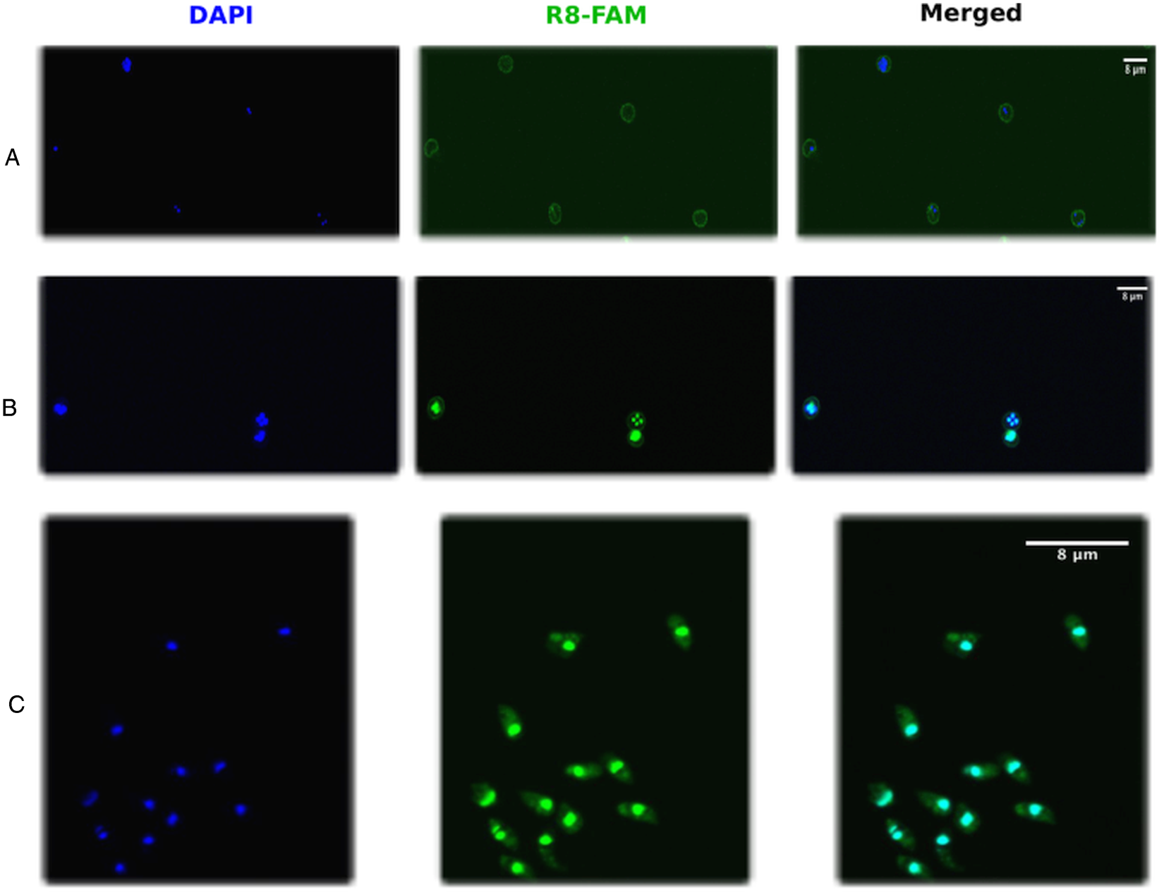
Before applying PEI and octaarginine as transfection agents for Cryptosporidium we first aimed as assessing the permeability of the parasite membrane for CPPs. For this reason, fluorescently labelled octaarginine (R8-FAM) was incubated with freshly excysted C. parvum sporozoites, intact oocysts, or STE oocysts and peptide uptake was monitored using confocal microscopy. We found that the membrane of intact oocysts was impermeable to octaarginine as shown by the green fluorescence staining only the cell wall of the oocysts (Fig. 1A). In contrast, free sporozoites, as well as STE oocsyts, were able to take up the peptide rather rapidly (Fig. 1B and C).
Permeation of Cryptosporidium membranes by octaarginine. Octaarginine labelled with 6-FAM (R8-FAM) was incubated with either intact oocysts (A), short-time excystation exposed oocysts (STE) oocysts (B) or free sporozoites (C). Intact oocysts are impermeable to octaarginine and the fluorescence stains only the oocysts wall. When the oocysts are exposed to excystation over a short period they become permeable to the peptide. Free sporozoites rapidly take up the peptide in general.

Condensation of plasmid DNA with PEI and formation of the transfection complex
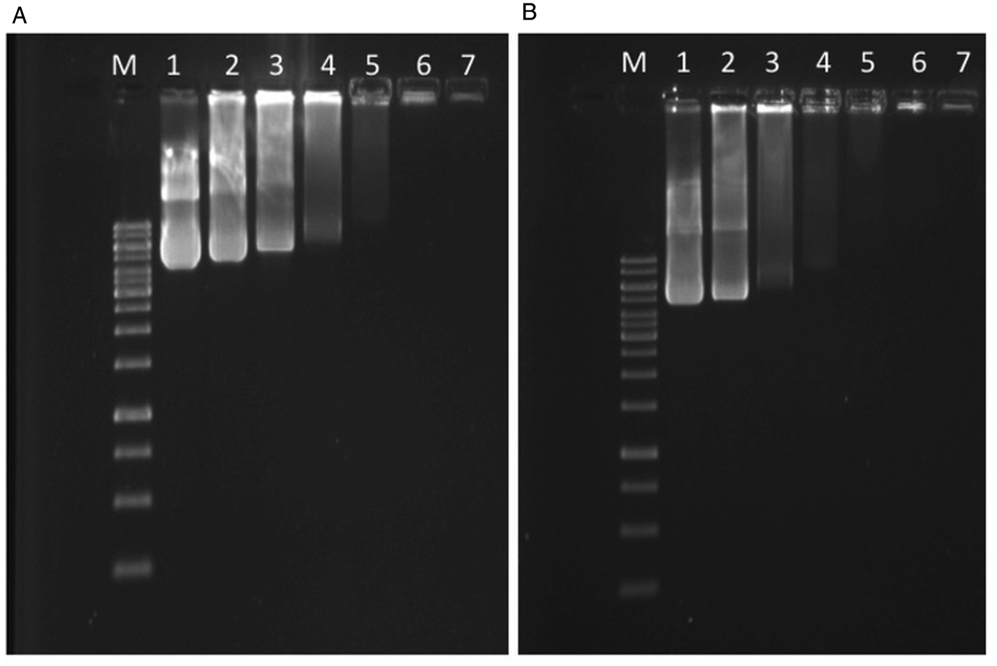
As next, we decided to investigate the optimal condition for condensing plasmid DNA with PEI and octaarginine using the gel retardation assay. For this purpose, we applied several combinations of PEI and plasmid DNA in order to identify the most suitable N/P ratio for full condensation of DNA where N represents the number of charged amine functions and P the number of charged phosphate groups of the DNA backbone. Typically, the optimal N/P ratio allows the total condensation of the DNA available. Using 5 μg plasmid DNA as starting point and combining with PEI we tested the following N/P ratios 2, 4, 6, 8, 12, 16 and 20 and observed that with an N/P ratio of 12 or more, the entire plasmid was condensed (Fig. 2A). In the next step, we added octaarginine to the generated DNA/PEI complex in order to form the final transfection complex. Interestingly, when octaarginine was added to the DNA/PEI complex the N/P ratio required to achieve full condensation appeared to be slightly lower (8) than when PEI alone (12) was used, as seen on the retardation assay (Fig. 2B).
Gel retardation assay for PEI-plasmid condensation. (A) DNA plasmid (5 μg) and PEI were combined at N/P ratios of 2, 4, 6, 8, 12, 16 and 20 (lanes 1-7, respectively). (B) The complex consisting of PEI and plasmid DNA at these N/P ratios was further incubated with a 10-fold excess of octaarginine. At the same concentration of plasmid DNA increasing concentrations of PEI lead to condensation of DNA and as a result, the DNA run into the gel with delay (A). Adding an excess of octaarginine to the plasmid/PEI complex further condenses the DNA and the retardation is even more pronounced (B).

Transfection of C. parvum with the DNA/PEI/octaarginine complex
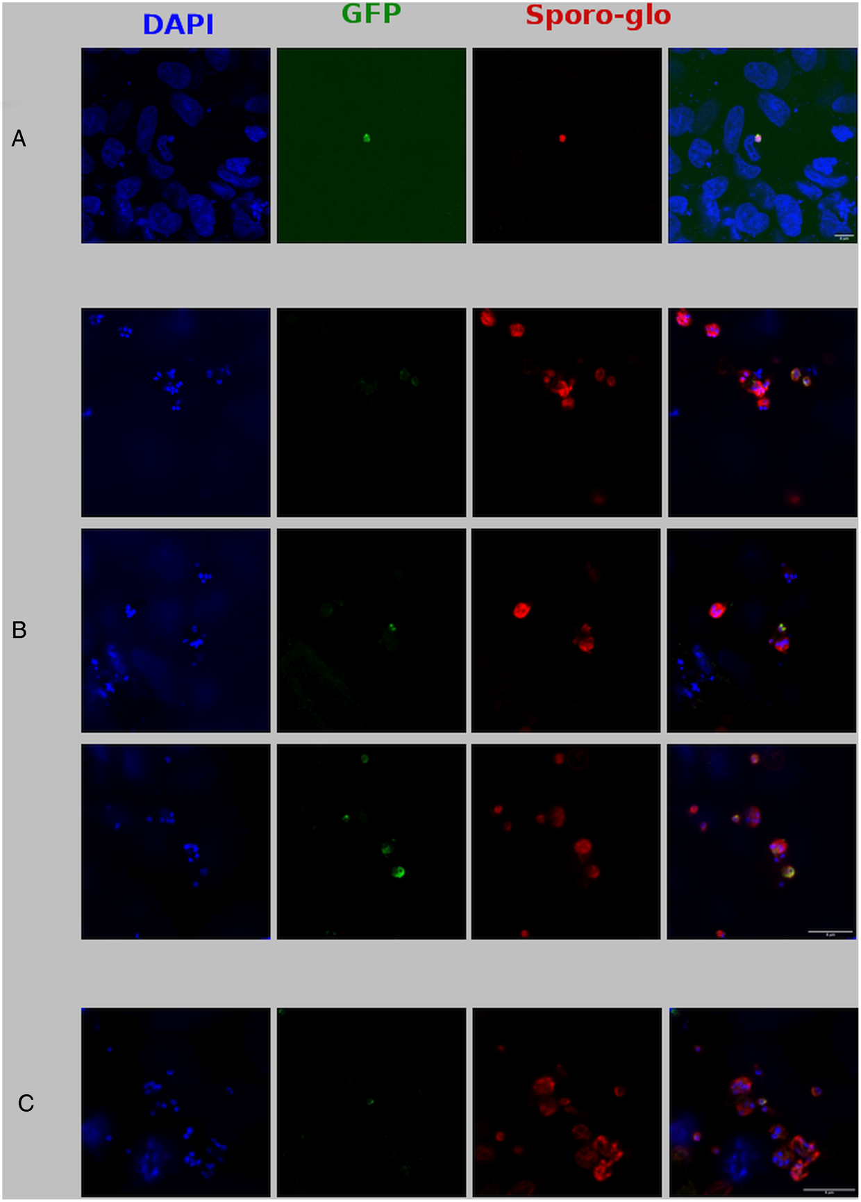
Next, we decided to assess whether the transfection complex could be successfully transferred to the parasite without electroporation. For this purpose, we used freshly excysted and purified sporozoites, STE oocysts as well as intact oocysts. The transfection complex consisting of plasmid DNA, PEI and octaarginine was incubated with each of the above-mentioned parasite materials for 1 hour at 37°C. Thereafter, these parasites were transferred to an exponentially growing culture of HCT-8 cells. Successful transfection was assessed by confocal microscopy after 24 h. We found, as expected, that with intact oocysts, no transfection could be observed (data not shown). This is in accordance with the impermeability of intact oocyst membranes to octaarginine as demonstrated with the fluorescently labelled peptide (Fig. 1A). Using excysted sporozoites, transfected parasites expressing the green fluorescent protein could be observed (Fig. 3A). However, the overall infection rate of transfected sporozoites was rather low and infected host cells could only be found sporadically. When STE oocysts were used, a higher infection rate was observed and transfected parasites in various developmental stages could be visualized (Fig. 3B). Importantly, when STE oocysts were transfected using only 1 μg of plasmid DNA we were also able to see few GFP-expressing parasites (Fig. 3C). Transfection with either PEI and plasmid or octaarginine and plasmid did not yield any GFP expressing parasite (data not shown).
Transfection of Cryptosporidium with PEI/Octaarginine/Plasmid. Transfection of excysted sporozoites (A) or short time excystation exposed oocysts (B) with the PEI/Octaarginine/plasmid complex consisting of 5 μg plasmid DNA at the N/P ratio of 16. Transfected parasites were incubated with exponentially growing HCT-8 cells and expression of the GFP was monitored microscopically 24 h post-infection. anti-Cryptosporidium red fluorescent antibody (Sporo-glo®) was used as control staining for the parasites. The experiment was repeated using 1 μg of plasmid DNA (C).

Discussion
It has been shown in the past that polymers with a high density of primary amine functions have the ability to facilitate gene delivery (Ou et al., Reference Ou, Xu, Kim, Bull and Kim2009). Because of its excellent DNA condensing properties, PEI is an attractive candidate for transfection. However, it is rather high molecular size brings toxicity issues for the host cells (Nam et al., Reference Nam, Jung, Nam and Kim2015) or decreases the cell metabolic activity (Fischer et al., Reference Fischer, Bieber, Li, Elsässer and Kissel1999; Godbey et al., Reference Godbey, Wu and Mikos2001; Florea et al., Reference Florea, Meaney, Junginger and Borchard2002). Although a CPP alone is unable to properly condense DNA plasmid (Jeong et al., Reference Jeong, Yoo, Lee and Kim2016), the combination of CPPs and PEI significantly improves the transfection efficiency. Moreover, some CPPs namely octaarginine can also inhibit the intracellular proteolytic systems like the proteasome (Kloß et al., Reference Kloß, Henklein, Siele, Schmolke, Apcher, Kuehn and Dahlmann2009) but it is unknown whether they can permanently inhibit cellular function. Certain CPPs can safely deliver their cargos such as DNA plasmid, therapeutics into the cells and are applied in in vitro and in vivo studies. For instance, when Glycosaminoglycan (GAG)-enhanced transduction (GET) peptides were combined to PEI, the overall efficiency of transfection of nanoparticles for lung gene therapy was significantly improved in the mouse model (Osman et al., Reference Osman, Rodriguez, Chan, Chisholm, Duncan, Kim and Dixon2018).
Importantly, the ability of octaarginine to deliver a cargo to a given compartment is not limited by the size of the corresponding cargo but rather by the membrane permeability of the corresponding compartment to octaarginine. Almost all biological membranes tested so far seem permeable to ocataarginine with the only exceptions recorded so far being the membranes of non-infected red blood cells and the parasitophorous vacuole membrane of T. gondii (Kamena et al., Reference Kamena, Monnanda, Makou, Capone, Patora-Komisarska and Seebach2011; Sparr et al., Reference Sparr, Purkayastha, Kolesinska, Gengenbacher, Amulic and Matuschewski2013). Our observation that the membrane of intact oocysts is completely impermeable to octaarginine while STE oocysts and free sporozoites efficiently take up the peptide, suggests that free sporozoites and STE oocysts would be more appropriate for plasmid delivery using the polycationic molecules.
In addition, our findings showed that the combination of PEI with octaarginine improve the DNA condensation thereby facilitating the plasmid delivery to Cryptosporidium for parasite transfection. Although a covalent conjugation of a cell-penetrating peptide to PEI seems to be more efficient for the delivery process, a non-covalent complex as applied in our experiments is also capable of delivering DNA to the cell (Kilk et al., Reference Kilk, El-Andaloussi, Järver, Meikas, Valkna, Bartfai and Langel2005). More importantly, the use of a non-covalent complex represents an easy to use alternative that does not require any specific chemical modification of the compounds involved and as such will be accessible to a wider range of scientists. It was reported previously that the use of a CPP alone is not sufficient for efficient plasmid DNA condensation (Kilk et al., Reference Kilk, El-Andaloussi, Järver, Meikas, Valkna, Bartfai and Langel2005) and although TP10 and not octaarginine was the CPP used in that report it is very likely that octaagrinine will display the same properties based on their structural similarities. The positive effect on transfection observed in this study is really the result of the combination of the two polycationic molecules PEI and octaarginine.
In conclusion, we have developed a simple protocol for the transfection of Cryptosporidium parvum. The novel protocol takes advantage of the DNA condensing properties of PEI and the cell-penetrating properties of octaarginine. The combination of these two polycationic molecules seems optimal for the delivery of plasmid DNA into Cryptosporidium. Importantly, successful transfection depends on the permeability of the parasite membrane to octaarginine albeit not exclusively as free sporozoites, although they can rapidly take up octaarginine, were unable to efficiently support transfection. This is probably due to the rapid loss of infectivity during the various incubation steps. Intact oocysts are totally impermeable to octaarginine and hence do not allow any plasmid uptake. In contrast, when oocysts are pre-incubated with excystation agents for a short time that does not allow complete excystation (STE oocysts), their wall is obviously less resistant to penetration by the transfection complex. Accordingly, successful transfection of the sporozoites inside these oocysts was demonstrated. It is plausible to think that sporozoites retain infectivity for a longer period if they remain inside the oocysts in comparison to fully excysted sporozoites. Therefore, preservation of infectivity is an essential factor in the whole transfection process. A major advantage of our protocol is the fact that it works without electroporation and does not require any other sophisticated equipment. It is thus much easier to perform, affordable and applicable by a wider range of scientist around the world.
Acknowledgement
We acknowledge the BioImaging Core Facility Universität Leipzig, Germany for enabling confocal microscopy.
Financial support
The work was supported by a doctoral scholarship of the Vietnamese Government (Project 911) to Tran Nguyen-Ho-Bao.
Conflict of interest
The authors declare there are no conflicts of interest.
Ethical standards
All animal work was conducted in accordance with the German ‘Tierschutzgesetz in der Fassung vom 22. Juli 2009,’ which implements Directive 2010/63/EU from the European Parliament and Council (On the Protection of Animals Used for Scientific Purposes). The protocol was approved by the Saxon federal authorities.