Ebstein's anomaly is a rare congenital heart disorder that occurs in about one to five per 200,000 live births, accounting for <1% of all congenital heart diseases.Reference Dearani and Danielson 1 The essence of the originally described Ebstein's anomaly is a displacement of the effective tricuspid valve orifice downward into the right ventricle, dividing the right ventricle into an atrialised and a functional portion. The severity of this malformation varies from mild, with few or no clinical findings, to severe defects incompatible with life. Presentation within the first 12 months of life is highly associated with poor prognosis, whereas in older children the long-term prognosis is superior. In this review, we summarise the current management related to the anatomical and electrophysiological comorbidities of Ebstein's anomaly, as well as surgical interventions and predictors of outcome in the first two decades of life.
Pathological features
In the normal heart, the tricuspid valve consists of the anterior, posterior, and septal leaflet. In Ebstein's anomaly, an abnormal development of the valvular components is seen. The leaflet tissue of the valves extends from the ventricular walls into a process called “delamination”. Owing to incomplete delamination, the hinge points of the septal and posterior leaflet are displaced. They are attached within the cavity of the ventricle itself, creating a functional tricuspid annulus below the true anatomic annulus. Furthermore, a rotation between 10° and 90° from the level of the atrioventricular junction often accompanies this apical displacement of the functional annulusReference Schreiber, Cook, Ho, Augustin and Anderson 2 (Fig 1). The leaflets have irregular shapes, are dysplastic and are tethered by short chordae and papillary muscles or have direct myocardial insertions. In a subset of patients, complete fusion makes it impossible to distinguish one leaflet from another. The malformed tricuspid valve may be incompetent, stenotic, or, rarely, imperforated.
Representation of the right ventricle. The ellipses demonstrate effective valvular orifices as estimated in 23 specimens by Schreiber et al. The solid lines represent cases with linear attachment; the ellipses with dotted lines and broken lines represent hearts with focal and hyphenated attachment of the anterosuperior leaflet, respectively. The vertical ellipse represents the solitary case with the origin parallel to the atrioventricular junction. The scale represents the maximal width of the orifice as a percentage of the distance between the atrioventricular junction and the apex of the right ventricle, measured along the acute margin. Source: Schreiber et al.Reference Schreiber, Cook, Ho, Augustin and Anderson 2

The intraventricular displaced functional tricuspid annulus divides the right ventricle into two parts: the “atrialised” and the functional right ventricle. The atrialised right ventricle, which can be seen as a continuation of the true right atrium, shows variable degrees of thinning of the wall; it is grossly fibrotic and contains only sparse muscular fibres. The functional right ventricle may be massively dilated, including the infundibulum. The wall may be thinned and fibrotic.
Classification
Classifying Ebstein's anomaly is difficult because of its wide spectrum of abnormalities. The first attempt was made by Carpentier et al.,Reference Carpentier, Chauvaud and Macé 3 proposing four grades of Ebstein's anomaly: type A, the volume of the true right ventricle is adequate; type B, there is a large atrialised component of the right ventricle but the anterior leaflet moves freely; type C, the anterior leaflet is severely restricted in its movement and may cause significant obstruction of the right ventricular outflow tract; and type D, almost complete atrialisation of the right ventricle with the exception of a small infundibular component. This classification, however, has its restrictions because the various components of the anomaly do not necessarily correlate with the functional severity of the disease, and a combination of types can be seen.
Celermajer et al.Reference Celermajer, Bull and Till 4 described an echocardiographic grading score for neonates with Ebstein's anomaly; the Glasgow Outcome Score Extended Score grade 1–4. Severity of Ebstein's anomaly was graded by calculating the ratio of the combined area of the right atrium and the atrialised right ventricle to that of the combined area of the functional right ventricle, left atrium, and left ventricle in a four-chamber view at end diastole. The ratio was used to define four grades of increasing severity: grade 1, ratio <0.5; grade 2, 0.5–0.99; grade 3, 1–1.49, and grade 4 ≥ 1.5.
Dearani et al.Reference Dearani and Danielson 1 proposed a new classification according to two different approaches. The first is based on echocardiographic appearance. Ebstein's anomaly is categorised into mild, moderate, or severe, similar to the classification used for echocardiographic quantification of mitral regurgitation. However, they describe this classification as imprecise and subjective. Their second approach considers the exact anatomy of all three leaflets of the tricuspid valve, the right ventricle, and the right atrium, resulting in the classification of Ebstein's anomaly type I–IV. The latter limits classification of Ebstein's anomaly to direct surgical observation.
Associated cardiac malformations
An interatrial communication is commonly present, either as a patent foramen ovale or as a (secondary) atrial septal defect. This communication is usually associated with a right-to-left shunt, although left-to-right shunt may be present in some young patients with mild forms of Ebstein's anomaly. A ventricular septal defect is often present, usually leading to right-to-left shunting in the presence of neonatal anatomic or functional severe pulmonic stenosis and pulmonary atresia. Furthermore, Ebstein's anomaly is often associated with distal right ventricle anomalies including pulmonary valve stenosis or atresia. Miscellaneous cardiac defects are reported as well.
Attenhofer et al.Reference Attenhofer Jost, Connolly, O'Leary, Warnes, Tajik and Seward 5 focused on the left side of the heart in Ebstein's anomaly patients, finding abnormalities involving the myocardium or valves in 39% of the patients, for example mitral valve prolapse or dysplasia, bicuspid aortic valve, and non-compaction. Therefore, Ebstein's anomaly should not be regarded as a disease of the right side of the heart alone.
Clinical presentation
Physical examination
Physical signs on examination vary markedly, but they are present in the majority of the patients. Growth, development, and body build of the patients are generally normal, although Kapusta et al.Reference Kapusta, Eveleigh and Poulino 6 found children with Ebstein's anomaly to be slightly shorter than the population average in addition to the mean body mass index standard deviation score being slightly decreased. Cyanosis is seen in all degrees along with various degrees of digital clubbing. A prominent third or even a fourth heart sound is often heard. In the study by Celermajer et al.,Reference Celermajer, Bull and Till 4 out of 88 neonates, 65 (74%) presented with cyanosis, nine presented with heart failure, eight neonates had an incidental murmur, five presented with arrhythmia, and one neonate was diagnosed after cardiac screening, performed because of the clinical suspicion of having trisomy 18. In the study of Arizmendi et al.,Reference Flores Arizmendi, Fernández Pineda and Quero Jiménez 7 18 out of 21 (86%) neonates presented with cyanosis, six with heart failure, and six with a murmur. No neonates were seen with arrhythmia. Older children are often referred for further examination after a heart murmur is heard.Reference Celermajer, Bull and Till 4 , Reference Flores Arizmendi, Fernández Pineda and Quero Jiménez 7 Celermajer et al. described a murmur as a presenting symptom in 33 out of 50 (66%) children aged up to 10 years; seven of the 50 patients had cyanosis, six had arrhythmias, and four had heart failure. In adolescence, arrhythmias, but also heart failure, murmurs, and cyanosis are reported as presenting symptoms.Reference Celermajer, Bull and Till 4 , Reference Flores Arizmendi, Fernández Pineda and Quero Jiménez 7
Electrophysiology
Atrial and ventricular arrhythmias are common in patients with Ebstein's anomaly. Owing to the fact that the anatomic tricuspid annulus represents an incomplete fibrous ring, it may allow direct muscular connections between the right atrium and right ventricle, providing the anatomic substrate for ventricular pre-excitation. Pre-excitation and Wolff–Parkinson–White syndrome are strongly associated with Ebstein's anomaly.
Delhaas et al.Reference Delhaas, Sarvaas and Rijlaarsdam 8 studied 93 children with Ebstein's anomaly for pre-excitation or arrhythmic events. Rhythm disturbances were exhibited in 17%. The median age at diagnosis of this subgroup (n = 16) was 0 days and the median age at first electrocardiogram abnormality was 3 months. There were five patients who presented with supraventricular tachycardia, three showed pre-excitation on routine electrocardiogram at initial diagnosis of Ebstein's anomaly, and seven presented later in life with rhythm disturbances. In all, two patients died, one because of intractable supraventricular tachycardia and one probably due to an arrhythmic event. All 14 surviving patients showed pre-excitations, in four intermittently. In addition, atrial flutter, atrial fibrillation, and also ventricular tachyarrhythmia may occur in young patients with Ebstein's anomaly.
Laboratory assessment
Echocardiography
Two-dimensional echocardiography is the most important diagnostic tool for children with congenital heart disease. It is portable, non-invasive, has no radiation risk, and provides immediate high-resolution anatomical and physiological information.
Prenatal echocardiography
Foetal echocardiography is an accurate predictor of congenital heart disease from the mid-trimester of pregnancy.Reference Sharland, Chita and Allan 9 Abnormalities of the tricuspid valve can be accurately defined prenatally, although there is much overlap between valvular dysplasia and Ebstein's malformation. Sharland et al.Reference Sharland, Chita and Allan 9 diagnosed by echocardiography 38 foetuses with abnormalities of the tricuspid valve. In all, 14 were diagnosed with Ebstein's anomaly and diagnosis was confirmed in 10 cases; three proved to have valve dysplasia only and one was lost to follow-up. Nevertheless, foetuses with severe Ebstein's anomaly might suffer from early intrauterine demise before the first ultrasound examination is made, resulting in underestimation of the disease diagnosed prenatally.
Echocardiography in infants and children
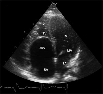
In patients with Ebstein's anomaly, the anatomy and function of the tricuspid and pulmonary valve, the right atrium, and the atrialised portion of the right ventricle, as well as the size and function of both ventricles, can be carefully evaluated by echocardiography (Fig 2). An apical displacement of the septal tricuspid valve leaflet from the insertion of the anterior leaflet of the mitral valve by more than 0.8 cm/m2 body surface area distinguishes Ebstein's anomaly from other malformations associated with tricuspid regurgitation.Reference Shiina, Seward, Edwards, Hagler and Tajik 10 However, the quality of images can be compromised in non-cooperative patients or when only poor acoustic windows are available.
Echocardiographic feature of a patient with Ebstein's anomaly demonstrating the apical displacement of the TV, dividing the right ventricle in an atrialised (aRV) and a functional portion (fRV). LA = left atrium; LV = left ventricle; MV = mitral valve; RA = right atrium; TV = tricuspid valve.

Real-time three-dimensional echocardiography (3DE) is a feasible method in addition to conventional two-dimensional echocardiography to evaluate the anatomy and the dynamic function of the abnormal tricuspid valve in young Ebstein's anomaly patients. It also permits clear differentiation from tricuspid valve dysplasia.Reference Bharucha, Anderson, Lim and Vettukattil 11 It can show extension of tricuspid regurgitation and adds information regarding septal leaflet insertion and right ventricle volumes. Furthermore, the size, location, and shapes of septal defects can be visualised clearly. Even foetal hearts can be examined by 3DE. However, this modality is not yet widely used as a bedside technique.
Electrocardiogram
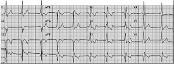
In most patients with Ebstein's anomaly, electrocardiogram findings are abnormal, especially in older patients, but they are not diagnostic. The rhythm is usually a normal sinus rhythm. P waves can be increased in amplitude as a sign of right atrium enlargement. P waves and the PR interval are often prolonged because of a delayed conduction in the enlarged right atrium. Small initial q waves may be found, occasionally seen in leads as far as V4, and are also considered to be due to right atrial enlargement. Signs of ventricular pre-excitation may be present. The QRS might show right axis deviation. It is usually of low voltage and prolonged, and it might exhibit a right bundle branch block pattern of various types in the right precordial leads (Fig 3). The right bundle branch block might be caused by pressure of the atrialised portion of the right ventricle onto the right septal surface. Left bundle branch block and atrioventricular block are also common.
A 12-lead electrocardiogram of an 8-year-old girl with Ebstein's anomaly. Signs of pre-excitation and repolarisation disorder are present.

Exercise testing
Exercise testing may assist in determining the severity of the disease. Children, adolescents, and adults with Ebstein's anomaly may have limited exercise tolerance, defined as maximal oxygen consumption (VO2) at peak exercise.Reference Driscoll, Mottram and Danielson 12 – Reference Müller, Kühn, Vogt, Schreiber, Hess and Hager 14 There is a strong negative correlation between blood oxygen levels and exercise tolerance. Observed heart rate at maximum exercise is lower than predicted.Reference MacLellan-Tobert, Driscoll, Mottram, Mahoney, Wollan and Danielson 13 The cardiac output at rest is within the lower range of normal for age. Driscoll et al.Reference Driscoll, Mottram and Danielson 12 showed that surgical repair improves exercise tolerance mostly because of the elimination of the right-to-left shunt in these patients. The exercise performance of patients without atrial septal defect was significantly greater than that of pre-operative patients with atrial septal defect, but was similar to that of patients who had atrial septal defect closure. This suggests that the presence and size of intracardiac right-to-left shunt might be a major contributing factor to exercise intolerance. The study of MacLellan-Tobert et al.Reference MacLellan-Tobert, Driscoll, Mottram, Mahoney, Wollan and Danielson 13 supports this. By shunting, only a portion of the systemic venous return reaches the lung for ventilator exchange of carbon dioxide. To maintain normal systemic arterial partial pressure of carbon dioxide, the patient must compensate by increasing ventilator exchange to allow for removal of carbon dioxide. Indeed, in Ebstein's anomaly patients minute ventilation at rest and ventilatory equivalent for oxygen (VE/VO2) at rest and during exercise are significantly higher before surgical repair than after.Reference Driscoll, Mottram and Danielson 12 – Reference Müller, Kühn, Vogt, Schreiber, Hess and Hager 14 In addition, improvements in tricuspid and consecutive right heart function might be another substrate for the improvement in exercise performance after surgery.Reference Müller, Kühn, Vogt, Schreiber, Hess and Hager 14
However, it is difficult to establish from which age children can safely participate in exercise tests. Furthermore, young children might be more difficult to encourage to exercise to exhaustion. Interpretation of results should be performed carefully, whereas sex and body surface area influence outcomes.Reference James, Kaplan, Glueck, Tsay, Knight and Sarwar 15
Chest radiography
The cardiac silhouette may vary from almost normal to a typical configuration of a box or balloon-shaped heart with a narrow waist. The greatly enlarged right atrium is mainly responsible, but also the dilated right ventricular outflow tract can be displaced outward and upward. The aorta cross-section is small, and the pulmonary trunk, which normally appears as a discrete convex bulge, is absent. The cardiothoracic ratio is an important factor influencing outcome of patients with Ebstein's anomaly. Pulmonary vascularity may be normal or decreased. When a newborn presents with systemic desaturation and the chest radiography shows cardiomegaly and decreased pulmonary vascularity, the diagnosis of Ebstein's anomaly should be considered.
Cardiac magnetic resonance imaging
Cardiac magnetic resonance imaging is a powerful tool providing anatomic and haemodynamic information that echocardiography alone cannot always provide. The extent of tricuspid valve regurgitation, the degree of right-to-left shunt, as well as the ventricular volumes and function can be well assessed. Tobler et al.Reference Tobler, Yalonetsky and Crean 16 even conclude that the volume of the atrialised right ventricle is a novel cardiac magnetic resonance imaging measure, which may express severity of disease. To fully characterise right heart size and function, the atrialised right ventricle, functional right ventricle, and total right ventricle volumes need to be measured by adequate cine view of a four-chamber, axial and short-axis view. The axial view, however, appears to provide the most reproducible data.Reference Yalonetsky, Tobler and Greutmann 17
Cardiac magnetic resonance imaging is also particularly helpful in cases with associated cardiac malformation and allows accurate planning of the best surgical approach. Attenhofer et al.Reference Attenhofer Jost, Edmister and Julsrud 18 compared the use of echocardiography and cardiac magnetic resonance imaging in patients with Ebstein's anomaly. They concluded that both provide complementary data. For appropriate risk stratification in Ebstein's anomaly, both examinations should be performed before surgery.
However, cardiac magnetic resonance imaging requires specific knowledge of anatomical variations and functional implications of Ebstein's anomaly, as well as high-resolution imaging, and extended patient compliance. General anaesthesia is commonly integrated into the examination protocol of younger children.
Medical management
Children with mild Ebstein's anomaly might not need any interventions during infancy, childhood, or adulthood. These patients, however, will need lifelong visits at the outpatient clinic. The frequency of these visits depends on their clinical condition. Others with moderate to severe types of Ebstein's anomaly – with or without associated malformations – might require medical treatment and/or intervention early in life.
Medical treatment
Little evidence is published about the use of medication in children with Ebstein's anomaly. In the large paediatric cohort of Kapusta et al.,Reference Kapusta, Eveleigh and Poulino 6 41% of the children with Ebstein's anomaly (n = 38) required medication at presentation. Patients diagnosed during the first year of life received significantly more medication as compared with children diagnosed later in life. Prostaglandin was required in 23 of 93 patients, all in the neonatal period. Neonates often require short-term prostaglandin therapy in case of severe tricuspid regurgitation and severe cyanosis, due to the functional pulmonary valve atresia and the high pulmonary arteriolar resistance. Other required medication at presentation included diuretics (n = 15), digitalis (n = 14), and inotropics (n = 5). At the end of their study, 19% of their children were classified as New York Heart Association class II for which only a few patients required – a combination of – medication.Reference Kapusta, Eveleigh and Poulino 6 Medication included diuretics (n = 5), angiotensin-converting enzyme blocker (n = 1), digitalis (n = 1), beta-blocker (n = 4), and aspirin (n = 2).
Reemtsen et al.Reference Reemtsen, Fagan, Wells and Starnes 19 reported on 16 pre-operative neonates of whom 88% were in need of prostaglandin and 56% required inotropics – dopamine, dobutamine, or epinephrine. Furthermore, inhaled nitric oxide may play an important therapeutic role in neonates with Ebstein's anomaly after discontinuation of prostaglandin.Reference Atz, Munoz, Adatia and Wessel 20
Rhythm anomalies, including re-entrant tachycardias, are comorbidities requiring medical intervention at any age in patients with Ebstein's anomaly. Supraventricular arrhythmias are the most common rhythm disturbance in children and tend to be recurrent and drug resistant. Currently, catheter ablation of accessory pathways is often performed and anti-arrhythmic drugs are less often prescribed. Delhaas et al.Reference Delhaas, Sarvaas and Rijlaarsdam 8 found in 16 out of 93 patients of their paediatric population rhythm disturbances of which nine out of 14 of the surviving patients underwent ablation. Of these patients, four were still on antiarrhythmic drugs after a mean follow-up of 13 years and 3 months.
Radiofrequency ablation procedures
Up to one-third of the Ebstein's anomaly patients are presenting accessory pathways. These are usually right sided and may even be multiple.Reference Delhaas, Sarvaas and Rijlaarsdam 8 , Reference Roten, Lukac and DE Groot 21 Indications for radiofrequency ablation include apparent life-threatening arrhythmias and drug-refractory tachycardia. Radiofrequency ablation procedures in Ebstein's anomaly patients are challenging because of the anatomical and functional variations. The right atrium may be severely dilated, the atrioventricular junction might be less well demarcated, abnormal electrocardiograms can be recorded along the atrialised right ventricle, and catheter stability may be impaired.Reference Roten, Lukac and DE Groot 21
Acute success rates for patients with Ebstein's anomaly are similar to those of patients with other congenital heart diseases, but Ebstein's anomaly patients are more likely to have a recurrence.Reference Chetaille, Walsh and Triedman 22 Supraventricular tachyarrhythmias can also be successfully ablated at the time of operative repair.Reference Bockeria, Golukhova and Dadasheva 23
Surgical management
The customary objectives of surgical intervention in patients with Ebstein's anomaly are: (1) to reduce the severity and haemodynamic sequelae of tricuspid regurgitation and/or stenosis; (2) to improve hypoxaemia; and (3) to assist in controlling intractable tachyarrhythmias.
The indications for operation in the first two decades of life are multiple and vary widely. In neonates, they include overt heart failure, cyanosis, acidosis associated with tricuspid regurgitation, depressed right ventricle function, severe cardiomegaly, persistent need for inotropic support, ventilator dependency, and prostaglandin-dependent circulation.Reference Reemtsen, Fagan, Wells and Starnes 19 , Reference Knott-Craig, Goldberg, Overholt, Colvin and Kirklin 24 After infancy, frequently reported indications for surgery include New York Heart Association class III or IV, progressive exercise intolerance, tachyarrhythmias refractory to drug treatment or not amenable to catheter-based intervention, recurrent palpitations or significant associated anomalies. Furthermore, patients with a cardiothoracic ratio >0.65 and persistent severe cyanosis may need surgical intervention.Reference Brown, Dearani and Danielson 25 – Reference Badiu, Schreiber and Hörer 28
Neonates
Symptomatic neonates with Ebstein's anomaly can present with severe heart failure, cyanosis, and acidosis, and without surgical intervention most of them will die.Reference Reemtsen, Fagan, Wells and Starnes 19 , Reference Knott-Craig, Goldberg, Overholt, Colvin and Kirklin 24 , Reference Starnes, Pitlick, Bernstein, Griffin, Choy and Shumway 29 Surgical intervention includes tricuspid valve repair, right ventricle exclusion, or transplantation. The first essential surgical decision that needs to be made is whether a two-ventricle repair is feasible, valve repair, or not, right ventricle exclusion.Reference Reemtsen, Fagan, Wells and Starnes 19 , Reference Knott-Craig, Goldberg, Overholt, Colvin and Kirklin 24 Right ventricle exclusion was first suggested by Starnes and associates in 1991,Reference Starnes, Pitlick, Bernstein, Griffin, Choy and Shumway 29 but the technique has been modified over the last 10 years. If the functional portion of the right ventricle is inadequate or if there is right ventricle outflow tract obstruction, one might choose for this right ventricle exclusion. This is performed by patching the tricuspid valve, preferably with a fenestration of the patch (Figs 4 and 5).Reference Reemtsen, Fagan, Wells and Starnes 19 , Reference Watanabe, Harada, Takeuchi, Satomi and Yasukouchi 30 A reduction right atrioplasty is performed and possibly also a right ventricle plication. Next, the right ventricle outflow tract is accessed. Finally, a Blalock–Taussig shunt provides pulmonary blood flow.
A glutaraldehyde-fixed autologous pericardial patch is sewn at the “anatomic” level of the tricuspid valve annulus. ASD = atrial septal defect. Source: Reemtsen et al.Reference Reemtsen, Fagan, Wells and Starnes 19

A fenestration in the patch is created by using a 4-mm coronary punch. Source: Reemtsen et al.Reference Reemtsen, Fagan, Wells and Starnes 19

A two-ventricle approach may include modifications in the anterior leaflet creating functionally monocuspid tricuspid valve architecture. A reduction atrioplasty is performed with partial closure of the atrial septal defect, leaving behind a small fenestration. Finally, other cardiac defects are simultaneously repaired.Reference Knott-Craig, Goldberg, Overholt, Colvin and Kirklin 24
The choice of surgical approach seems to be partially dependent on the clinic's experience. Reemtsen et al.Reference Reemtsen, Fagan, Wells and Starnes 19 in 2006 described 16 critically ill neonates undergoing surgery. There were 12 patients who underwent right ventricle exclusion as initial palliation; in three patients, the tricuspid valve was repaired and one patient underwent heart transplantation. The overall survival was 69% with no late deaths reported at a mean follow-up of 27 months. Knott-Craig et al.Reference Knott-Craig, Goldberg, Overholt, Colvin and Kirklin 24 described in 2007 surgical intervention in 22 neonates and five young infants. In all but two patients, two-ventricle repair was performed. They reported a 74% survival rate with a median follow-up of 5.4 years. Boston et al. reported in 2011 the long-term results of 23 neonates and nine young infants. A biventricular repair was achieved in 91% of the patients. The early survival was 78% with one late death. The mean follow-up was 5.9 years.Reference Boston, Goldberg and Ward 31
Nevertheless, some neonates and young infants with a primary biventricular repair will eventually need a bidirectional cavopulmonary shunt because the right ventricle fails to support the pulmonary circulation. This technique is called a “one and a half ventricle repair”. To the best of our knowledge, there are no data available about the number of neonates who underwent a biventricular repair in the neonatal period, but were eventually in need of a bidirectional cavopulmonary shunt later in life. Surgical management of critically ill neonates and young infants remains challenging.
Child, adolescent, and adult
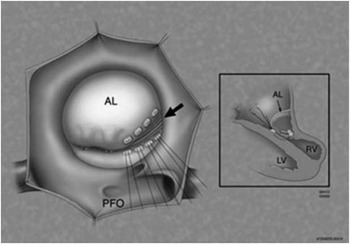
Surgical treatment after infancy is much more promising. A biventricular repair is usually possible. Dearani et al.Reference Dearani, Oleary and Danielson 32 described their experience in surgical treatment up to 2006. The repair was predominantly based on a satisfactory arrangement of the anterior leaflet (Figs 6–8). Some patients will have a large enough posterior leaflet permitting bicuspid reconstruction, and rarely a trifoliate repair is possible involving the septal leaflet as well. Various modifications in the technique of repair have been advanced with growing experience.
The manoeuvres are designed to progressively bring the leading edge of the anterior leaflet (AL) closer to the ventricular septum (VS), or septal leaflet, in order to optimise leaflet coaptation and establish competence of the valve. The base of the intact major papillary muscle(s), which arises from the free wall of the right ventricle, is moved towards the ventricular septum at the appropriate level with pledgeted horizontal mattress sutures. Inset, coronal view of the right ventricle (RV) and the right atrium demonstrating a small dimple effect that occurs in the anterior free wall of the right ventricle after this manoeuvre is completed. CS = coronary sinus; IL = inferior leaflet; LV = left ventricle; PFO = patent foramen ovale; PV = pulmonary valve. Source: Dearani et al.Reference Dearani, Oleary and Danielson 32

The inferior angle of the tricuspid orifice is closed by bringing the right side of the anterior leaflet down to the septum and plicating the non-functional inferior leaflet in the process (arrow). Inset, after all of the mattress sutures are secured, improved proximity of the leading edge of the anterior leaflet with the ventricular septum is noted. Source: Dearani et al.Reference Dearani, Oleary and Danielson 32

Plication of the inferior angle of the annulus with pledgeted mattress sutures (arrow). An anterior purse-string annuloplasty (arrowhead) may be performed to further narrow the tricuspid annulus. This annuloplasty may begin at the anteroseptal commissure, anterior to the membranous septum, and ends beyond the inferoseptal commissure adjacent to the coronary sinus. Alternatively, the annuloplasty can be performed posterolaterally to reduce the size of the annulus, which also brings the free wall closer to the septum. Source: Dearani et al.Reference Dearani, Oleary and Danielson 32

In 2004, another technique for valve repair was described by Wu et al.Reference Wu and Huang 33 This repair restores to near normal anatomical and physiological functioning of the tricuspid valve. The displaced posterior and septal leaflets with some chordae tendinae and corresponding papillary muscle are detached from the annulus and ventricular wall. The leaflets are then reattached to the native posterior annulus with re-implantation of the papillary muscle. Most of the atrialised portion of the ventricular wall is excised and the tricuspid annulus is plicated. In case of a severely hypoplastic septal leaflet, autologous pericardial tissue is used to create a new leaflet.
In 2009, Malhotra et al.Reference Malhotra, Petrossian and Reddy 34 described a technique with reconstruction performed at the functional rather than the anatomic orifice. Detachment and re-implantation of valve leaflets is avoided and only limited plication is performed at the level of the displaced valve.
Da Silva et al.Reference Silva, Baumgratz and Fonseca 35 proposed a conic reconstruction of the tricuspid valve in 2004. This technique consists of (almost) total detachment of the anterior tricuspid megaleaflet from the ventricular wall and valvular ring. This leaflet is transformed into a cone rotating it clockwise. The vertex remains fixed in the right ventricular tip. Then the atrialised right ventricle is plicated to exclude its thin part. The true tricuspid annulus is plicated to make up the new annulus at the anatomically correct level. After its plication, the base of the cone is sutured to the tricuspid ring. The end result of this cone reconstruction includes 360° of leaflet tissue surrounding the right atrioventricular junction.
In general, the “one and a half ventricle repair” using a bidirectional cavopulmonary shunt is preserved for Ebstein's anomaly patients with a severely enlarged and poorly functioning right ventricle.Reference Dearani, Oleary and Danielson 32 , Reference Wu and Huang 33 This shunt reduces the preload of the right ventricle but optimises the preload of the left ventricle. It therefore reduces both the risk of post-repair tricuspid stenosis and of potential progression of residual tricuspid regurgitation.
Valve reconstruction is preferable to replacement given the attendant risks of thromboembolism and the need for re-intervention with patient's growth. Tricuspid valve replacement in adulthood is also a risk factor for 30-day mortality and for complications.Reference Hörer, Vogt and Wottke 36 However, in case of extreme hypoplasia of the anterior leaflet and severe right ventricular dysfunction, valve replacement may be the only option. The type of implanted prosthesis, mechanical or biological, seems not to affect the outcome.Reference Garatti, Nano and Bruschi 37 When replacing the valve, the conduction tissue and the right coronary artery should be protected. All leaflet tissue should be removed to avoid obstruction.Reference Dearani, Oleary and Danielson 32 In case of severe dysfunction of the left ventricle, cardiac transplantation should be considered.
Outcome of surgical intervention
Surgical outcomes vary with an early survival rate of 86–96% and a 10-year survival rate up to 96%.Reference Brown, Dearani and Danielson 25 , Reference Sarris, Giannopoulos and Tsoutsinos 26 , Reference Badiu, Schreiber and Hörer 28 Studies included different surgical procedures and a subset of included patients had prior surgical intervention. Although re-operations were associated with a trend towards higher mortality, this did not reach significance level.Reference Sarris, Giannopoulos and Tsoutsinos 26 No significant difference was found between valve repair and valve replacement groups. Commonly reported post-operative complications include atrial and ventricular arrhythmias, low cardiac output, and respiratory insufficiency.Reference Brown, Dearani and Danielson 25 , Reference Sarris, Giannopoulos and Tsoutsinos 26 , Reference Badiu, Schreiber and Hörer 28 In all, 77–82% are free from re-operation 10 years after surgery.Reference Brown, Dearani and Danielson 25 , Reference Badiu, Schreiber and Hörer 28
Pre-operative risk factors for mortality include an increased pre-operative right ventricle size with decreased systolic function, pre-operative decreased left ventricle systolic function, cardiothoracic ratio >0.65, and variables associated with right ventricle outflow tract obstruction or narrow pulmonary arteries.Reference Brown, Dearani and Danielson 25 In addition, younger age and corresponding low bodyweight and body surface area <0.5 m2 is associated with higher mortality rates.Reference Sarris, Giannopoulos and Tsoutsinos 26 Pre-operative risk factors for re-operation are pulmonary valve stenosis, hypoplastic or stenotic pulmonary arteries, prior cavopulmonary shunt, and younger age at the time of operation.Reference Brown, Dearani and Danielson 25
Factors associated with poor prognosis of Ebstein's anomaly
The prognosis of patients diagnosed in the neonatal period is generally poor. Survival rates vary from 56–83% after 1 month to 55–71% after 1 year.Reference Flores Arizmendi, Fernández Pineda and Quero Jiménez 7 , Reference Chang, Wang and Chiu 38 In older children and adolescents, the outcome is superior. Multiple factors associated with poor outcome have been described over the last two decades. They include presentation in foetal or neonatal life, echocardiographic grade of severity 3 or 4 by Celermajer index, New York Heart Association class III or IV, associated anomalies, and a cardiothoracic ratio of ≥0.65.Reference Celermajer, Bull and Till 4 , Reference Flores Arizmendi, Fernández Pineda and Quero Jiménez 7 , Reference Gentles, Calder, Clarkson and Neutze 39 Furthermore, male sex and the absence of Wolff–Parkinson–White syndrome are reported as negative predictors of long-term survival, in contrast to what might be expected in a larger cohort.Reference Gentles, Calder, Clarkson and Neutze 39
Kapusta et al.Reference Kapusta, Eveleigh and Poulino 6 also found that young age at presentation (<12 months) is strongly associated with death. The mortality rate in these children is 7.9 times the risk in the group of children ≥12 months. In addition to hepatomegaly, the need for medication and mechanical ventilation at presentation were associated with death. Furthermore, patients with a patent arterial duct, ventricular septal defect, or pulmonary valve defects have a higher mortality rate.
Conclusion
Ebstein's anomaly is a rare and complex congenital disorder characterised by a rotational displacement of at least one of the leaflets of the tricuspid valve. Clinical features vary widely from mild symptoms presenting in adolescence to severe neonatal cyanosis and heart failure resulting in death. New diagnostic methodologies are available for establishing severity of disease. Many patients develop arrhythmias and may need recurrent radiofrequency ablation procedures. Severely cyanotic neonates on mechanical ventilation with persistent need for inotropic support often require immediate surgical treatment in order to survive, but these interventions remain extremely challenging. Therapeutic options after infancy are more promising. New surgical techniques have been developed over the last decade and they seem to alter both prognosis and quality of life in the first two decades of life.
Acknowledgements
None.
Financial Support
This research received no specific grant from any funding agency, commercial or not-for-profit sectors.
Conflicts of Interest
None.