1. Introduction
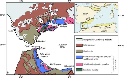
Located in the western tip of the Mediterranean, the Gibraltar arc consists of the Alboran Basin surrounded by the Betic (in Spain) and the Rif (in Morocco) orogenic belts built, since late Mesozoic time, in the context of Africa and Eurasia plate convergence (Fig. 1). Today, the geodynamic evolution of the internal zone of these cordilleras (i.e. the Alboran domain) is considered to be the result of subduction followed by slab rollback, which produced the opening of a back-arc basin (e.g. Royden, Reference Royden1993; Lonergan & White, Reference Lonergan and White1997; Faccenna et al. Reference Faccenna, Piromallo, Crespo-Blanc, Jolivet and Rossetti2004, Reference Faccenna, Becker, Auer, Billi, Boschi, Brun, Capitanio, Funiciello, Horvàth, Jolivet, Piromallo, Royden, Rossetti and Serpelloni2014; Spakman & Wortel, Reference Spakman, Wortel, Cavazza, Roure, Spakman, Stampfli and Ziegler2004; Vergés & Fernàndez, Reference Vergés and Fernàndez2012; Bezada et al. Reference Bezada, Humphreys, Toomey, Harnafi, Dávila and Gallart2013; Van Hinsbergen et al. Reference Van Hinsbergen, Vissers and Spakman2014). Such a scenario with a retreating slab, migrating behind the orogenic wedge is also assumed in other domains of the Mediterranean such as the Calabrian arc (e.g. Faccenna et al. Reference Faccenna, Becker, Lucente, Jolivet and Rossetti2001, Reference Faccenna, Speranza, Caracciolo, Mattei and Oggiano2002) or the Aegean arc (e.g. Jolivet & Brun, Reference Jolivet and Brun2010; Jolivet et al. Reference Jolivet, Faccenna, Huet, Labrousse, Le Pourhiet, Lacombe, Lecomte, Burov, Denèle, Brun, Philippon, Paul, Salaün, Karabulut, Piromallo, Monié, Gueydan, Okay, Oberhänsli, Pourteau, Augier, Gadenne and Driussi2013).
(a) Location of the Gibraltar arc and Alboran Basin in the framework of the Western Mediterranean and peri-Mediterranean orogenic belts (light green). (b) Structural map of the Gibraltar arc.

Numerous geological, petrological, geochemical and geochronological studies were carried out on the internal zones of the Betic and the Rif belts in order to constrain the geodynamic evolution of the Alboran domain (e.g. Beccaluva et al. Reference Beccaluva, Bianchini and Wilson2007; Duggen et al. Reference Duggen, Hoernle, Klügel, Geldmacher, Thirlwall, Hauff, Lowry and Oates2008; Conticelli et al. Reference Conticelli, Guarnieri, Farinelli, Mattei, Avanzinelli, Bianchini, Boari, Tommasini, Tiepolo, Prelević and Venturelli2009; Lustrino et al. Reference Lustrino, Duggen and Rosenberg2011; Carminati et al. Reference Carminati, Lustrino and Doglioni2012). However, in spite of these detailed studies, no expression of arc-related calc-alkaline magmatism has ever been documented so far in the Internal Rif. In this paper we unravel for the first time the occurrence of subduction-related magmatism in the Ceuta Peninsula, located in the internal zone of the Rif belt.
2. Geological context
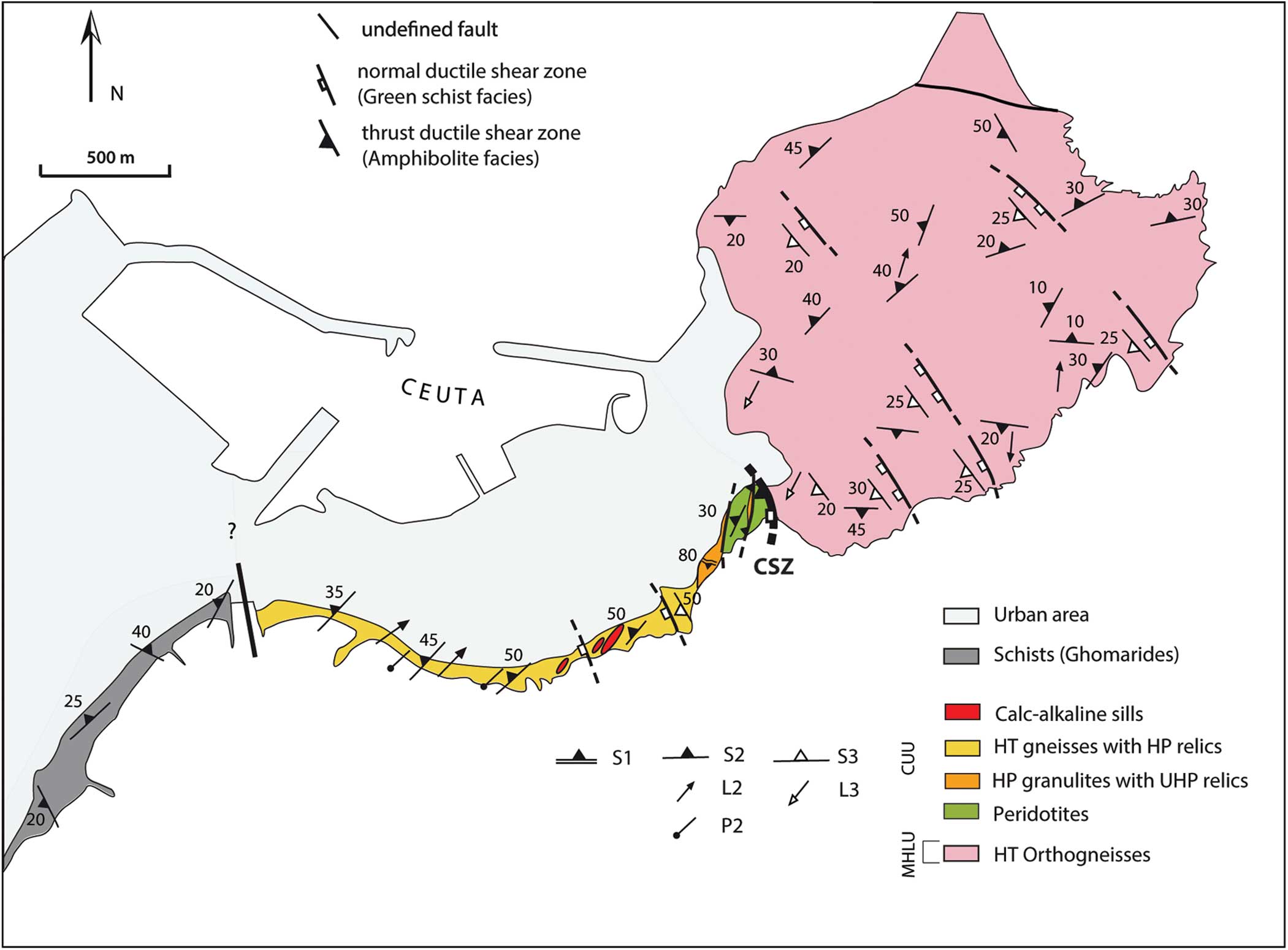
The Rif Cordillera, located in the northern part of Morocco, consists of stacked sedimentary and metamorphic units thrust onto foreland basins. Classically, three main structural domains are recognized in this orogenic system (Fig. 1b): (i) the External zone, (ii) the Flysch zone and (iii) the Internal zone (or Alboran domain, see Michard et al. Reference Michard, Goffe, Chalouan and Saddiqi1991; Chalouan et al. Reference Chalouan, Michard, Kadiri, Negro, Frizon de Lamotte, Soto, Saddiqi and Michard2008). The Internal Rif consists of three units, namely, from top to bottom, the Dorsale calcaire, the Ghomarides unit and the Sebtides unit (Fig. 1b, e.g. Chalouan et al. Reference Chalouan, Michard, Feinberg, Montigny and Saddiqi2001). The Sebtides unit is composed of a stack of high- to low-grade metamorphic rocks, subdivided into the pre-Permian Lower Sebtides and the Palaeozoic to Triassic Upper Sebtides (Chalouan et al. Reference Chalouan, Michard, Kadiri, Negro, Frizon de Lamotte, Soto, Saddiqi and Michard2008). In the Ceuta area, two sub-units are distinguished in the Lower Sebtides: the Ceuta Upper Unit (CUU) and the Monte Hacho Lower Unit (MHLU). The CUU is composed of migmatitic metapelites containing peridotite lenses and boudins of high- to ultra-high-pressure granulites at its base (Kornprobst, Reference Kornprobst1974; Ruiz-Cruz & De Galdeano, Reference Ruiz-Cruz and De Galdeano2012, Reference Ruiz-Cruz and De Galdeano2013), overthrust upon the migmatitic orthogneisses of the MHLU (Fig. 2, Kornprobst, Reference Kornprobst1974; Homonnay et al. Reference Homonnay, Corsini, Lardeaux, Romagny, Münch, Bosch, Cenki-Tok and Ouazzani-Touhami2018). Thrust tectonics developed under intermediate-pressure granulite-facies metamorphism before Miocene time, probably during Oligocene time (Homonnay et al. Reference Homonnay, Corsini, Lardeaux, Romagny, Münch, Bosch, Cenki-Tok and Ouazzani-Touhami2018). In this domain, three ductile deformation stages (D1–D3) have been identified, where S1–S3 foliations are associated with distinctive metamorphic assemblages (Homonnay et al. Reference Homonnay, Corsini, Lardeaux, Romagny, Münch, Bosch, Cenki-Tok and Ouazzani-Touhami2018). In both Lower Sebtides units, an S2 composite foliation, bearing a well-developed stretching lineation L2, is the dominant regional fabric whatever the lithology. In the uppermost unit, pre-D2 fabrics (i.e. D1/S1) are locally preserved in volumes (i.e. lozenge-shaped blocks) that escaped overprinting by the pervasive S2. In both units, D3 structures consist of localized normal ductile shear zones associated with an S3 foliation. Detailed microstructural, mineralogical and geochronological investigations (Homonnay et al. Reference Homonnay, Corsini, Lardeaux, Romagny, Münch, Bosch, Cenki-Tok and Ouazzani-Touhami2018) demonstrate that:
(1) D1 was contemporaneous with high-pressure/high-temperature metamorphic conditions (high-pressure granulite facies) of unknown age;
(2) D2 developed, contemporaneously with thrust tectonics, under intermediate-pressure granulite-facies conditions (7–10 kbar and 780–820 °C) before 28 Ma;
(3) D3 is characterized by metamorphic re-equilibration at the boundary between amphibolite- and greenschist-facies conditions (400–550 °C and 1–3 kbar) during early Miocene time (i.e. c. 21 Ma).
Geological map showing the location of the magmatic sill within the structural framework of the Ceuta Peninsula (modified after Homonnay et al. Reference Homonnay, Corsini, Lardeaux, Romagny, Münch, Bosch, Cenki-Tok and Ouazzani-Touhami2018). CUU – Ceuta Upper Unit; MHLU – Monte Hacho Lower Unit; CSZ – Ceuta Shear Zone. See Figure 1b for location.

In the most western part of the Ceuta area, the Upper Sebtides (Federico units) underwent an Alpine, probably Oligocene, high-pressure/low-temperature metamorphism typical of subduction zone conditions (Bouybaouene et al. Reference Bouybaouene, Goffé and Michard1995; Goffe et al. Reference Goffe, Azanon, Bouybaouene and Jullien1996; Michard et al. Reference Michard, Goffe, Bouybaouene and Saddiqi1997; Janots et al. Reference Janots, Negro, Brunet, Goffé, Engi and Bouybaouène2006).
3. Evidence for high-K calc-alkaline magmatism in the Lower Sebtides
3.a. Field observations
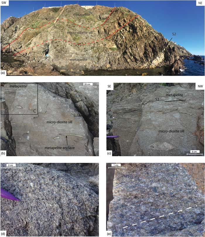
Metapelites from the CUU present a main NE-striking (N30 to N50) S2 foliation plane, gently to moderately dipping (20 to 50°) towards the NW (Fig. 2). This foliation is marked by the alternation of quartz–feldspars and garnet–biotite–sillimanite-rich levels as well as by the alternation of centimetric quartzo-feldspathic leucosomes and biotite–sillimanite mesosomes (Fig. 3a). This main D2 deformation episode, synchronous with partial melting of the metapelites, is also contemporaneous with the thrusting of the CUU over the MHLU (Homonnay et al. Reference Homonnay, Corsini, Lardeaux, Romagny, Münch, Bosch, Cenki-Tok and Ouazzani-Touhami2018).
Field observations. (a) Decametric sill parallel to the main S2 metapelite foliation plane. (b) Edge of the sill with magmatic fluidality and enclave alignments parallel to the main S2 foliation plane. (c) Contact between the micro-diorite sill and the metapelite. (d) Microgranular porphyric texture preserved in the core of the sill. (e) Magmatic mineral orientation (underlined by white dashed lines) within the micro-diorite sill.

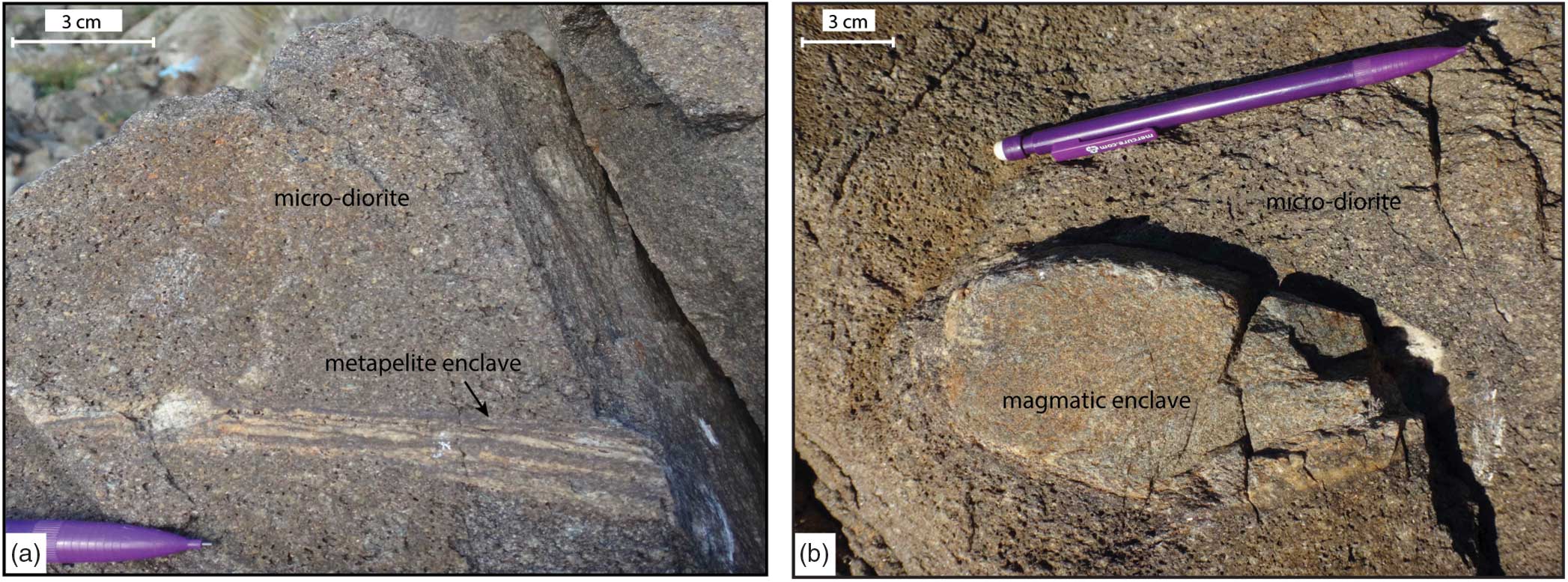
In these metapelites we found several pluricentimetric to plurimetric magmatic sills parallel to the main S2 foliation plane (Fig. 3a,b,c). In these ‘micro-granitoid’ sills we recognized two types of enclaves: (1) enclaves of migmatitic metapelites (i.e. host rock, Fig. 4a) and (2) granular to microgranular magmatic enclaves (Fig. 4b). The cores of the magmatic sills are clearly not deformed and contain a well-preserved microgranular porphyric texture (Fig. 3d). On the other hand, the edges of the sills display preferred mineral orientation interpreted as a magmatic flow-fabric (Fig. 3b,c,e). This interpretation is supported by enclave alignments parallel to the main foliation of the surrounding rocks (Fig. 3b). These structural observations are typical for syn- to late-tectonic (i.e. D2 deformation event) magma emplacement. In such a case, the magmatic sills were emplaced in thermal equilibrium with the surrounding metapelites.
Enclaves observed within the micro-diorite sill. (a) Metapelite enclave. (b) Magmatic enclave with microgranular texture.

3.b. Petrographic observations
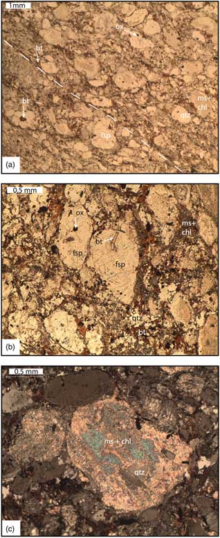
The magmatic sills present a microgranular porphyric texture, while magmatic fluidality is visible in thin-sections from the edges of the sills (Fig. 5a). The sills contain a primary magmatic assemblage essentially composed of feldspars, quartz, biotites and oxides (ilmenite and magnetite). Feldspars occur as subhedral to anhedral large-sized grains (1–5 mm) (Fig. 5b) and present a magmatic zonation. They are predominantly plagioclases (An7–An25), which exhibit polysynthetic twinning. K-feldspars (orthoclase, microcline) are observed in minor amounts and orthoclase shows Carlsbad twinning. In the groundmass, xenomorphic quartz grains show wavy extinctions. Biotite appears as small (< 1 mm) brown, strongly pleochroic grains in the groundmass or as minute inclusions in large plagioclase grains (Fig. 5b). In the largest biotite grains, needle-shaped rutile developed along cleavage planes. In addition, we frequently observed a ‘ghost’ mineral with a rectangular or rhomboidal shape (around 5 mm), which is totally replaced by an association of chlorite, muscovite and epidote (Fig. 5c). Although the nature of this mineral cannot be definitely determined, the secondary mineral association together with the mineral shapes strongly suggests a primary magmatic amphibole or a primary magmatic pyroxene. Indeed, in one thin-section we recognized one fragment of a rectangular-shaped pyroxene within the pseudomorphic aggregate. Magnetite occurs as more or less rhombohedral isotropic crystals in the groundmass, while ilmenite is found as elongated dark brown frequently altered grains. Accessory minerals are titanite, apatite, monazite and zircon.
Thin-section photomicrographs of the micro-diorite sills. (a) Magmatic mineral fluidality marked by white dashed lines (plane-polarized light). (b) Microgranular porphyric texture (plane-polarized light). (c) Typical ‘ghost’ mineral totally replaced by muscovite and chlorite ± quartz (cross-polarized). fsp – feldspar; bt – biotite; ox – oxide; qtz – quartz; ms – muscovite; chl – chlorite.

This primary magmatic mineral association of plagioclase + quartz + K-feldspar + biotite + Fe–Ti oxide + Fe–Mg-mineral (amphibole and/or pyroxene) indicates a moderately differentiated magmatic rock (micro-granodiorite or micro-diorite). The lack of primary magmatic muscovite or aluminium silicates together with the presence of amphibole or pyroxene, Fe–Ti oxides and titanite in the magmatic association is typical for calc-alkaline magmas, i.e. Hybrid Late Orogenic (HLO) or Hybrid Continental Arc (HCA) magmatic series in the mineralogical/petrogenetic classification of Barbarin (Reference Barbarin1990) and thus of mixed (crustal + mantle) origin.
Most magmatic minerals underwent secondary metamorphic transformations (Fig. 5c). Feldspars are partly or totally replaced by patches of albite and white mica. White micas also occur as radial aggregates along magmatic grain boundaries. Chlorite forms pseudomorphic patches within magmatic phases. In some samples, epidote developed at the expense of primary Fe–Mg magmatic minerals.
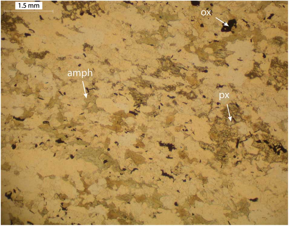
The magmatic enclaves display a magmatic mineral association of plagioclase, amphibole, pyroxene and minor quartz (Fig. 6). Plagioclase occurs as lath-shaped crystals that show polysynthetic twinning. Amphibole is anhedral, green coloured and pleochroic with, when observable, an extinction angle of about 20°. Millimetre-sized pyroxene grains occur as anhedral crystals and are largely replaced by secondary metamorphic minerals. Quartz grains, with irregular grain boundaries, are scattered in the matrix. Accessory magmatic minerals include magnetite and/or ilmenite, titanite and zircon. Secondary metamorphic minerals are well developed; plagioclases are frequently converted to fine-grained white mica, while amphibole and pyroxene are partly or completely pseudomorphosed by muscovite, chlorite and epidote.
Thin-section photomicrograph of a magmatic enclave (plane-polarized light). The micro-porphyric texture is underlined by crystals of amphibole (amph), pyroxene (px) and oxides (ox). The domains in white correspond to plagioclase and minor quartz.

The development of a secondary metamorphic mineral association (i.e. white mica + chlorite + quartz + albite ± epidote assemblage) in the magmatic sills and their enclaves attests to a severe metamorphic re-equilibration at the boundary between amphibolite- and greenschist-facies conditions (i.e. synchronous with the D3 tectonic event; see Homonnay et al. Reference Homonnay, Corsini, Lardeaux, Romagny, Münch, Bosch, Cenki-Tok and Ouazzani-Touhami2018 for discussion). Thus, both the magmatic sills and surrounding metapelites are affected by D3 metamorphism, and consequently, field observations together with petrographic analysis are compatible with syn- to late-D2 and pre-D3 magmatic sill emplacement.
3.c. Geochemical investigations
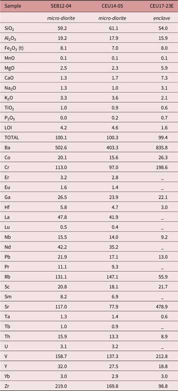
Because the magmatic sills are significantly affected by D3 metamorphism, in order to investigate the geochemical characteristics of these magmatic rocks we sampled the magmatic rocks only in the largest and best-preserved sill. In this sill, in view of outcrop quality, we found only two samples of micro-granitoids with well-preserved magmatic texture (SEB12-04 and CEU14-05) and one enclave (CEU17-23E), also very well preserved. These three samples were selected for major- and trace-element chemical analyses. Bulk rock compositions were obtained by inductively coupled plasma atomic emission spectrometry (ICP-AES) and inductively coupled plasma mass spectrometry (ICP-MS), for major and trace elements, respectively, on rock powders at the Geochemical and Petrographical Research Centre in Nancy (SARM laboratory, CNRS-CRPG; Table 1) following the procedure described in Carignan et al. (Reference Carignan, Hild, Mevelle, Morel and Yeghicheyan2001).
Chemical analyses: major elements (wt %), minor and trace elements (ppm)

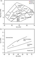
In the total alkalis versus silica classification (i.e. TAS diagram after Le Maitre et al. Reference Le Maitre, Bateman, Dudek, Keller, Lameyre, Le Bas, Sabine, Schmid, Sorensen, Streickeisen, Woolley, Zanettin and Le Maitre1989), the ‘micro-granitoid’ samples are micro-diorites, while the microgranular enclave is a less differentiated micro-gabbrodiorite (Fig. 7a). Moreover, with respect to SiO2 and K2O (i.e. in the classification of Peccerillo & Taylor, Reference Peccerillo and Taylor1976), all these magmatic rocks have a geochemical signature typical for high-K calc-alkaline magmas (Fig. 7b).
Rock classification diagrams using major elements. (a) TAS diagram for igneous rocks after Cox et al. (Reference Cox, Bell and Pankhurst1979). (b) Rock series discriminant diagram from Peccerillo & Taylor (Reference Peccerillo and Taylor1976).

However, the identification of the geochemical characteristics of magmatic rocks re-equilibrated during regional metamorphism is still delicate because of the mobility of the chemical elements during fluid-assisted metamorphism (Bridgwater, Reference Bridgwater1989; Ferry, Reference Ferry1994). Numerous studies have already shown that major elements as well as some trace elements, mainly large ion lithophile elements (LILE) (e.g. Rb, Cs, Sr, Ba) and light rare earth elements (LREE) (e.g. La, Ce, Nd), are considered to be the most mobile elements under greenschist- and amphibolite-facies conditions, whatever the considered lithology (Bridgwater, Reference Bridgwater1989; Potdevin et al. Reference Potdevin, Cotonian and Bertrand1989; Lawrie, Reference Lawrie1992; Ague, Reference Ague1994, Reference Ague2003; Roser & Nathan, Reference Roser and Nathan1997; Brewer & Menuge, Reference Brewer and Menuge1998; Masters & Ague, Reference Masters and Ague2005; Zheng et al. Reference Zheng, Chen and Zhao2009). Therefore, in order to improve the characterization of the geochemical signature of the studied samples, we selected diagrams suitable for magmatic rocks, using only minor and trace elements considered immobile during metamorphic processes.
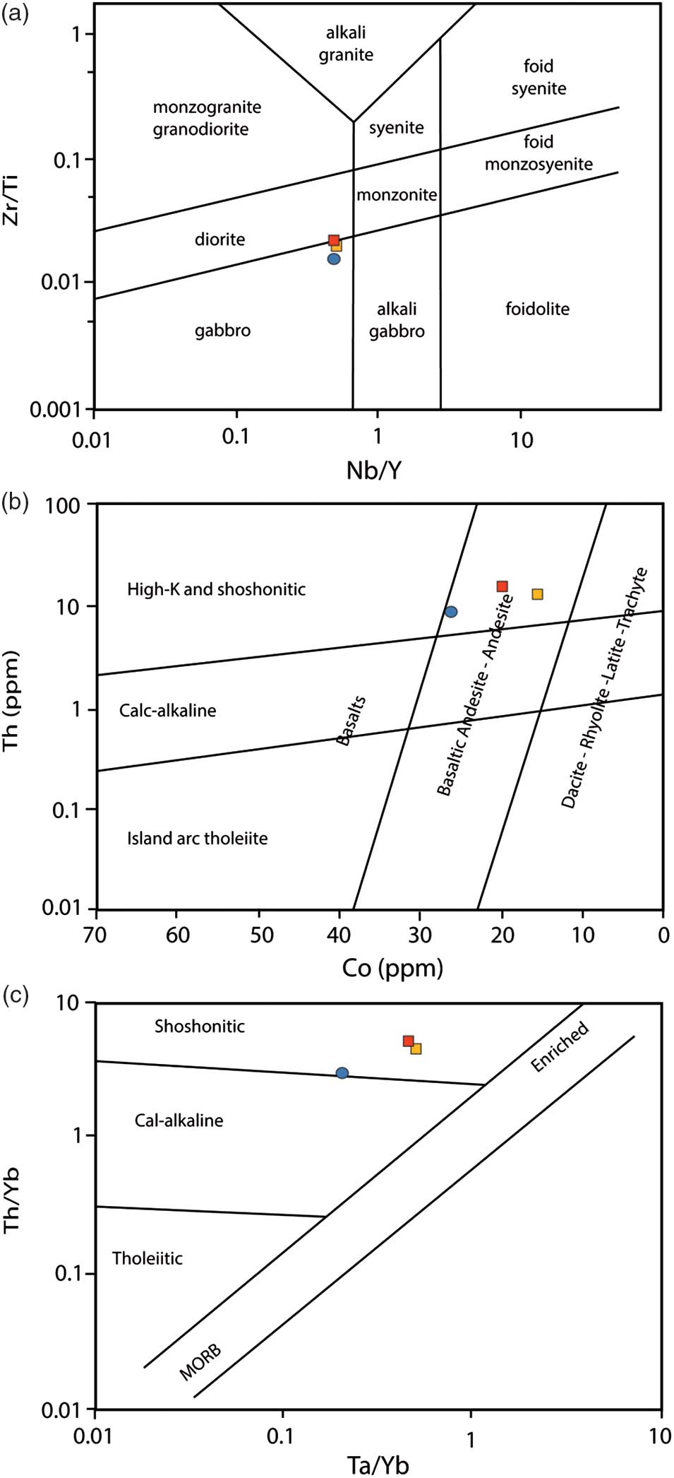
First, in the Winchester & Floyd (Reference Winchester and Floyd1977) magmatic rock classification including its modification by Pearce (Reference Pearce and Wyman1996), based on immobile element ratios such as Zr/Ti versus Nb/Y, the studied samples range in the field of gabbroic and dioritic rock types (Fig. 8a). Second, we used the accurate Th–Co diagram proposed by Hastie et al. (Reference Hastie, Kerr, Pearce and Mitchell2007) for magmatic rocks transformed under greenschist-facies conditions (Fig. 8b) and the Th/Yb versus Ta/Yb diagram (Fig. 8c) from Pearce (Reference Pearce, Hawkesworth and Norry1983). In these diagrams, the three samples are micro-gabbrodiorites and diorites, whereas their compositions still have a geochemical signature typical for the high-K to shoshonitic series.
(a) Zr/Ti versus Nb/Y diagram from Pearce (Reference Pearce and Wyman1996) modified after Winchester & Floyd (Reference Winchester and Floyd1977). (b) Th–Co discriminant diagram for altered volcanic island arc rocks, using immobile trace elements, from Hastie et al. (Reference Hastie, Kerr, Pearce and Mitchell2007). (c) Th/Yb versus Ta/Yb diagram after Pearce (Reference Pearce, Hawkesworth and Norry1983).

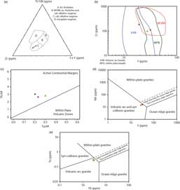
Trace-element discriminant diagrams are often considered an important way to determine the tectonic environment of magma formation (Pearce, Reference Pearce, Hawkesworth and Norry1983; Pearce et al. Reference Pearce, Nigel, Harris and Tindle1984). However, trace-element compositions are also a function of source components and the crystallization history of the melts (Rollinson, Reference Rollinson1993; Frost et al. Reference Frost, Barnes, Collins, Arculus, Ellis and Frost2001). Since trace-element diagrams can discriminate various tectonic settings, the composition of the studied samples was plotted on some of these diagrams (Fig. 9). In the discriminant diagrams proposed by Pearce & Cann (Reference Pearce and Cann1973) and Pearce (Reference Pearce and Thorpe1982) (Ti/Zr/Y in Fig. 9a and Cr versus Y in Fig. 9b, respectively), the three samples plot in the calc-alkaline and volcanic arc fields. In the Th/Hf versus Ta/Hf diagram (Schandl & Gorton, Reference Schandl and Gorton2002), samples plot in the active continental margin field (Fig. 9c). Moreover, the two micro-diorites typically have an intermediate rock composition. Consequently, we felt it was also interesting to confront their chemical signatures with discriminant diagrams established for granitoids. In the Nb versus Y diagram from (Pearce et al. Reference Pearce, Nigel, Harris and Tindle1984), the two samples plot in the volcanic arc and syn-collisional granitoid field (Fig. 9d), while using the more accurate Ta versus Yb diagram proposed by Pearce et al. (Reference Pearce, Nigel, Harris and Tindle1984) to distinguish between volcanic arc and syn-collisional granitoids, our samples plot in the volcanic arc granitoid field (Fig. 9e).
(a) Ti/Zr/Y discriminant diagram from Pearce & Cann (Reference Pearce and Cann1973). A – field of island arc tholeiites; B – field of MORB, arc tholeiites and calc-alkaline magmas; C – field of calc-alkaline magmas; D – field of within-plate magmas. (b) Cr versus Y discriminant diagram between MORB, volcanic arc basalts (VAB) and within-plate basalts (WPB) from Pearce (Reference Pearce and Thorpe1982). (c) Th/Hf versus Ta/Hf diagram from Schandl & Gorton (Reference Schandl and Gorton2002). (d) Nb versus Y tectonic discriminant diagram for granitoid rocks (Pearce et al. Reference Pearce, Nigel, Harris and Tindle1984). (e) Ta versus Yb tectonic discriminant diagram for granitoid rocks (Pearce et al. Reference Pearce, Nigel, Harris and Tindle1984).

Altogether, the geochemical characters of the studied sills are consistent with a high-K calc-alkaline magma formed within a continental magmatic arc.
4. U–Pb dating
4.a. Analytical method
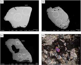
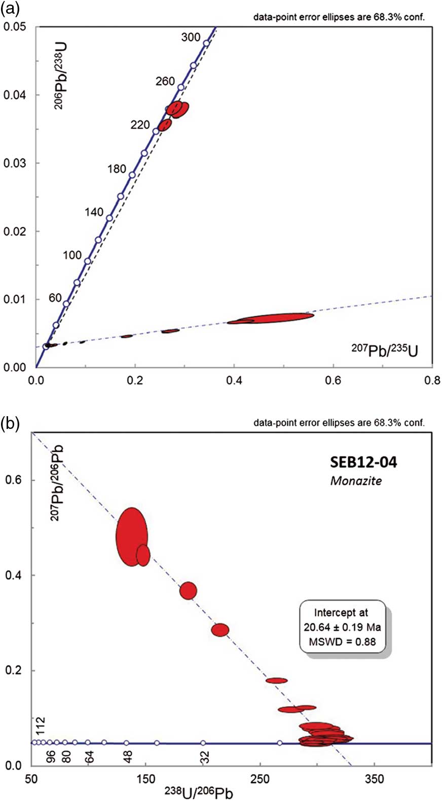
U–Th–Pb dating was performed on monazites and one xenotime from the micro-diorite SEB12-04. In situ analyses were achieved on thin-sections, in order to keep the microstructural context of the analysed minerals. Prior to LA-ICP-MS in situ U–Th–Pb analyses, minerals were examined with backscattered electrons (BSE) (Fig. 10), using a scanning electron microscope (SEM) at the CRHEA in Sophia Antipolis (Valbonne, France), to reveal the internal structure of the minerals. The operating conditions were 15 kV accelerating voltage and a 17 nA beam current. In situ U–Th–Pb analyses were performed at Geosciences Montpellier (University of Montpellier, France) by laser ablation (LA)-ICP-MS using a Lambda Physik CompEx 102 Excimer laser coupled to an Element XR sector field ICP-MS at the AETE-ISO regional facility (OSU OREME). Laser spot sizes were 15 μm. Before analysis, the laser was first fired to clean the sample surface (ten pulses). The targeted mineral was subsequently ablated under ultrapure helium in a 15 cm3 circular-shaped ablation cell with an energy density of 12 J/cm2 at a frequency of 2 Hz during 45 s. A gas blank was measured during 15 s before each analysis. Analyses of unknown monazite grains were bracketed by analyses of the Manangotry monazite standard (Poitrasson et al. Reference Poitrasson, Chenery and Shepherd2000). Repeated analyses of this reference mineral were used to correct for inter-element fractionation and for mass bias factor corrections (see details in Bosch et al. Reference Bosch, Garrido, Bruguier, Dhuime, Bodinier, Padròn-Navarta and Galland2011). Quoted ratios (see Table 2) correspond to measured ratios corrected for background and mass discrimination (+ elemental fractionation for the 206Pb/238U ratios). All ages have been calculated using the U and Th decay constants recommended by Steiger & Jäger (Reference Steiger and Jäger1977). Analytical data were plotted and ages were calculated using the IsoplotEx program (Ludwig, Reference Ludwig2003). Individual analyses in Table 2 and in Concordia plots (Fig. 11) are ± 1σ errors, and uncertainties in ages are quoted in the text at the 2σ level.
Backscattered electron (BSE) and microscopic images of monazite grains in the micro-diorite sill. Locations of U–Th–Pb analyses are circled in BSE images. (a) Homogeneous monazite grain. (b) Monazite grain exhibiting light grey core and dark grey rim. (c) Monazite grain with light grey patches and inclusion. (d) Microscopic photo of a monazite (mnz3) surrounded by chlorite (chl) and muscovite (ms).

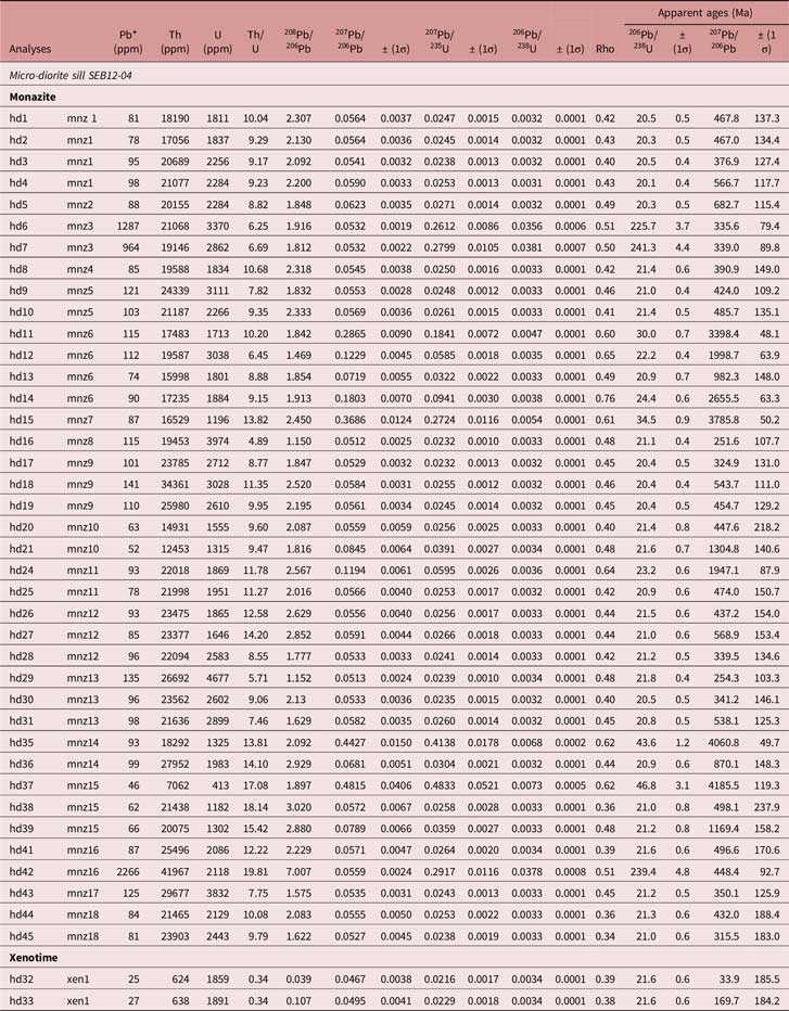
Results of the U–Th–Pb LA-ICP-MS analysis of monazite and xenotime from the micro-diorite sill SEB12-04
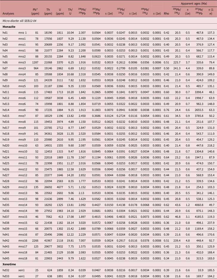
Pb* – radiogenic lead.
(a) Concordia diagram for U–Pb analyses on monazites and xenotime. (b) Tera-Wasserburg diagram close-up of young U–Pb analyses.

4.b. Results
Eighteen monazites and one xenotime were analysed from the sample SEB12-04. Under BSE imaging, 11 monazites display a homogeneous structure (Fig. 10a), whereas seven show either a light grey core surrounded by a dark grey rim (Fig. 10b; mnz3, mnz12, mnz13) or irregular light grey patches (Fig. 10c; mnz 2, mnz9, mnz16, mnz18) suggesting recrystallization of pre-existing minerals (e.g. Bosch et al. Reference Bosch, Hammor, Bruguier, Caby and Luck2002). The monazites sometimes contain inclusions (mnz1, 3–6, 15–18; Fig. 10c) and have a rounded or elongated shape. The shape of the monazite, and lack of oscillatory zoning, is not consistent with crystallization from a melt, as observed for magmatic monazite (e.g. Bruguier et al. Reference Bruguier, Hammor, Bosch and Caby2009). The xenotime is homogeneous, rounded in shape with undulant rims. Thirty-nine analyses were performed on 18 monazite grains (see Table 2) and two analyses were performed on the xenotime grain identified in the thin-section. Despite the observed differences in internal structures, most monazite analyses display a single alignment in the Concordia diagram and provide an intercept age of 20.7 ± 0.19 Ma (Fig. 11b). The xenotime analysis also plots along this alignment, suggesting that the monazite and xenotime are coeval. Two grains (mnz3 and mnz16) are significantly older with apparent 206Pb–238U ages ranging from 241.3 ± 8.8 Ma to 225.7 ± 7.4 Ma (Table 2; Fig. 11a). It is noteworthy that mnz16 displays a heterogeneous age distribution in which Miocene and older domains are present. The BSE imaging of this grain is not consistent with a core-like structure but, on the contrary, suggests recrystallization processes. On that basis, the c. 21 Ma age is interpreted as reflecting monazite growth or resetting of the U–Pb system of pre-existing minerals under metamorphic conditions. Older ages are interpreted as partial updating of minerals that were most likely snatched from deep-seated material by the dioritic magma.
4.c. Age interpretations
Most of the dated minerals yield an age of c. 21 Ma. In the Ceuta Peninsula, this age is now well documented and is associated with the D3 tectonic event, developed at the boundary between amphibolite- and greenschist-facies conditions, and which corresponds to the development of ductile extension (Homonnay et al. Reference Homonnay, Corsini, Lardeaux, Romagny, Münch, Bosch, Cenki-Tok and Ouazzani-Touhami2018). Interestingly, in the investigated micro-diorite, the dated monazites and xenotime are systematically in textural equilibrium with the secondary metamorphic phases in the groundmass as well as in inclusions within magmatic minerals, the latter being totally replaced by muscovite and chlorite (Fig. 10d). Consequently, monazite ages at c. 21 Ma cannot be interpreted as the ages of magma crystallization but, on the contrary, are related to the late-D3 stage of metamorphism characterized by metamorphic re-equilibration, associated with fluid circulation, during early Miocene time. Resetting of monazite ages during greenschist-facies metamorphism and related fluid activity is clearly demonstrated (e.g. Williams et al. Reference Williams, Jercinovic, Harlov, Budzyń and Hetherington2011; Seydoux-Guillaume et al. Reference Seydoux-Guillaume, Montel, Bingen, Bosse, de Parseval, Paquette, Janots and Wirth2012) and was already outlined on metamorphic rocks from the Internal Rif (Montel et al. Reference Montel, Kornprobst and Vielzeuf2000; Janots et al. Reference Janots, Negro, Brunet, Goffé, Engi and Bouybaouène2006; Michard et al. Reference Michard, Negro, Saddiqi, Bouybaouene, Chalouan, Montigny and Goffé2006; Negro et al. Reference Negro, Beyssac, Goffe, Saddiqi and Bouybaouene2006; Chalouan et al. Reference Chalouan, Michard, Kadiri, Negro, Frizon de Lamotte, Soto, Saddiqi and Michard2008; Rossetti et al. Reference Rossetti, Theye, Lucci, Bouybaouene, Dini, Gerdes, Phillips and Cozzupoli2010; Gueydan et al. Reference Gueydan, Pitra, Afiri, Poujol, Essaifi and Paquette2015; Homonnay et al. Reference Homonnay, Corsini, Lardeaux, Romagny, Münch, Bosch, Cenki-Tok and Ouazzani-Touhami2018) and also in the Internal Betic (Acosta-Vigil et al. Reference Acosta-Vigil, Rubatto, Bartoli, Cesare, Meli, Pedrera, Azor and Tajčmanová2014).
5. Geological consequences
We report for the first time the occurrence of calc-alkaline magmatic sills in the Ceuta Peninsula (Internal Rif). Sampled micro-diorites and micro-gabbrodiorites show a primary magmatic mineralogy and a geochemical signature diagnostic for high-K calc-alkaline to shoshonitic island arc magmatism. Field observations based on geometric relationships between the micro-diorite sills and the main regional foliation S2, developed under intermediate-pressure granulite-facies conditions before 28 Ma (Homonnay et al. Reference Homonnay, Corsini, Lardeaux, Romagny, Münch, Bosch, Cenki-Tok and Ouazzani-Touhami2018), demonstrate that the magmatic sills emplaced during the late stage of the D2 tectonic event. Furthermore, these magmatic rocks are fully metamorphosed under greenschist-facies metamorphic conditions. The latter, depicted at the regional scale, is coeval with an early Miocene D3 extensional tectonic event (Homonnay et al. Reference Homonnay, Corsini, Lardeaux, Romagny, Münch, Bosch, Cenki-Tok and Ouazzani-Touhami2018). Hence, syn- to late-D2 and pre-D3 magmatic sill emplacement is consistent with the magmatic monazite U–Pb age resetting at c. 21 Ma.
The significance of this pre-Miocene high-K calc-alkaline magmatism must now be envisioned and discussed within the framework of the geodynamic context presently held for the Alboran domain. Following geophysical investigations, and particularly tomographic studies (Blanco & Spakman, Reference Blanco and Spakman1993; Gutscher et al. Reference Gutscher, Malod, Rehault, Contrucci, Klingelhoefer, Mendes-Victor and Spakman2002; Timoulali et al. Reference Timoulali, Djellit, Hahou, Jabour and Merrouch2014; Villaseñor et al. Reference Villaseñor, Chevrot, Harnafi, Gallart, Pazos, Serrano, Córdoba, Pulgar and Ibarra2015), the Betic–Rif orogen that surrounds the Alboran Basin is regarded as an emblematic example of a subduction-controlled orogeny followed by slab rollback leading to the opening of the Alboran back-arc basin (e.g. Faccenna et al. Reference Faccenna, Piromallo, Crespo-Blanc, Jolivet and Rossetti2004; Spakman & Wortel, Reference Spakman, Wortel, Cavazza, Roure, Spakman, Stampfli and Ziegler2004; Vergés & Fernàndez, Reference Vergés and Fernàndez2012; Bezada et al. Reference Bezada, Humphreys, Toomey, Harnafi, Dávila and Gallart2013; Van Hinsbergen et al. Reference Van Hinsbergen, Vissers and Spakman2014). In this geodynamic framework, slab rollback of the Tethyan oceanic lithosphere initiated at c. 23–21 Ma, while the timing of the Alpine subduction is Eocene up to Oligocene. The occurrence of pre-Miocene high-K calc-alkaline to shoshonitic arc-related magmatism is clearly consistent with the nowadays-proposed geodynamic context and represents the last geological component, so far unknown, of the Alboran subduction system. In such a framework, the discovery of calc-alkaline magmatic sills allows the magmatic arc, and thus the upper plate of the subduction system, to be identified. In the case of the Rif belt, the Lower Sebtides should correspond to the upper plate of the subduction system. Such a pattern is consistent with the scenario recently proposed by Homonnay et al. (Reference Homonnay, Corsini, Lardeaux, Romagny, Münch, Bosch, Cenki-Tok and Ouazzani-Touhami2018) for the development of high-temperature/intermediate- to low-pressure metamorphism within the lowermost Ceuta units (i.e. Lower Sebtides) interpreted as an orogenic wedge located, in a backward position, in the upper plate of the Alboran subduction zone.
Our structural and geochronological data reveal that, in the Ceuta area, high-temperature metamorphism and calc-alkaline magmatism are, at least in part, synchronous. This is consistent with the thermal behaviour of supra-subduction magmatic arcs. Indeed, in numerous examples of active or extinct arcs, regional metamorphism is coeval with arc building, and heat input is related to continuous or episodic emplacement of magmatic intrusions (Miyashiro, Reference Miyashiro1961; Ernst, Reference Ernst1975; Schiffman et al. Reference Schiffman, Elders, Williams, McDowell and Bird1984; Aguirre et al. Reference Aguirre, Robinson, Bevins, Morata, Vergara, Fonseca and Carrasco2000; Willner et al. Reference Willner, Glodny, Gerya, Godoy and Massonne2004; Willner, Reference Willner2005). The small volume of magmatic rocks identified up to now could be the result of a weakly productive magmatic arc or be the result of a difficulty in identifying these rocks in the field.
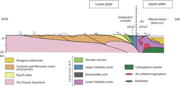
Furthermore, in the northern Rif, the Upper Sebtides located westward of the Ceuta Peninsula underwent an Oligocene high-pressure/low-temperature metamorphism (Bouybaouene et al. Reference Bouybaouene, Goffé and Michard1995; Goffe et al. Reference Goffe, Azanon, Bouybaouene and Jullien1996; Michard et al. Reference Michard, Goffe, Bouybaouene and Saddiqi1997; Janots et al. Reference Janots, Negro, Brunet, Goffé, Engi and Bouybaouène2006) and thus must be considered as the lower plate subducted beneath the Lower Sebtides (Fig. 12). The tectonic coupling between the Upper and Lower Sebtides defines a typical example of ‘paired metamorphic belts’ (Brown, Reference Brown2010 and references therein). Indeed, island arc and back-arc regions are recognized as a suitable tectonic setting for the development of regional high-temperature/low-pressure metamorphism (Miyashiro, Reference Miyashiro1961; Oxburgh & Turcotte, Reference Oxburgh and Turcotte1970; Ernst, Reference Ernst1973). This metamorphism developed in the upper plate of the subduction zone is the counterpart of the high-pressure/low-temperature metamorphism widespread within the down-going lower plate (Fig. 12).
Schematic cross-section of the Rif belt (modified from Chalouan et al. Reference Chalouan, Michard, Kadiri, Negro, Frizon de Lamotte, Soto, Saddiqi and Michard2008). HP/LT: high pressure/low temperature. HT/LP: high temperature/low pressure.

After the development of the paired metamorphic belts, during early Miocene time, the Upper Sebtides underwent normal faulting associated with back-arc extension and opening of the Alboran Basin (Fig. 12; Comas et al. Reference Comas, Platt, Soto, Watts, Zahn, Comas and Klaus1999; Platt et al. Reference Platt, Argles, Carter, Kelley, Whitehouse and Lonergan2003; Martínez-García et al. Reference Martínez-García, Comas, Lonergan and Watts2017; Homonnay et al. Reference Homonnay, Corsini, Lardeaux, Romagny, Münch, Bosch, Cenki-Tok and Ouazzani-Touhami2018). Post-subduction deformation is thus observed in the studied area; however, this deformation is not strong enough to reverse the initial relationships between the Upper and Lower Sebtides.
At the scale of the Betic–Rif orogen, published magmatic studies mostly deal with recent magmatic episodes within the Neogene volcanic province widespread in the eastern part of the Alboran domain (Duggen et al. Reference Duggen, Hoernle, Klügel, Geldmacher, Thirlwall, Hauff, Lowry and Oates2008; see Lustrino et al. Reference Lustrino, Duggen and Rosenberg2011 for a review). Up to now, earlier magmatic events were only described in the Betic Cordilleras (e.g. Bellon et al. Reference Bellon, Bordet and Montenat1983; Torres-Roldan et al. Reference Torres-Roldan, Poli and Peccerillo1986; Turner et al. Reference Turner, Platt, George, Kelley, Pearson and Nowell1999; Duggen et al. Reference Duggen, Hoernle, van den Bogaard and Harris2004; Lustrino et al. Reference Lustrino, Duggen and Rosenberg2011; Esteban et al. Reference Esteban, Tubía, Cuevas, Seward, Larionov, Sergeev and Navarro-Vilá2013) and more precisely in the Malaga area (SW Betic), as tholeiitic and calc-alkaline basaltic andesite dykes. K–Ar and Ar–Ar geochronological investigations on these rocks reveal two age groups: (1) 30–38 Ma, which corresponds to calc-alkaline dyke emplacement during the subduction process, and (2) 17.4–19.8 Ma, interpreted as K–Ar system resetting due to a wide-scale thermal event. Furthermore, a high-K to shoshonitic magmatic episode occurred between 16 and 8 Ma, in the eastern Rif and Betics and in the Alboran Sea. It is related to the slab rollback and marks the position of the deep subducted lithosphere beneath the Alboran Basin (Gill et al. Reference Gill, Aparicio, El Azzouzi, Hernandez, Thirlwall, Bourgois and Marriner2004; Duggen et al. Reference Duggen, Hoernle, van den Bogaard and Garbe-Schönberg2005).
The new data we obtained in the northern Rif are in agreement with this interpretation proposed for the Betic chains, and all together support a model of subduction-related orogeny for the Betic/Rif Cordilleras.
6. Conclusions
We described for the first time a high-K calc-alkaline magmatism in the Lower Sebtides (Internal Rif). Our structural and petrographic observations with geochronological data demonstrate that: (i) pre-Miocene high-K calc-alkaline to shoshonitic micro-diorite sills emplaced in the Ceuta Peninsula within the Lower Sebtides (Internal Rif) during high-temperature/medium-pressure metamorphism (i.e. D2 deformation event); (ii) these newly recognized magmatic rocks have a primary magmatic mineralogy and geochemical signature diagnostic of island arc magmatism; (iii) the micro-diorite sills display a secondary medium-temperature/low-pressure metamorphic episode (i.e. D3 deformation event), which led to monazite U–Pb age resetting at c. 21 Ma.
The presence of arc-related magmatism, probably Oligocene in age, is not surprising in the nowadays widely accepted subduction system for the Gibraltar arc formation. Therefore, in such a framework, the so-called Lower Sebtides correspond to the upper plate of the subduction system, whereas the Upper Sebtides may be considered as the lower plate of the subduction system.
Acknowledgements
The field work was supported by the FP7-IRSES-MEDYNA project.