Introduction
Malignant melanoma is the most common melanocytic tumour. It originates in the skin but can also arise in uveal, gastrointestinal or vulvar mucosae. Cutaneous melanoma can be subdivided into specific groups, based on the anatomic site and pattern of growth, namely: (i) the superficial melanoma, which is the most common form (70%), mainly occurring on the trunk or legs, with a good prognosis; (ii) the nodular melanoma (10–25%); (iii) the lentigo maligna melanoma (<1%) and the lentiginous melanoma (5%), which primarily originate in the head and neck region, either associated or not with chronic sun exposure, respectively; and (iv) the non cutaneous melanoma (5%). Besides the mentioned melanoma subtypes, other less frequent variants such as desmoplastic, nevoid, spitzoid, angiotropic melanomas, malignant blue nevi and animal type have been described. For the extremely low recurrence, their clinical features, however, are still not well defined (Ref Reference LeBoit, Burg, Weedon and Sarasin1).
Animal type melanoma (ATM) is a very rare form of malignant melanoma and is characterised by the presence of largely homogeneous blue-black hyperpigmented cutaneous nodules with melanised epithelioid cells and a deep exo-endophytic growth pattern (Refs Reference Crowson, Magro and Mihm2, Reference Massi3). Due to its deep rarity, to date, there is still a remarkable lack of uniformity in its clinical and histological characterisation even including the name itself of this disease (Ref Reference Vyas4).
The first report dates back to 1832 when William Dick primarily described a pigmented melanocytic neoplasm characterised by the presence of nodules of highly melanised cells in old grey horses (Ref Reference Dick5). In 1925, Darier described the ‘melanosarcoma’, a similar lesion in human subjects and noticed the occurrence of common histologic and clinical features between this human lesion and the ‘equine melanotic disease’ (Ref Reference Darier6). In 1982 Tuthill claimed the similarity between the ‘melanosarcoma’ previously described by Darier and the ‘malignant blue nevus’ (Ref Reference Tuthill, Clark and Levene7), but it was only in 1990 that Clark used for the first time the definition of ATM to describe the peculiarity of this skin lesion (Ref Reference Clark, Elder and Guerry8).
The term ‘pigmented epithelioid melanocytoma’ (PEM) was proposed by Zembowicz in 2004 for both the ‘animal-type melanoma’ and the ‘epithelioid blue nevus (EBN)’ occurring in patients affected by the Carney Complex (Ref Reference Zembowicz, Carney and Mihm9) (OMIM # 160980) which is an autosomal dominant syndrome characterised by a wide phenotypic spectrum of lesions of both skin and mucosae associated with cardiac, endocrine, cutaneous and neural myxomatous tumours. The syndrome is caused by mutations in the protein kinase A regulatory subunit-1-alpha gene (PRKAR1A) on chromosome 17q24.2 (Ref Reference Stratakis10).
Both ATM and EBN types were then included in a unique nosography entity corresponding to a low-grade melanoma or borderline melanocytic tumour with metastatic potential. Despite the thin thickness, the absence of ulceration and the low mitotic index that are the usual features of this lesion, metastases to lymph nodes are frequently described (Ref Reference Zembowicz, Carney and Mihm9). On the other hand, survival analysis of ATM revealed its lower tendency to produce distant metastases with respect to the superficial melanoma, thus resulting in a more favourable prognosis. This is clearly controversial with observations from accumulating clinical practice showing cases of ATM with aggressive behaviour despite the initial interpretation of ‘indolent disease’ according to common prognostic criteria for melanoma (Ref Reference Gavriilidis11).
In the present study, we describe a case of ATM with aggressive behaviour in a Southern Italian patient. Furthermore, here we revisit the major aspects of ATM and particularly its immunogenicity and genomic aspects which have significantly improved the treatment and prognosis of melanomas in the era of targeted therapies and immune checkpoint inhibitors.
Review of the literature
We carried out an extensive systematic search in the international literature using the PubMed, Scopus and ISI-Web of Science databases assessed up to 30 June 2017. To this, we used the following keywords, alone or in combination: ‘animal type melanoma’, ‘melanosarcoma’, ‘equine melanotic disease’, ‘malignant blue nevus’, ‘epithelioid blue nevus’, ‘pigmented epithelioid melanocytoma’. A further search was performed by reviewing the references of the retrieved articles. Despite this extensive research, in most cases, the available information on ATM was derived from isolated case-reports and from retrospective studies on short numbers of patients with time-limited follow-ups. The largest meta-analysis was provided by Vyas and co-workers on 190 ATM cases that collected data from 39 small studies in which, when available, specific clinical features, histology, clinical management and outcome were carefully revised (Ref Reference Vyas4).
Epidemiology
ATM usually raises de novo and with no association with pre-existing melanocytic lesions. There is neither a correlation with familial dysplastic nevus syndrome nor with a family history of malignant melanoma (Refs Reference Crowson, Magro and Mihm2, Reference Vyas4). According to published available studies, the disease is diagnosed in all age groups, including children and elderlies, with a prevalence in young adults with a median age of 27 years at the onset (Refs Reference Vyas4, Reference Battistella12, Reference Sestini13, Reference AL-Johani14). In contrast with conventional melanomas that predominantly affect males with respect to females, ATM is equally distributed in both sexes at a similar frequency. Although previous studies excluded ethnic differences in its incidence, (Ref Reference Zembowicz, Carney and Mihm9), the above-mentioned meta-analysis of Vyas suggested highest recurrence in Caucasians (54%), followed by Hispanics (18.5%), Asians/Persians (18.5%) and blacks (9.3%) (Ref Reference Vyas4). However, these data refer only to a minority of the population evaluated in the meta-analysis since the consistency of the ethnic evaluation of ATM in this study included no more than 25% of the full cohort of patients, namely 54/190 cases (Ref Reference Vyas4).
Clinical features and histology
ATM usually arises as a homogeneous blue, or blue-black plaque or nodule, sharply bordered, of approximately 1–4 cm in diameter showing a slow growth since childhood over years, although a couple of case reports describe the presence of this malignancy even at birth (Ref Reference Battistella12). In up to 40% of patients, the lesion recurs at the extremities, then on trunk (30%), and head and neck (30%), and with no correlation with the sun exposure (Refs Reference Vyas4, Reference Sabah, Leader, Fahy and Kay15, Reference Lim16, Reference Cheng17, Reference Urso18, Reference Akita19, Reference Mandal20, Reference Marques21). Rare cases of ATM have been described on the skin of genitalia as reported on a newborn on left major labium (Ref Reference Ranabhat22). Mucosal localisation of ATM has also been described on the tarsal conjunctive (Ref Reference Bissig23) as oral mucosal lentiginous melanoma with prominent pigment synthesis (Ref Reference Kumar24), or similar to the typical Ota nevus in the sclera (Ref Reference Nitta25).
At the dermoscopic evaluation, the ATM appears as a homogeneously pigmented blue-black area, because of heavily pigmented dermal melanocytes, with irregular whitish structures as grey-white dots and lines for the epidermal hyperplasia and polymorphous vascular pattern composed of large irregular vessels. With the exception of the blue pigmentation and respect to other melanocytic malignancies, ATM does not show specific dermoscopic patterns and a careful histology evaluation is necessary for its diagnosis (Ref Reference Aviles-Izquierdo, Leis-Dosil and Lazaro-Ochaita26).
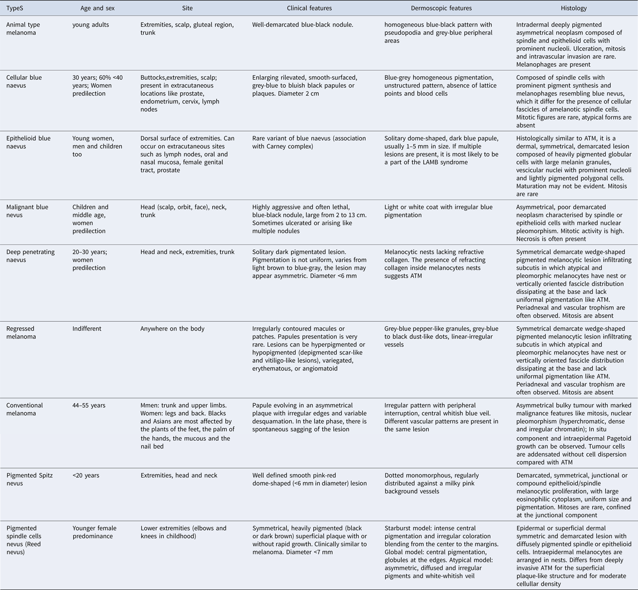
ATM is histologically characterised by a deeply pigmented, nodular, dermal proliferation of three kinds of cells: (i) peripheral spindle cells with pseudodendrites drifting apart from the lesion in the collagen; (ii) central, small size, epithelioid cells with heavily pigmented cytoplasm; and (iii) large sized, nested, epithelioid cells with big and eosinophilic nucleoli and large cytoplasm showing a rim of granular pigmentation. Spindled cells are often disposed in fascicles, whereas rounded or polygonal cells show nodular arrangement (Refs Reference Massi3, Reference Moscarella27, Reference Sevilla Chica and Bernett28, Reference Moyano de Fossati29, Reference Howard, Ragsdale and Lundquist30, Reference Lee31). The neoplasm is typically intradermal and subverts the normal dermis architecture while it often infiltrates subcutaneous tissue although it is separated from the epidermidis. It is possible to find pseudoepitheliomatous pagetoid hyperplasia without epidermal component (Ref Reference Crowson, Magro and Mihm2). Histiocytes and macrophages with fine granular pigment can be present in the centre of the tumour (Ref Reference Sadayasu32). Massive numbers of melanophages similar to epithelioid pigmented cells are generally mixed with inflammatory lymphocytic infiltrate. The pigmentation pattern can range from a fine to a granular light-brown or course dark-brown melanin. Because of the heavy pigmentation, pre-treatment with hydrogen peroxide of samples allows to evaluate the cellular morphology and reveal a typically moderate anisonucleosis with oval or round nuclear shape and small nucleoli, without fulfilling cytological criteria of malignancy (Ref Reference Magro, Crowson and Mihm33). Based on these aspects, both confluent melanocytic growth and altered dermal architecture support the diagnosis of melanoma (Ref Reference Crowson, Magro and Mihm2). Mitotic activity is usually low and ulceration, necrosis, regression and intravascular activity are generally rare. In contrast with the primary lesion, more cytological features of atypia are detectable in metastatic sites, where the mitotic index is generally higher and the malignant melanocytes appear pleomorphic. In this contest, it has been also described a rare case of ATM with aggressive histological features and mild mitotic activity, namely one single mitosis per mm2, tumour necrosis, focal ulceration and both perineural and vascular invasion (Ref Reference Russo34). For its association with follicular sheath, some authors named this neoplasm as pilar neurocristic hamartoma (Refs Reference Crowson, Magro and Mihm2, Reference Pathy35) whereas, based on histological features, Ludgate described it as equivocal or unequivocal melanoma. Patients with equivocal ATM were younger with a median age 12 versus 53 years, and had a lower rate of positive lymph nodes compared with unequivocal ATM thus suggesting molecular differences to justify differential clinical behaviour (Ref Reference Ludgate36). Immunoistochemistry of ATM is positive for S-100 protein, HMB-45, vimentin and Melan-A (Refs Reference Vyas4, Reference Ludgate36, Reference Neelam37, Reference Posch38, Reference Aung39) and testing CD68 is useful to differentiate pigmented melanocytic cells from melanophages. The principal differential diagnosis of ATM or PEM are summarised in Table 1 (Refs Reference Crowson, Magro and Mihm2, Reference Vyas4, Reference Sabah, Leader, Fahy and Kay15, Reference Lim16, Reference Magro, Crowson and Mihm33, Reference Aung39, Reference Antony40).
Differential diagnosis between ATM and other cutaneous lesions: clinical, dermoscopic and histological features

Contrary to conventional melanomas, ATM is not immunogenic. Therefore, peritumoral inflammation is rarely described and no mention regarding the host response has been provided. In six clinical cases of malignant melanoma with prominent pigment synthesis (Ref Reference Crowson, Magro and Mihm2), one of them included a mild lymphocytic host response, while the others were defective of the host inflammatory response and with no mention on TIL in the last three cases. No histological evidence of regression nor host lymphocytic response was present in the case described by Sabah (Ref Reference Sabah, Leader, Fahy and Kay15), whereas Sadayasu and co-workers (Ref Reference Sadayasu32) analysed immunohistochemical profiles of tumour-infiltrating histiocytes on a specimen of pigmented epithelioid melanocytoma to evaluate the content of regulatory T cells or CD163+ M2 macrophages implicated in promoting angioinvasion and immunosuppression in tumour microenvironment. They found only tumour-infiltrating histiocytes and CD68+/CD163−/MMP9− macrophages associated with a small number of T-regulatory cells (Tregs) and suggested that the defective infiltration by immunosuppressive cells as Tregs and MMP9+ cells may correlate with the indolent behaviour of this tumour. However, considering the extremely low number of clinical studies evaluating TILs in ATM, more studies focusing on this aspect are needed.
Clinical course: prognostic role of the sentinel lymph node biopsy
Given the rarity of this subtype of melanoma and the paucity of description in literature, its clinical behaviour is undefined although the tumour is associated with satellitosis (Refs Reference Vyas4, Reference Sestini13, Reference Aviles-Izquierdo, Leis-Dosil and Lazaro-Ochaita26, Reference Pathy35, Reference Posch38, Reference Antony40) and nodal involvement (Refs Reference Bagny41, Reference Matthey-Gie42). Several studies have reported a more benign clinical course of ATM if compared with conventional melanoma with the same histological parameters (Refs Reference Antony40, Reference Mentzel43). Since ATM shows high metastatic potential, a wide excision with the sentinel node biopsy (SLNB) is recommended (Ref Reference Crowson, Magro and Mihm2). To this regard, previous reports found that up to 46% of patients affected by ATM that underwent SLNB showed regional lymph node metastases confirming the higher lymph node metastatic rate with respect to the conventional melanomas (Ref Reference Morton44), which may be partially explained by the deeper average of Breslow's thickness at the first ATM diagnosis (Ref Reference Ludgate36). In contrast with these findings, other Authors testify a low incidence of nodal involvement (Refs Reference Orlandi45, Reference Scolyer46) although these studies have included cases with a more indolent biological behaviour in whom a low mitotic and cellular atypia rate were histologically described. However, ATM seems to have a greater propensity to metastasize to regional lymphatic nodes than other melanomas, and low likelihood to develop distant metastatic spread (Ref Reference Zembowicz, Carney and Mihm9) although the positive lymph node status is not a good marker of poor prognosis (Ref Reference Posch38). In fact, ATMs show a 5-year survival rate up to approximately 95% of patients whereas this parameter is lower than 50% in conventional melanomas at a similar stage (Ref Reference Balch47). Melanocytic cells in loco-regional nodes should not be considered as metastatic localisation, rather benign melanocytes, similar to those found in corresponding regional lymph nodes of congenital nevi although further studies by FISH are required to define the benign or malignant nature of these melanocytic cells. Based on these reports, ATM should be considered as a dermal melanocytic borderline tumour that is similar to equine melanotic disease, is characterised by a long indolent phase which in several instances may undergo to aggressive evolution and behaviour.
To date, ATM is treated with a surgical cutaneous excision including appropriate margins of healthy tissue, with SLNB in case of Breslow thickness higher than 0.75 mm, or in presence of unfavourable prognostic parameters as regression, ulceration, high mitotic rate and vertical growth phase. However, in the case of positive SLNB, a complete lymph node dissection is recommended (Refs Reference Ludgate36, Reference Posch38). To this regard, it has been also proved that more aggressive surgical and/or systemic therapies do not provide a major benefit for patients (Refs Reference Mandal20, Reference Scolyer48) although further studies are required to fully determinate the biological nature and the long-term prognosis of ATMs. Observation of indolent clinical behaviour in ATM with either satellitosis or nodal involvement makes these important prognostic factors in conventional melanoma as uncertain in ATM. In fact, it is not clear the reason by which in these patients the nodal involvement does not correlate with higher risk of recurrence. Intrinsic genomic characterisation, more than clinical prognostic factors, may probably be useful in understanding these peculiar features.
Genomic characterisation
Recently The Cancer Genome Atlas (TCGA) Research Network published a massive study performed on 333 melanomas from 331 patients using whole-exome, whole-genome, RNA and microRNA sequencing, DNA copy-number, SNPs array, DNA methylation and protein array. This integrative analysis allowed the identification of four subtypes clinically useful for the adoption of targeted therapies (TCGA 2015). The largest genomic subtype was characterised by the presence of BRAF gene hotspot mutations, namely in decreasing frequency order as V600E, V600K, V600R and K601E. The second major subtype revealed the presence of N-, K- and H-RAS classical hotspot mutations, essentially in codons 12, 13 and 61. An additional subtype was characterised by loss-of-function mutations in the NF1 gene, while the fourth cluster included a heterogeneous group of tumours identified as triple wild-type for the absence of typical molecular alterations of the first three subtypes (TCGA 2015).
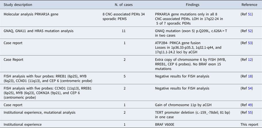
ATM is a rare tumour and only in a very limited number of cases, it is possible to find specific studies concerning its molecular characterisation, mostly in the context of extended clinical studies of ‘classic’ melanomas and nevi (Ref Reference Dadras49). To date, the largest study was performed by Zembowicz who analysed 34 sporadic and 8 Carney complex-associated PEMs for PRKAR1A mutations. This gene locates on chromosome 17q22-24 and encodes for the R1α protein, which is essential for the melanocytic function, since it regulates the pathway of cyclic AMP and protein kinase A. The molecular analysis demonstrated the PRKAR1A gene mutations in all 8 Carney Complex (CNC)-associated PEMs, and loss of heterozygosity (LOH) in 17q22-24 in 5 of the 7 analysed sporadic PEMs. Loss of expression of the R1α protein, measured by immunohistochemistry, was identified in all the 8 EBN – associated with CNC and in 28 of 34 sporadic PEMs (Refs Reference Zembowicz and Scolyer50, Reference Zembowicz51) suggesting that PEM and Carney complex PEMs nevi are closely related subtypes of melanoma (Ref Reference Cheng17). Sporadic and non-significant cases of PRKAR1A gene mutations are also reported in the TCGA study (Table 2).
Summary of molecular testing performed on Animal Type Melanoma

In an extended mutational analysis sequencing HRAS, GNAQ and GNA11 genes in a heterogeneous group of cellular blue nevi (CBN), common blue nevi (BN), deep penetrating nevi (DPN) and epithelioid blue nevi (EBN), Bender et al., found the same c.626A > T point (p.Q209L) mutation in the exon 5 of GNAQ gene in 2 out of 11 cases. Although, in the presence of limited data, the authors argued that EBN may be considered a histologic variant of the blue nevi, with a specific mutational pattern of more the 80% of mutations in exon 5 of GNAQ (Ref Reference Bender52). GNAQ Q209P mutation was also found in a single case of melanoma classified in the triple wildtype subtype by the TCGA study.
Recently, a case report described a novel gene fusion transcript between the ATP2B4 (ATPase, Ca2+ transporting, plasma membrane, 4) and PRKCA (Protein Kinase C, alpha) genes in a 5-month-old girl with a large scalp pigment-synthesizing melanocytic neoplasm. The array comparative genomic hybridisation (aCGH) analysis showed the loss of 1p36.33-p35.3, 1q32.1-q44 and 17q11.1-24.2 loci, and the mutational screening for BRAF, NRAS, GNAQ, GNA11, PRKAR1A and TERT genes, failed to identify pathogenic further sequence variants (Ref Reference Bahrami53).
Further cytogenetic studies by FISH (fluorescent in situ hybridisation) of chromosomal aberrations in RREB1 (RAS-Responsive Element Binding Protein 1), MYB (V-MYB Avian Myeloblastosis Viral Oncogene Homolog), CCND1 (Cyclin D1) and CEP6 (Centrosomal Protein for chromosome 6) genes completed on two congenital cases of PEM, revealed in a single case an extra copy of chromosome 6 (3 MYB, 3 RREB1, 3 CEP6 and 2 CCND1 signals) per nucleus. In both cases the analysis for BRAF V600 mutation was negative (Ref Reference Battistella12), whereas in additional seven cases, the same FISH tumour analysis resulted negative (Refs Reference Urso18, Reference Muhlbauer54) while the evaluation by comparative genomic hybridisation for a single case demonstrated a gain of chromosome 11p, a molecular feature similar to a Spitz nevus (Ref Reference Dadras49).
More recently, the primary lesions from paraffin blocks of two PEM patients were screened for mutational hotspot codon in BRAF, GNAQ, GNA11, SF3B1, RAC1, NRAS, HRAS, KRAS, TP53 and TERT genes. The only mutation identified was a 81 bp deletion in the promoter region of TERT gene (c.-159-78del), previously reported in basal cell carcinoma and squamous cell carcinoma, but not in melanoma (Ref Reference Bax55). However, while it is necessary to specify that point mutations of the gene TERT promoter are found in about 80% of NF1, 70% of BRAF and RAS and 6% of triple-WT subtypes, it is essential to emphasise that in our study, we have identified for the first time a BRAF variant V600E identified in both primitive cancer and circulating tumour cells (Table 2).
Case report
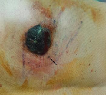
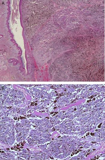
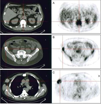
A 55-year-old-man was admitted to our Department of Medical Oncology for a rapidly enlarging, elevated, bleeding, itchy and darkly pigmented cutaneous lesion of the back. No correlation with CNC was documented. At the clinical observation, the lesion was highly suspicious for melanoma. It was indeed exophitic, 3.8 × 3.7 cm2 in its main diameters and adjacent to pigmented lesions of 2–5 mm of larger diameter, suggestive for satellitoses (Fig. 1). A wide excision was performed and the histological analysis described the cutaneous exophytic lesion as ulcerated ATM infiltrating the reticular dermis (Clark level IV) and showing 11 mm in Breslow's depth with hypermelanic aspects and typical metastatic malignant epidermotrope melanoma satellitoses. However, residual naevi, endolymphatic and vessels permeation, intratumoral or peritumoral lymphocytic infiltration were undetected (Fig. 2) whereas a small nodular melanoma was revealed in the subcutaneous adipose tissue. SLNB was not performed because of a rapidly growing right axillar adenopathy confirmed by CT scan, whereas the PET/CT scan showed multiple bone metastases at vertebrae and pelvis bones (Fig. 3).
Animal type melanoma lesion detected in our patient which appeared as black and ulcerated nodule of 4 × 3.5 cm2 in diameter, with a multiple satellitosis (arrow).

(a) Microscopical description of the lesion showed a voluminous polypoid animal-type melanoma, with Breslow depth equal to 11 mm and dermal proliferation of pigmented elements associated with epidermal hyperplasia (Haematoxylin & Eosin, 20x). (b) Higher magnification revealed heavily pigmented epithelioid and spindle melanocytes characterised by voluminous and atypical nuclei with prominent nucleoli and scarce eosinophilic cytoplasm (Haematoxylin & Eosin, 200x).

Pretreatment staging PET scan depicting axial images with highly increased FDG uptake on (a) thoracic vertebra; (b) ilium; and (c) right axillary adenopathy.

Molecular analysis by the paraffin-embedded tumour sections was carried out from DNA extracted using the QIAamp DNA FFPE Tissue Kit (Qiagen) and subsequent DNeasy PowerClean Pro Cleanup Kit (Qiagen) to improve the detection of pigmented DNA refractory to polymerase chain reaction (PCR) amplification.
BRAF exon 15 and all flanking intron-exon borders junctions for the PRKAR1A gene were amplified using intronic primers (Refs Reference Palmirotta56, Reference Kirschner57), and PCR products were analysed by direct sequencing reactions performed on a 3500 Genetic Analyser (Applied Biosystems). In order to exclude preanalytical and analytical errors, all sequencing analyses were carried out on both strands and were repeated on PCR products obtained from new nucleic acid extractions and purifications. The PRKAR1A molecular analysis did not reveal any pathogenetic mutation, but only three heterozygous polymorphisms (rs1033764683, rs1018453628 and rs8080306) in the 5'UTR of the gene for which there is no clinical evidence of pathogenicity.
Instead, the molecular analyses on primary FFPE tumour sample identified the V600E mutation of the BRAF gene and the patient thus started a combined anti-BRAF and anti-MEK targeted therapy.
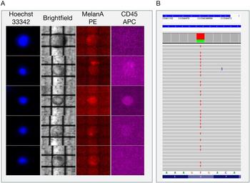
To explore potential mutational events responsible for the resistance to the combined anti-BRAF/anti-MEK targeted therapy, several molecular analyses on circulating tumour cells (CTC) were addressed in line with our described protocol (Ref Reference Palmirotta58). Briefly, peripheral blood mononuclear cells were separated by Ficoll (Sigma) gradient separation from 15 ml of blood. The cell sample was depleted of CD45+ and Glycophorin+ cells by immunomagnetic sorting (AUTOMacs, Miltenyi). Negatively selected cells including CTCs and residual lymphocytes, were then stained with monoclonal antibody phycoerythrin (PE)-conjugated anti-MelanA (Miltenyi) and with Hoechst 33342 (Sigma) to reveal the nucleated cells, and with anti-CD45 allophycocyanin (APC) –conjugated antibody (EBioscience) to exclude the lymphocytes. The fluorescent cell suspension was further sorted by DEPArray System (Silicon Biosystems). The combined sorting procedures were allowed to identify 65 CTCs with CD45-negative/MelanA-positive phenotype. This small amount of cells was thus recovered in 200 µl tubes and processed for subsequent molecular analysis (Fig. 4).
(a) Recruitment of CTCs through the DEPArray system. The image shows the fluorescence panel including five CTCs of variable size (from up to down), negative for MelanA (red) and CD45 (purple) as marker of leukocytes. Cell viability was confirmed by morphology integrity of nuclei stained with DAPI (blue). (b) NGS analysis on single cells detecting the pathogenic variant c.1799T > A in the BRAF gene responsible for the pathogenic p.Val600Glu (V600E, rs113488022) missense mutation.

For the mutational analysis, we used a protocol developed in our laboratory that performs the sequencing assay by next-generation sequencing (NGS) on a single CTC avoiding both DNA extraction and whole-genome amplification (WGA) (Ref Reference Palmirotta58). After cell lysis with Ampli1™ WGA Kit (Silicon Biosystems), the sample was barcoded and the library was prepared using the Ion Xpress™ barcode adapters and the Ion AmpliSeq™ Library kit 2.0 (Life Technologies). Reaction sequence was completed in the Ion Torrent PGM™ system (Life Technologies) using an Ion AmpliSeq™ Cancer Hotspot Panel v2 (Life Technologies) primer pool kit. All results were analysed using the Torrent Suite Software 5.0.4 and variant calling was performed running the Torrent Variant Caller plugin version 5.0.4.0. NGS analysis on single cells allowed the detection of the pathogenic variant c.1799T > A in the BRAF gene responsible for the pathogenic p.Val600Glu (V600E, rs113488022) missense mutation, confirming the molecular data obtained from the analysis of the primitive neoplastic lesion (Fig. 4). In addition, we found a c.215C > G transversion responsible for a TP53 p.Pro72Arg missense mutation (rs1042522) reported by ClinVar as a drug response variant and a c.1173A > G substitution determining a benign p.I391M missense variant (rs2230461) in the PIK3CA gene.
However, the disease underwent rapid progression as showed by CT scan after 5 months as a remarkable involvement of the liver and subcutaneous tissues. No other treatments were then addressed because of the rapid decline of the performance status, with severe liver and kidney damage while the death occurred after only 1 month from the clinical progression.
Conclusions and future perspectives
In this paper, we described a case of an atypically aggressive ATM form, showing a different evolution from the typical indolent behaviour associated with this pathology. Aggressive forms have already been described sporadically in the literature. As reported in this review, several cases with visceral metastases, or aggressive behaviour and fatal outcome, or even with metastatic disease have occurred in our revisiting of the literature. Unfortunately, the limited recoveries from case reports and retrospective studies lead to a considerable difficulty in defining the clinical profile of this histotype and still today some controversies are described even in the histological diagnosis, given the heterogeneity of the available observational studies (Ref Reference Vyas4). In this misty landscape, it is even more difficult to identify prognostic factors that could accurately and promptly distinguish forms of aggressive disease from the indolent ones. Probably even the unusually high melanin content in ATM could be a protective factor against the effects of free radicals, which are abundant in melanoma and shown to be contributing to its aggressive behaviour (Ref Reference Margaryan59). Thus, molecular characterisation might help to better classify this rare histotype of melanoma and provide a more accurate assessment of outcomes in the era of immunotherapy and targeted therapy. The few molecular data currently available and summarised in Table 2 suggest that the ATM is not classifiable in any of the four molecular subtypes described by TCGA (Ref Reference Cancer Genome Atlas60). On the other hand, no specific patterns of alterations can be attributed to the ATM.
The most homogeneous data showing the mutational properties of the PRKAR1A gene undoubtedly refer to the Carney Syndrome (Ref Reference Zembowicz, Carney and Mihm9), while in sporadic forms we find a remarkable heterogeneity with the presence of mutational events ranging from fusion genes and chromosomal deletions to promoter mutations of the TERT gene (Refs Reference Dadras49, Reference Bahrami53, Reference Bax55).
Remarkably, our case is the first description in which the variant V600E of the BRAF gene is identified both in primary tumour tissue and in CTC of the patient.
Therefore, the data from our review underline the need for future studies selectively focusing on a massive DNA/RNA analysis and a subsequent use of bioinformatic tools for the interpretation of the results. Given the rarity of this neoplasm, achieving this goal should certainly include multicenter collaborations and a wide work of clinical data collection and patient follow-up for desirable genotype-phenotype correlations. Only in this way, we would be able to identify the real nosography nature of this tumour and define further subtypes useful in defining the correct therapy to adopt and to predict the life expectancy of these patients.
Acknowledgements and funding
This work was partially supported by a grant from the Italian Association for Cancer Research (AIRC, IG 17536) (Franco Silvestris) and from the Apulia Region (‘Oncogenomic Project’ and ‘Jonico-Salentino Project’).
Conflict of interest
The authors confirm that there are no conflicts of interest.
Ethical standards
The authors assert that all procedures contributing to this work comply with the ethical standards of the relevant national and institutional committees on human experimentation and with the Helsinki Declaration of 1975, as revised in 2008.