Introduction
Nematodes of the genus Serpentirhabdias Tkach, Kuzmin & Snyder, 2014 are mostly lung-dwelling parasites of snakes. Up to 20 nominal species of the genus are known worldwide, nine of them occur in the Neotropical realm: S. atracti Kuzmin, Melo & Santos, 2014; S. atroxi Kuzmin, Giese, Melo, da Costa, Maschio & Santos, 2016; S. filicaudalis (Barrella, dos Santos & da Silva, 2009); S. cf. fuscovenosa (Railliet, 1899); S. labiata (Pereira, 1927); S. lamothei (Martínez-Salazar & León-Régagnon, 2006); S. moi Machado, Kuzmin, Tkach, Santos, Gonçalves & Melo, 2018; S. vellardi (Pereira, 1928); and S. viperidicus Morais, Aguiar, Müller, Narciso, Silva & Silva, 2017 (Machado et al., Reference Machado, Kuzmin, Tkach, Santos, Gonçalves and Melo2018). Two of them, namely S. atroxi and S. viperidicus, are parasitic in snakes of the family Viperidae, while the other seven species are parasites of colubroidean snakes.
Clelia clelia (Daudin, 1803), commonly known as ‘Mussurana’, is distributed from southern Mexico to South America, reaching as far south as Argentina. In Brazil it is widely distributed in the Amazon basin and Pantanal wetlands (Fraga et al., Reference Fraga, Lima, Prudente and Magnusson2013). Clelia clelia is primarily terrestrial, nocturnal and carnivorous; it is usually ophiophagous and feeds almost exclusively on snakes, especially venomous snakes of the genera Bothrops and Crotalus (Vitt & Vangilder, Reference Vitt and Vangilder1983; Pinto & Lema, Reference Pinto and Lema2002).
During the field collecting trip in the vicinities of Lábrea, Amazonas State, Brazil, we collected and examined for parasites one specimen of the Mussurana. The snake harboured lung nematodes, which corresponded to the genus Serpentirhabdias based on their morphology as well as the host and the site of infection. Further morphological studies and molecular investigations demonstrated that the nematode specimens from C. clelia differed from all known species of the genus. Thus, these nematodes are described herein as a new species, S. mussuranae n. sp. Morphological differentiation is supported by molecular evidence based on two mitochondrial genes and nuclear rDNA region. The description of adult nematodes is complemented with the information on the experimental life cycle and the morphology of free-living stages.
Material and methods
One specimen of C. clelia was collected in the vicinities of Lábrea, Amazonas State, Brazil, in June 2016, and examined for parasites (license permission SISBIO: 53557/8039).
Live nematodes removed from lung or isolated from experimental cultivation were rinsed in saline and fixed with hot 70% ethanol. For morphological studies, the worms were cleared in lactophenol and studied under an Olympus BX53 compound microscope (Olympus America, Center Valley, Pennsylvania) equipped with differential interference contrast optics and a digital imaging system. The drawings were made on an Olympus BX41 equipped with a drawing tube. Details of the anterior-end morphology were examined in the apical section made manually and observed en face. In some infective larvae, the sheath was removed by gentle movements of the cover slip on temporary mounts in order to examine the structure of the anterior end and the tail of the larva. All measurements in the text are in micrometres, except otherwise stated.
The free-living stages were obtained through experimental cultivation. Faeces obtained directly from the cloacal region of the snake were used as the culture medium, and one parasitic female was placed in the culture medium in a small Petri dish with saline solution that was kept inside a humidity chamber. The cultivation was performed at the natural ambient temperature of the Amazonian rainforest (varying from 24 to 28°C). Samples were collected 12, 24, 36, 60 and 72 h after the beginning of cultivation. The hosts were also examined for the presence of intestinal nematodes, but no other species of parasites were found.
Due to the low number of available specimens, genomic DNA was extracted from a single individual of the new species as well as from specimens of S. elaphe (Sharpilo, 1976) and S. eustreptos (McCallum, 1921) collected in Ukraine and the US, respectively. Based on the results of a previous broader phylogenetic study (Tkach et al., Reference Tkach, Kuzmin and Snyder2014), we also obtained DNA from a specimen of Rhabdias bufonis (Schrank, 1788) collected in Ukraine (see table 1 for hosts and localities). DNA was extracted according to Tkach & Pawlowski (Reference Tkach and Pawlowski1999). Amplification and sequencing of the nuclear ribosomal DNA region spanning the 3′ end of the 18S nuclear rRNA gene, internal transcribed spacer (ITS) region (ITS1 + 5.8S + ITS2) and 5′ end of the 28S gene, as well as partial mitochondrial 12S and cox1 genes, were amplified and sequenced according to the protocols and using primers described by Machado et al. (Reference Machado, Kuzmin, Tkach, Santos, Gonçalves and Melo2018). Contiguous sequences were assembled and edited using Sequencher™ version 4.1.1 (GeneCodes Corp., Ann Arbor, MI, USA), and deposited in GenBank (for accession numbers, see table 1).
Hosts, geographic origin and GenBank accession numbers for new 12S and cox1 sequences of nematodes obtained in this study.

Despite multiple attempts, we were unable to sequence the ITS region of S. eustreptos, although Polymerase chain reaction was successful. Therefore, we did not conduct phylogenetic analysis of the nuclear rDNA sequences because such analysis was presented recently (Machado et al., Reference Machado, Kuzmin, Tkach, Santos, Gonçalves and Melo2018) and included all available sequences other than the new species described herein. We ran phylogenetic analyses based on mitochondrial genes 12S and cox1 using newly obtained sequences as well as all currently available matching sequences of Serpentirhabdias. Newly obtained matching cox1 and 12S sequences of R. ranae Walton, 1929 were used as outgroups based on the results of previous larger phylogenetic analysis (Tkach et al., Reference Tkach, Kuzmin and Snyder2014). The sequences were aligned using MEGA7 software (Kumar et al., Reference Kumar, Stecher and Tamura2016) saved in Nexus format. No positions were excluded from the analysis due to the relatively high similarity between sequences included in 12S and cox1 alignments. The phylogenetic analyses were carried out using Bayesian inference, as implemented in the MrBayes program, version 3.2.6 (Ronquist & Huelsenbeck, Reference Ronquist and Huelsenbeck2003). Nucleotide substitution models were determined using MEGA7 software. Cox1 gene analysis was done using the Hasegawa–Kishino–Yano model and gamma-distributed among-site rate variation; 12S analysis used the general time-reversible model, including estimates of the proportion of invariant sites and gamma-distributed among-site rate variation. Posterior probabilities were approximated over 3,000,000 generations in 12S analysis and 1,000,000 generations in cox1 analysis; in both cases, only the final 75% of trees were used to produce the consensus trees. The resulting phylogenetic trees were visualized using FigTree version 1.4 software (Rambaut, Reference Rambaut2009).
To differentiate between Serpentirhabdias species, we compared newly obtained mitochondrial sequences from the new species and matching sequences of other Serpentirhabdias species obtained in this work or available in the GenBank. Genetic identity matrices for cox1 and 12S sequences were generated using MEGA7 software.
Results
Systematics
Family: Rhabdiasidae Railliet, 1915
Genus: Serpentirhabdias Tkach, Kuzmin & Snyder, 2014
Serpentirhabdias mussuranae n. sp. (figs 1 and 2)
Taxonomic summary
Type host. Clelia clelia (Daudin, 1803) (Reptilia: Serpentes: Colubroidea: Dipsadidae).
Site in host. Lungs.
Type locality. Lábrea municipality, Amazonas State, Brazil (07°15′32″S 64°47′52″W).
Type material. Holotype MPEG.NEM 00210; six paratypes MPEG.NEM00211; deposited in the Museu Paraense Emilio Goeldi, Belém, Pará, Brazil.
ZooBank registration. The Life Science Identifier (LSID) for S. mussuranae n. sp. is urn:lsid:zoobank.org:act:A7C1A2B7-926B-466B-8218-41DE15F81FA4.
Etymology. The species is named after the common name of its type host, Mussurana (C. clelia).
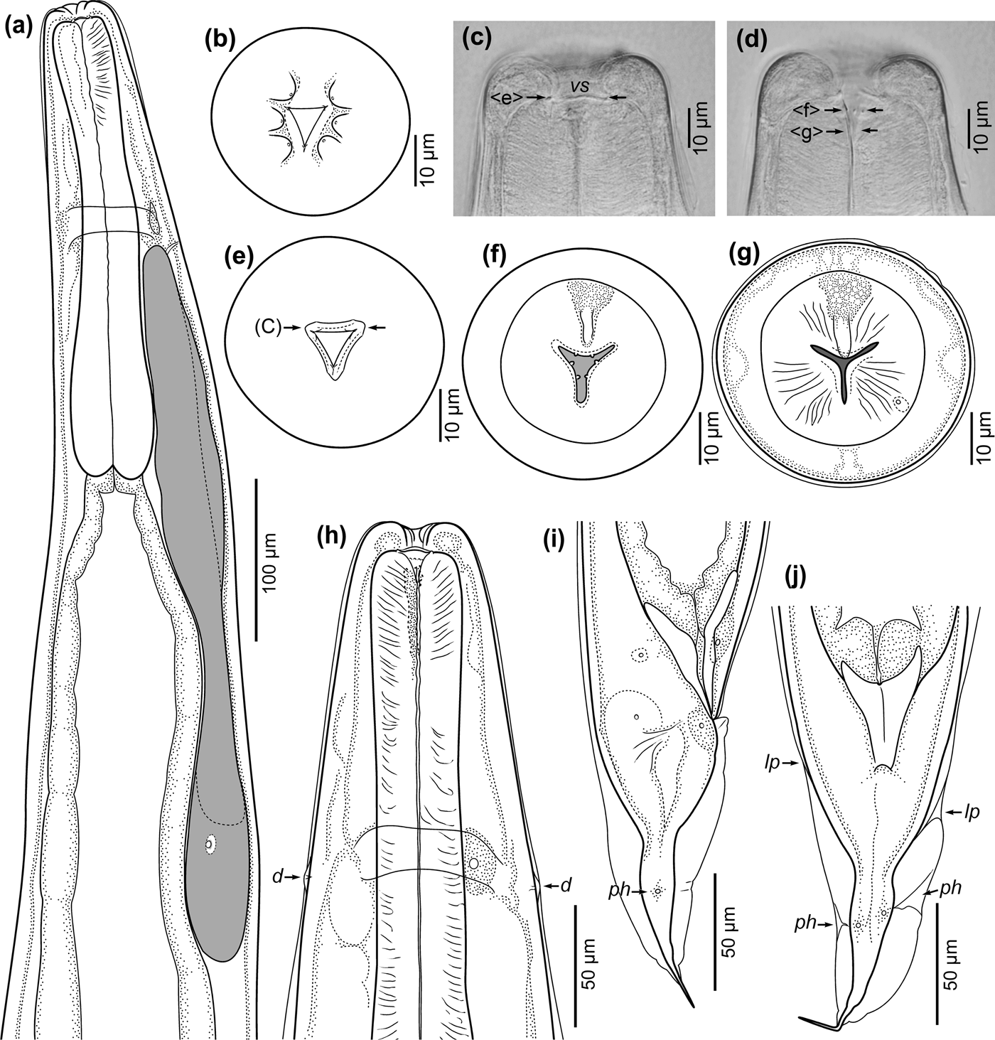
Serpentirhabdias mussuranae n. sp., adult hermaphrodites. (a) Anterior part of body, lateral view; (b) en face view of anterior end showing lips and oral opening; (c) lateral view of anterior end, longitudinal optical section at level of sclerotized anterior edge of oesophastome; (d) lateral view of anterior end, longitudinal optical section at level of mid-width of oesophastome; (e) apical view, transverse optical section at level of anterior edge of oesophastome (arrows and letters in parentheses indicate levels of optical sections on corresponding images); (f) apical view, transverse optical section at level of mid-depth of oesophastome showing onchia; (g) apical view, optical section at posterior part of oesophastome showing duct of dorsal oesophageal gland (asterisk); (h) anterior part of body in dorso-ventral view; (i) and (j) posterior part of body in lateral (i) and ventral (j) view. Abbreviations: vs, vestibulum; d, deirid; ph, phasmid; lp; lateral pore.

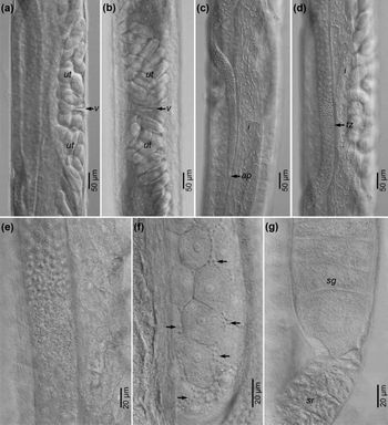
Serpentirhabdias mussuranae n. sp., details of the reproductive system morphology in adult hermaphrodites. (a) Mid-region of body showing vulva and uteri with eggs, lateral view; (b) same in ventral view; (c) proximal part and apex of anterior syngonium; (d) anterior syngonium with zone of spermiogenesis; (e) testis zone; (f) part of syngonium near testis zone, showing sperm cells (arrows) between oogonia; (g) part of genital tube at connection between syngonium and seminal receptacle. Abbreviations: v, vulva; ut, uteri; ap, apex; tz, testis zone; sg, syngonium; sr, seminal receptacle; i, intestins.

Description
Based on seven specimens, parasitic hermaphrodites; measurements are given for the holotype, followed by mean values and ranges for the whole type series in parentheses.
Body 4.2 (4.0; 3.7–4.2) mm long, gradually tapering from mid-region towards anterior and posterior extremities. Anterior end rounded, posterior end pointed. Width at mid-body 207 (192; 172–225), width at oesophago–intestinal junction 124 (110; 101–124). Body cuticle slightly inflated in anterior and posterior parts (fig. 1a, i, j), thin in mid-region, smooth, without transverse folds. Lateral pores are present along body, more conspicuous in inflated part of cuticle, with ducts directed anteriorly in anterior part and posteriorly in posterior part (fig. 1j).
On apical surface, six rounded lips arranged in two lateral groups of three, with prominent gaps on dorsal and ventral sides (fig. 1b). Lips almost similar in size, lateral lips somewhat smaller than sub-median ones. Each lip bearing minute papilla on top. Oral opening triangular, leading to short vestibulum (fig. 1c). Buccal capsule absent. Lumen of oesophagus in anterior part widened, forming thin-walled cavity (oesophastome), funnel-shaped in lateral view (fig. 1d) and triangular with concave sides and rounded corners in apical view (fig. 1f), 15 (14; 13–15) wide and 12 (11; 8–12) deep. Anterior edge of oesophastome sclerotized (fig. 1c, e). Six minute onchia located at mid-depth of oesophastome, two onchia on each sector of oesophagus (fig. 1d, f).
Oesophagus rounded on anterior and posterior ends (fig. 1a), slightly dilated in anterior part 37 (36; 34–39) wide, constricted in mid-region 35 (32; 30–35) wide, widening posteriorly to oval bulb 57 (53; 50–67) wide. Length of oesophagus 289 (284; 274–295), or 6.9 (7.1; 6.7–7.4)% of body length. Muscular fibres visible mostly in anterior part of oesophagus. Posterior bulb containing three unicellular oesophageal glands with large nuclei. Duct of dorsal gland entering posterior part of oesophastome (fig. 1g, h), ducts of sub-ventral glands entering oesophageal lumen at its mid-length. Lumen of oesophagus tri-radial on transverse sections (fig. 1g), thin, cuticularized, somewhat wavy in anterior part. Nerve ring surrounding oesophagus near its mid-length (fig. 1a), at 157 (139; 128–157) from anterior extremity 54 (49; 45–54)% of oesophagus length. Deirids small, flattened, located on lateral sides approximately at level of nerve ring (fig. 1h), 138 and 150 from anterior end (measured in holotype).
Excretory pore small, oval, transversely elongated, located ventrally at 160 (168; 160–185) from anterior end. Excretory duct short, thin-walled. Excretory glands unicellular, similar, large, elongated, rounded on anterior and posterior ends (fig. 1a). Shorter gland 468 (401; 334–468) long, longer gland 483 (453; 405–489) long and length difference measuring 15 (52; 8–123). Ratio of longer excretory gland length to oesophagus length 1.7 (1.6; 1.5–1.7). Large oval nucleus in posterior part of each gland. Two coelomocytes located in anterior part of body, on ventral side, close to anterior limb of reproductive system. One or two single coelomocytes observed in mid-region and/or in posterior part of body.
Intestine thin-walled in anterior part (fig. 1a), thick-walled in mid-region, with dilated lumen in posterior part. Three flattened cells forming oesophago–intestinal sphincter; other intestinal cells polygonal, convex towards intestinal lumen. Nuclei in intestinal cells prominent. Rectum short, funnel-shaped, strongly cuticularized (fig. 1i).
Reproductive system typical of Rhabdiasidae. Vulva slightly salient (fig. 2a), situated at 2.0 (1.9; 1.8–2.0) mm from anterior end 47.8 (48.5; 47.5–49.7)% of body length. Vagina short, transverse, lined with cuticle (fig. 2a). Uteri similar, amphidelphic, joined, thin-walled (fig. 2b). Eggs in uteri at early cleavage stages. Number of eggs 32 (28; 20–35) in both uteri. Egg size 38 (33–45) × 83 (70–95) (n = 15; measured in uteri of holotype and two paratypes). Seminal receptacles short, with crimped walls (fig. 2g). Syngonia long, often wavy (fig. 2c, d), each syngonium U-bending in its mid-region, 790 (692; 638–790) from body anterior end and at 961 (865; 784–961) from posterior end, often curved and with additional bends. Testis zones found in all specimens, at least in one syngonium (fig. 2d, e). Sperm cells small, oval, observed in testis zones and between oogonia (fig. 2f) usually closer to syngonial walls. Apices of syngonia narrow, rounded (fig. 2c), located at about level of seminal receptacle of opposite limb of reproductive system.
Tail conical, abruptly narrowing posteriorly from anus to mid-region, and then gradually tapering in posterior half; in some specimens slightly dilated at level of phasmids (fig. 1i, j). Short cuticular thread 22 (23; 21–28) long present on tail tip, separated from inflated body cuticle. Tail length 128 (131; 120–150), or 3.1 (3.3; 3.1–3.8)% of body length. Phasmids pore-shaped, situated just posterior to tail mid-length (fig. 1i, j), 61 and 71 from posterior end (measured in holotype).
Observations on routes and rate of free-living development in culture
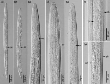
A sample collected 12 h after beginning of cultivation contained early-stage larvae (L1–2). All larvae (fig. 3a–c) had typical rhabditiform stoma and oesophagus, though differed slightly in the body shape and size, and more prominently in the shape and size of the genital primordium. The larvae with smaller genital primordium and more elongated body (fig. 3a) were identified as homogonic larvae of the hermaphroditic generation, whereas those with larger genital primordium and shorter body (fig. 3b) were identified as developing juveniles of the gonochoristic generation. The ratio of homogonic:heterogonic larvae was approximately 1.5:1.
Serpentirhabdias mussuranae n. sp., free-living larvae. (a) Early-stage homogonic larva of hermaphroditic generation, lateral view; (b) early-stage larva of amphimictic generation, lateral view; (c) anterior part of body in early-stage homogonic larva of hermaphroditic generation, lateral view; (d) anterior part of body in homogonic infective L3, lateral view; (e) anterior part of body in heterogonic infective L3, lateral view; (f) genital primordium in homogonic L3, ventral view; (g) genital primordium in heterogonic L3, lateral view. Abbreviations: gp, genital primordium; nr, nerve ring; sh, sheath.

In a sample collected 24 h after beginning of cultivation, third-stage homogonic larvae were observed, while the gonochoristic generation was represented mostly by fourth-stage larvae and several adult males.
In a sample collected 36 h after beginning of cultivation, all homogonic individuals were the ensheathed third-stage infective larvae. In the gonochoristic generation, adult males were present, along with juvenile females lacking eggs in uteri, and fourth-stage female larvae. The male:female ratio calculated in the sample was about 5:1 (27 males and 5 females).
By 60 h from the beginning of cultivation, the majority of males were found dead, and the free-living population consisted mostly of homogonic infective larvae and females with 0–3 eggs in uteri. No signs of larval development inside females were observed; instead, L1–3 were found free in the culture medium. The third-stage larvae in the progeny of the gonochoristic generation were ensheathed heterogonic infective larvae of the hermaphroditic generation (fig. 3e, g). The ratio of homogonic:heterogonic infective larvae in the sample was 1:4.
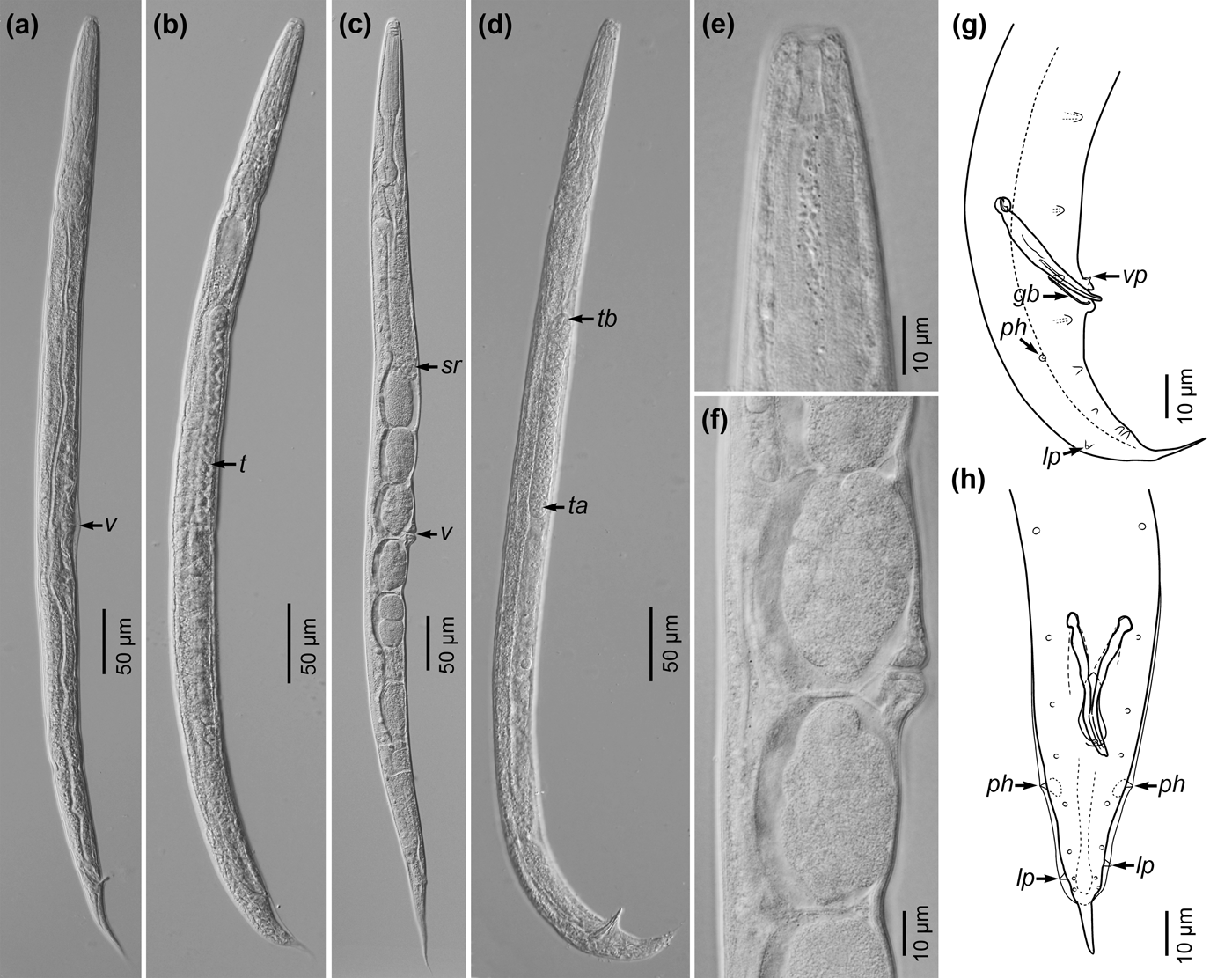
By the end of day three (sample collected 72 h after beginning of cultivation) no males were found in the sample collected. Females were alive and contained 4–6 eggs in uteri (fig. 5c). Ensheathed infective larvae and developing heterogonic larvae of the hermaphroditic generation were predominating in the sample.
Morphology of free-living stages
Measurements are given as mean values followed by ranges in parentheses.
Early-stage rhabditiform homogonic larvae. Based on ten specimens (fig. 3a, c), 475 (402–508) long, with maximum body width at mid-length, 24 (18–27); length:width ratio 20 (18–22):1. Body gradually tapering towards anterior and posterior extremities. Anterior end rounded, posterior end pointed. Stoma rhabditiform, 10 (7–14) long. Oesophagus 111 (94–124) long, 23 (20–25)% of body length, with distinct corpus, isthmus and posterior bulb. Nerve ring encircling isthmus. Intestine wide, with distinct lumen. Genital primordium 42 (33–46) long, lens-shaped in lateral view, oval in ventral view, located at level of intestine mid-length. Tail length 67 (64–71), or 14 (13–16)% of body length; tail conical, with sharply pointed tip.
Rhabditiform larvae of gonochoristic generation. Based on ten specimens (fig. 3b), 456 (428–483) long, 27 (24–30) wide at mid-length; length:width ratio 16 (15–19):1. Anterior part of body and intestine similar to those in homogonic rhabditiform larvae. Stoma 9 (7–12) long; oesophagus 109 (89–133) long, 24 (20–28)% of body length. Genital primordium 131 (96–151) long. Tail conical, 58 (49–64) long, tail length corresponding to 13 (11–14)% of body length. Tail end sharply pointed.
Homogonic infective larvae. Based on 16 specimens (fig. 3d, f), generally retaining the shape and structure of early-stage rhabditiform larvae. Body length 699 (624–755), width at mid-length 29 (23–35); length:width ratio 24 (21–27):1. Stoma rhabditiform, 10 (7–14) long, with cylindrical lumen and walls gradually thickening posteriorly. Oesophagus rhabditiform, 128 (110–153) long, 18 (15–20)% of body length, with distinct corpus, narrow isthmus and oval bulb. Nerve ring encircling isthmus at 103 (98–111) from anterior end (measured in six specimens). Intestine narrow, its lumen indistinct. Genital primordium small, lens-shaped in lateral view, pear-shaped in ventral view (fig. 3f), 40 (32–45) long, located at 385 (338–419) from anterior end; distance from anterior end to middle of genital primordium corresponding to 55 (53–57)% of body length. Sheath tightly covering body in larvae examined during second day of cultivation; on third day some space observed between larva and sheath (fig. 3d, f). Outer surface of sheath longitudinally striated, inner surface of sheath and surface of cuticle in larva transversely striated. Narrow lateral alae present along body of larva, wider and more prominent in posterior part. Tail conical, 80 (73–88) long, 11 (10–12)% of body length, tail tip sharply pointed.
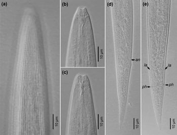
Heterogonic infective larvae. Based on 15 specimens (figs 3e, g and 4), similar to homogonic ones in shape and size. Sheath with characteristic longitudinal outer striation and transverse inner striation (fig. 4a). Body surface of larva finely transversely striated. Lateral alae present, more prominent in posterior part, not reaching tail tip (fig. 4e). Body length 753 (708–786), width at mid-length 30 (27–35); length:width ratio 25 (22–29):1. Dorsal and ventral pseudolabia present on anterior end, triangular, with rounded tips (fig. 4b). Stoma narrow, funnel-shaped in lateral view (fig. 4b), almost cylindrical in ventral view (fig. 4c), 10 (8–13) long. Oesophagus with less distinct corpus and more elongated posterior bulb (fig. 3e) than in homogonic IL. Length of oesophagus 195 (166–227), 26 (22–30)% of body length. Nerve ring 119 (102–142) from anterior end, surrounding isthmus in its anterior part (fig. 3e). Intestine narrow, lacking apparent lumen. Rectum thin, elongated (fig. 4d). Genital primordium oval (fig. 3g), 45 (35–53) long. Distance from anterior end to middle of genital primordium 412 (386–435) or 55 (52–56)% of body length. Tail conical, gradually tapering posteriorly, with rounded tip (fig. 4d, e). Phasmids located at tail mid-length (fig. 4e). Tail length 75 (67–87), 10 (9–12)% of body length.
Serpentirhabdias mussuranae n. sp., heterogonic infective larvae. (a) Structure of sheath in anterior part of body; (b) anterior end of larva with removed sheath, lateral view; (c) same, median view; (d) tail of larva, lateral view; (e) tail of larva, ventral view. Abbreviations: an, anus; la, lateral ala; ph, phasmid.

Females. Based on 14 specimens (fig. 5a, c, e, f). Body 845 (688–939) long, 43 (36–66) wide at mid-length; length:width ratio 20 (14–24):1. Anterior end rounded, posterior end pointed. Stoma rhabditiform (fig. 5e), 11 (7–13) long. Oesophagus with distinct corpus, isthmus and bulb, 156 (140–171) long, 18 (17–20)% of body length. Nerve ring encircling isthmus at its mid-length, 117 (102–134) from anterior end of body. Intestine wide, its lumen dilated in anterior and posterior parts. Reproductive system amphidelphic, with equal limbs. Vulva salient (fig. 5f), located at 447 (376–477) from anterior end; distance to vulva corresponding to 53 (51–56)% of body length. Uteri tubular, thin-walled, containing up to six eggs arranged in one row. Eggs in uteri at early cleavage stages (fig. 5c). Seminal receptacles prominent, short and wide, containing distinct spermiae (fig. 5c). Apices of ovaries at level of vulva. Tail narrow, gradually tapering, sharply pointed. Tail length 98 (81–112), 12 (10–13)% of body length.
Serpentirhabdias mussuranae n. sp., amphimictic generation. (a) Moulting female L4, lateral view showing formation of vulva; (b) male L4, lateral view showing testis; (c) adult female, lateral view; (d) adult male, lateral view; (e) anterior end of female; (f) mid-region of female body, lateral view; (g) posterior part of male, lateral view; (h) same, ventral view. Abbreviations: v, vulva; t, testis; sr, seminal receptacle; tb, bending point of testis; ta, apex of testis; vp, ventral unpaired papilla; ph, phasmid; lp, lateral papilla; gb, gubernaculum.

Males. Based on ten specimens (fig. 5b, d, g, h). Body narrow, posterior part curved ventrally (fig. 5d). Anterior end rounded, posterior end pointed. Body length 685 (665–702), width at mid-length 34 (32–37); length:width ratio 20 (19–21):1. Anterior part of body similar to that in females. Stoma 8 (7–10) long. Oesophagus length 119 (108–131), 17 (16–19)% of body length. Nerve ring 91 (78–103) from anterior end. Intestine wide, lumen of intestine dilated in anterior part. Total length of genital tube 586 (551–613). Testis with reversed anterior part (fig. 5d), already prominent in fourth-stage juveniles (fig. 5b), 158 (146–169) long in adult males; bending point located 200 (190–228) from anterior end. Reversed part of testis occupying 27 (26–29)% of genital tube length. Narrow lateral alae and eight pairs of ventro-lateral genital papillae present in posterior part of body: three pairs pre-cloacal and five pairs post-cloacal (fig. 5g, h). Two posteriormost pairs sub-terminal, located close to each other, free from lateral alae. One lateral pair of papillae located just anterior to two posterior ventro-lateral pairs. One unpaired ventral papilla just anterior to cloacal aperture. Spicules 37 (32–39) long, joined in distal parts. Gubernaculum 15 (12–18) long, slightly curved ventrally in posterior part (fig. 5g). Tail conical, abruptly tapering, with needle-shaped cuticular tip. Tail length 57 (52–65), 8.3 (7.7–9.3)% of body length. Phasmids papilla-shaped, located anterior to tail mid-length, connected to lateral alae (fig. 5h).
Remarks
Serpentirhabdias mussuranae n. sp. is assigned to the genus Serpentirhabdias Tkach, Kuzmin & Snyder, 2014 based on the arrangement of lips in two lateral groups, body cuticle only slightly inflated in anterior and posterior parts of body, absence of the buccal capsule and parasitism in the lung of a snake. Among the Neotropical representatives of the genus, three species are known to have the onchia in the oesophastome: S. atroxi, S. moi and S. viperidicus (Kuzmin et al., Reference Kuzmin, Giese, Melo, Costa, Maschio and Santos2016; Morais et al., Reference Morais, Aguiar, Müller, Narciso, Silva and Silva2017; Machado et al., Reference Machado, Kuzmin, Tkach, Santos, Gonçalves and Melo2018). However, due to insufficient details provided in some of the descriptions it cannot be excluded that some other Neotropical Serpentirhabdias may possess onchia. Therefore, we differentiate the new species from all Serpenthirhabdias species known from the Neotropics.
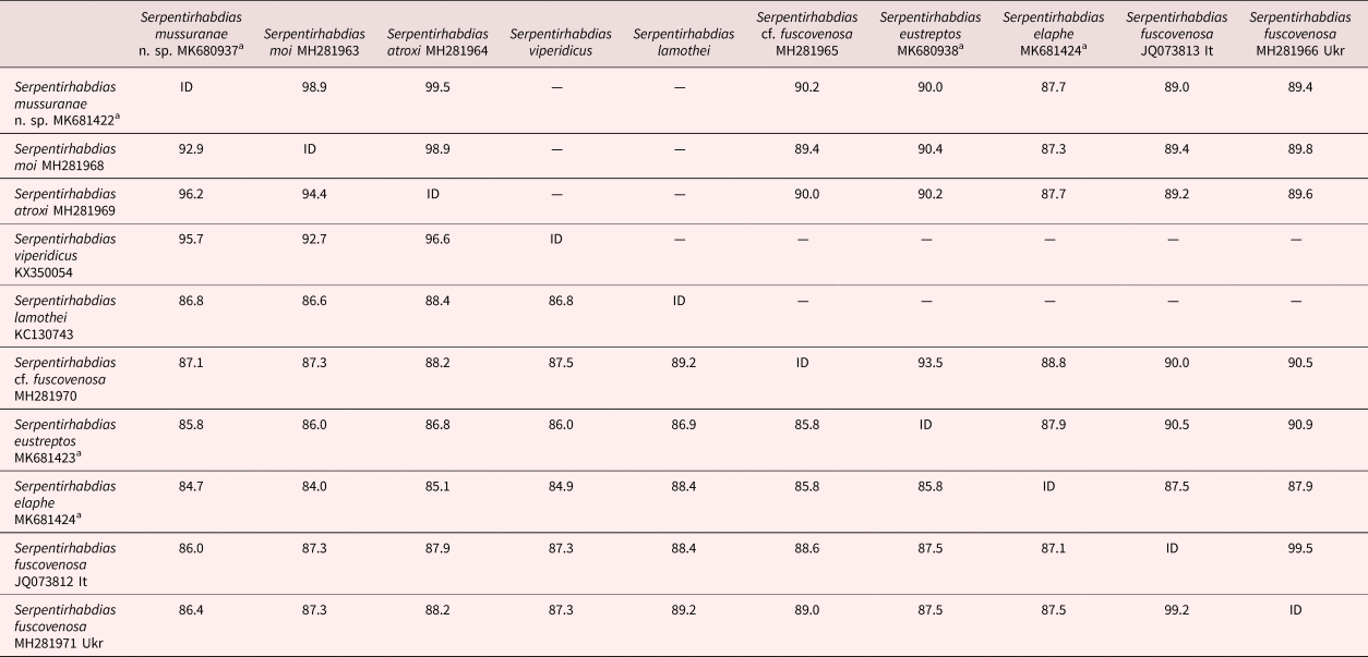
Serpentirhabdias mussuranae n. sp. differs from S. atroxi parasitic in Bothrops atrox (Linnaeus, 1758) (Viperidae) (Kuzmin et al., Reference Kuzmin, Giese, Melo, Costa, Maschio and Santos2016) in the triangular shape of the oral opening and the oesophastome in apical view, vs. round in S. atroxi, the absence of longitudinal thickenings of the buccal cavity walls, the shape of the tail (abruptly tapering in anterior half and gradually tapering in posterior half vs. gradually tapering in S. atroxi) and a lesser number of eggs in uteri (20–35 vs. >100). Nucleotide sequence divergence between the two species was 3.8% in cox1 gene and 0.5% in 12S gene (table 2).
Pairwise sequence identity levels (%) among sequences of Serpentirhabdias. Names, GenBank accession numbers and sequence identities in mitochondrial 12S gene are above diagonal; names, GenBank accession numbers and sequence identities in mitochondrial cox1 gene are below diagonal.

a New sequences obtained in this study. Ukr, Ukraine; It, Italy.
The new species differs from S. viperidicus parasitic in B. moojeni (Hoge, 1966) (Viperidae) in several morphological characters. Unlike the new species, S. viperidicus has a round oral opening, numerous eggs in uteri and a gradually tapering tail (Morais et al., Reference Morais, Aguiar, Müller, Narciso, Silva and Silva2017). Besides, S. viperidicus is larger than the new species (body 4.25–6.91 mm long) and has a larger oesophastome, 13–22 wide and 13–23 deep. Sequence divergence in cox1 gene between S. mussuranae n. sp. and S. viperidicus was 4.3% (table 2).
Serpentirhabdias mussuranae n. sp. is morphologically similar to S. moi from colubroidean snakes in Brazil (Machado et al., Reference Machado, Kuzmin, Tkach, Santos, Gonçalves and Melo2018). However, S. moi is shorter (2.2–2.6 mm long) than S. mussuranae n. sp. and has fewer eggs in uteri (6–13). In the new species, the excretory glands are relatively longer than those in S. moi; the average ratio of the longer gland length to the oesophagus length in S. mussuranae n. sp. is 1:6, whereas in S. moi it is 1:06 (recalculated from Machado et al., Reference Machado, Kuzmin, Tkach, Santos, Gonçalves and Melo2018). The sequences of the two species differed by 7.1% in cox1 gene and by 1.1% in 12S gene (table 2).
Serpentirhabdias mussuranae n. sp. differs from S. atracti parasitic in Atractus major Boulenger, 1894 (Colubridae, Dipsadinae), described by Kuzmin et al. (Reference Kuzmin, Melo and Santos2014a), because the new species have distinct lips and onchia in the oesophastome. Besides, the excretory glands are shorter than the oesophagus in S. atracti, whereas they are significantly longer than the oesophagus in the new species. The tail is narrow, gradually tapering in S. atracti, while the tail in S. mussuranae n. sp. is abruptly narrowing in its anterior half and gradually tapering in posterior half.
The new species differs from S. filicaudalis parasitic in Spilotes pullatus (Linnaeus, 1758) (Colubridae, Colubrinae) (Barrella et al., Reference Barrella, dos Santos and da Silva2009) in the presence of onchia, triangular shape of the oral opening (round in S. filicaudalis), shape of the tail (gradually tapering in S. filicaudalis) and lack of the buccal capsule.
Serpentirhabdias mussuranae n. sp. is somewhat larger than S. lamothei described from Leptodeira maculata (Hallowell, 1861) (Colubridae) in Mexico, according to Martínez-Salazar & León-Régagnon (Reference Martínez-Salazar and León-Régagnon2006); the later species is 2.51–3.43 mm long. In contrast to S. mussuranae n. sp., S. lamothei has the buccal capsule, and its oral opening is rounded. Excretory glands in S. lamothei are shorter than oesophagus (Martínez-Salazar & León-Régagnon, Reference Martínez-Salazar and León-Régagnon2006, Fig. 2a), whereas in the new species they are longer than the oesophagus. Eggs in S. lamothei are smaller than in S. mussuranae n. sp. (34–65 × 27–58 vs. 70–95 × 33–45). Sequence divergence in cox1 gene between S. mussuranae n. sp. and S. lamothei was 13.2% (table 2).
Serpentirhabdias cf. fuscovenosa was originally described from colubroidean snakes in the Nearctic (Baker, Reference Baker1978; Kuzmin et al., Reference Kuzmin, Tkach and Snyder2003). However, it was also reported from colubroidean snakes in Neotropical southern Mexico (Martínez-Salazar & León-Régagnon, Reference Martínez-Salazar and León-Régagnon2006). The new species differs from S. cf. fuscovenosa from Mexico in the shape of the oral opening, which is rounded in S. cf. fuscovenosa (in Martínez-Salazar & León-Régagnon, Reference Martínez-Salazar and León-Régagnon2006, Fig. 4a) vs. triangular in the new species. In contrast to S. mussuranae n. sp., S. cf. fuscovenosa from Nearctic has excretory glands shorter than the oesophagus and a tail gradually tapering. Sequence divergence between S. mussuranae n. sp. and S. cf. fuscovenosa was 12.9% in cox1 gene and 9.8% in 12S gene (table 2).
Serpentirhabdias mussuranae n. sp. is slightly larger than S. vellardi, which is 3.0–3.3 mm long (according to Pereira, Reference Pereira1928). Serpentirhabdias vellardi was originally described from two snake species: Philodryas schotti (= P. patagoniensis (Girard, 1858)) and Oxyrhopus trigeminus Dumeril, Bibron & Dumeril, 1854 (Colubridae, Dipsadinae) (Pereira, Reference Pereira1928). It is evident from Figs 1 and 2 in Pereira (Reference Pereira1928) that the two forms from separate hosts have a different shape of the tail: gradually tapering in the specimen from P. patagoniensis and abruptly tapering until the mid-length in the specimen from O. trigeminus. By this character, S. vellardi from O. trigeminus is close to S. mussuranae n. sp. However, the syngonia in S. vellardi form more bends and loops than in S. mussuranae n. sp., and the distance from the posterior bend of the reproductive system to the posterior end of the body in S. vellardi is only 610–670 vs. 784–961 in S. mussuranae n. sp.
The form recently identified as S. cf. vellardi from Xenodon merremii (Wagler, 1824) (Colubridae, Dipsadinae) in São Paulo State of Brazil (Emmerich et al., Reference Emmerich, Morais and Silva2018) differs from S. mussuranae n. sp. in the rounded shape of oral opening. The excretory glands in S. cf. vellardi are of about the same length as the oesophagus, whereas in the new species they are 1.6 times longer than oesophagus.
Serpentirhabdias mussuranae n. sp. is larger than S. labiata from X. merremii (Wagler, 1824) (Colubridae, Dipsadinae); the latter species is only 2.2–2.4 mm long (Pereira, Reference Pereira1927). At the same time, S. labiata has a longer tail (150–190) than S. mussuranae n. sp. (120–150). Besides, S. labiata has eight lips, whereas S. mussuranae n. sp. has six lips.
Molecular phylogeny
Nuclear ribosomal DNA sequence data from Neotropical Serpenthirhabdias are limited to the recently published sequence of S. moi (Machado et al., Reference Machado, Kuzmin, Tkach, Santos, Gonçalves and Melo2018), and the two species showed only a two-nucleotide difference in this DNA region. Therefore, we did not conduct a phylogenetic analysis based on this DNA region and focused on better represented mitochondrial cox1 and 12S genes. Upon trimming to the length of the shortest available sequences, the cox1 alignment was 538 bp long and the 12S alignment was 466 bp long. All sequences in the cox1 alignment were of equal length, while the 12S alignment needed only a few gaps due to minor length differences (2–3 bp) between South American species and the rest of the included taxa. All three 12S sequences of South American taxa were of equal length.
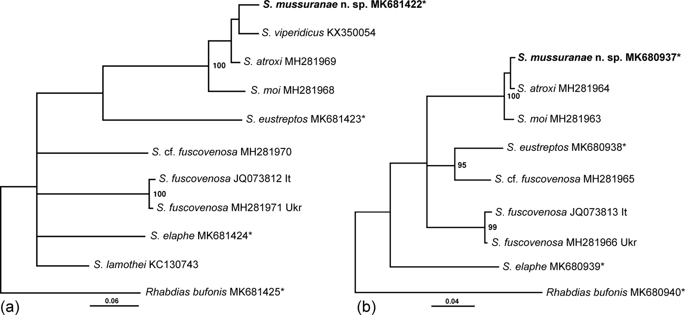
The species composition in the cox1 and 12S alignments was not fully congruent with cox1 analysis including nine species of Serpentirhabdias and 12S analysis including only seven species. Nevertheless, both analyses revealed equally well supported clades of closely related South American Serpentirhabdias species (fig. 6a, b). At the same time, the interrelationships within this clade were not resolved due to relatively similar genetic distances among these species (table 2). Similarly, the trees were characterized by having very little to no resolution when it comes to the relationships among remaining Serpentirhabdias species from North America and Europe.
Phylogenetic relationships among Serpentirhabdias including S. mussuranae n. sp. New sequences obtained in this study are indicated by an asterisk. Posterior probabilities greater than 0.9 are presented at internodes as percentages. The scale bar indicates the number of substitutions per site. (a) Interrelationships among nine species based on partial cox1 sequences inferred from Bayesian analysis (1,000,000 generations); (b) interrelationships among seven species based on partial 12S sequences inferred from Bayesian analysis (3,000,000 generations).

Discussion
Serpentirhabdias mussuranae n. sp. is the tenth Neotropical species of the genus and the fourth species possessing onchia in the oesophastome. Previously, the onchia were reported in three Neotropical species – namely, S. atroxi, S. moi and S. viperidicus (Kuzmin et al., Reference Kuzmin, Giese, Melo, Costa, Maschio and Santos2016; Morais et al., Reference Morais, Aguiar, Müller, Narciso, Silva and Silva2017; Machado et al., Reference Machado, Kuzmin, Tkach, Santos, Gonçalves and Melo2018). Several distinct morphological characters separate these species from each other. Each of them has a peculiar structure of the anterior end, shape and size of the oesophastome, length of the excretory glands, shape of the tail, etc.
Both the pairwise sequence comparison and phylogenetic analyses demonstrated close affinities among four species of Serpentirhabdias from South America, including the new species (table 2, fig. 6a, b). Considering that our phylogenetic analyses included species from Neotropical and Holarctic parts of North America as well as from Europe, our results provided evidence of the formation of a unique, endemic and actively speciating fauna of Serpentirhabdias in South America (fig. 6a, b). We expect that future studies including higher diversity of Serpentirhabdias species will support this hypothesis.
Despite the obvious close phylogenetic affinities among the South American Serpentirhabdias, the pairwise comparison between cox1 sequences of the new species and other available sequences of Serpentirhabdias has demonstrated significant differences between S. mussuranae n. sp. and all other species present in the dataset (table 2). Sequence divergence between the new species and geographically distant species from South America (S. viperidicus), North America (S. lamothei, S. cf. fuscovenosa, S. eustreptos) and Europe (S. fuscovenosa, S. elaphe) was very high at 12.9–15.3%. At the same time, the divergence from the closely related and geographically much closer South American species was lower at 4.3–7.1%. Considering that the previously reported intraspecific differences within some Serpentirhabidas species (S. lamothei in North America, S. fuscovenosa in Europe) were very low and varied between 0 and 0.8% (table 2; Machado et al., Reference Machado, Kuzmin, Tkach, Santos, Gonçalves and Melo2018), molecular data confirm the status of S. mussuranae n. sp. as independent species.
Our observations on the free-living development of S. mussuranae n. sp., though preliminary, allowed us to characterize the life cycle of the species as typical of Serpentirhabdias, as suggested by Tkach et al. (Reference Tkach, Kuzmin and Snyder2014). In the present study, both homogony and heterogony were ascertained by the presence of homogonic infective larvae and amphimictic generation in culture. Interestingly, the male-to-female ratio in the amphimictic generation was 5:1, thus demonstrating a very strong bias towards males. We suppose that the decrease in female numbers could have been somehow caused by extreme conditions of cultivation during the first few hours, when the culture was occasionally exposed to direct sunlight. A part of female first-stage larvae might have shifted to the direct development and thus became the homogonic larvae. Similar influences of environmental conditions on the development of free-living females were reported for the species of the nematode genus Strongyloides (Streit, Reference Streit2008). Further experimental studies might confirm or disprove our assumption. It is noteworthy that S. mussuranae n. sp. is a useful model object for these kind of studies because the ratio of homogonic:heterogonic types of development in this species is close to 1:1. In most other species of the genus Serpentirhabdias studied experimentally so far – for example, S. fuscovenosa, S. elaphe and S. eustreptos (Langford & Janovy, Reference Langford and Janovy2009; Kuzmin, Reference Kuzmin2013) – homogony is strongly predominant and a deviation from optimal cultivation conditions tends to eliminate all individuals of the amphimictic generation in the cultures.
Ensheathed third-stage infective larvae are the final stage of free-living development in rhabdiasid nematodes. Homogonic and heterogonic infective larvae in S. mussuranae n. sp. were similar in sheath structure, body size and size and shape of the genital primordium, and differences were observed only in the morphology of the anterior part of the body. Homogonic infective larvae had typical rhabditoid structures of stoma and oesophagus, whereas heterogonic larvae had modified stoma and oesophagus, similar to those in infective larvae of Rhabdias and Entomelas Travassos, 1930 (Kuzmin et al., Reference Kuzmin, Junker and Bain2014b). It is not clear whether both kinds of infective larvae, homogonic and heterogonic, participate in transmission of the species. Langford & Janovy (Reference Langford and Janovy2009) reported an inability of heterogonic infective larvae of S. eustreptos and S. fuscovenosa to infect the hosts under experimental conditions. In these species, homogony was strongly dominant and amphimictic generation accounted for less than 2% of the free-living population (Langford & Janovy, Reference Langford and Janovy2009), and heterogonic infective larvae are less numerous than homogonic ones. In S. mussuranae n. sp., despite the slight predomination of homogony observed in the present study, heterogonic infective larvae were numerous due to the active reproduction of free-living females. It may be presumed that both types of infective larvae participate in transmission of the species; however, this assumption needs experimental confirmation.
Author ORCIDs
F.T. de Vasconcelos Melo, 0000-0001-8935-2923
Financial support
This work was supported by the National Council for Scientific and Technological Development (CNPq) (grant number 431809/2018-6; Productivity Scholarship grant to FTVMelo CNPq Process: 304955/2018-3).
Conflicts of interest
None.
Ethical standards
All applicable institutional, national and international guidelines for the care and use of animals were followed. The collection of vertebrates for the present study was approved by Instituto Chico Mendes de Conservação da Biodiversidade (ICMBio), Brazil.