Introduction
Since the first transcatheter closure of a patent ductus arteriosus was performed in 1967, this technique has undergone significant improvements and has become an established safe alternative to the surgical closure.Reference Porstmann, Wierny and Warnke1 Among various transcatheter devices, coils have been proven to be safe and effective ductus-occluding devices with limited complications and minimal haemodynamic consequences.Reference Magee, Huggon, Seed, Qureshi and Tynan2,Reference Elmarsafawy, Matter and Abo-Haded3 Several complications have been reported in the literature, which include coil migration, coarctation of the aorta, left pulmonary artery stenosis, and, very rarely, haemolysis following coil implantation.Reference Tomita, Uemura and Haneda4,Reference Galal5 Despite the extensive clinical experience with coil occlusion devices over several decades, new and unexpected complications may still occur.
Case report
A 1.5-year-old female patient presented to our paediatric cardiology clinic following the incidental detection of a cardiac murmur during routine clinical examination. Her past medical history and family history were unremarkable. A physical examination revealed a girl weighing 9.3 kg, with a continuous machinery murmur audible in the left infraclavicular area and without an associated thrill. Trans-thoracic echocardiography demonstrated mild left ventricular dilatation and a moderate-sized patent ductus arteriosus with a minimal diameter of 2 mm at the pulmonary end, resulting in left-to-right shunting from the aorta to the main pulmonary artery.
Based on these findings, transcatheter closure of the patent ductus arteriosus was planned. The procedure was performed under general anaesthesia with vascular access obtained via the right femoral artery and vein. A 5F arterial sheath and a 6F venous sheath were placed, respectively. Anticoagulation was achieved with intravenous heparin (50 units/kg), and antibiotic prophylaxis with cefazolin (100 mg/kg) was administered.
Cardiac catheterisation was performed using a 5F pigtail catheter advanced from the femoral artery to the left ventricle via the ascending aorta and a 5F multipurpose catheter positioned in the main pulmonary artery via the venous route through the right heart chambers. Aortic angiography confirmed the presence of a moderate-sized patent ductus arteriosus measuring 2 mm in diameter at the pulmonary end and 7.5 mm at the ampulla.
The patent ductus arteriosus was successfully crossed with a guidewire from the pulmonary artery to the descending aorta. A 9x6 Nit Occlud® PDA coil (PFM Medical, Cologne, Germany) was then deployed through the delivery sheath. The coil was initially positioned with two loops in the aortic ampulla before being withdrawn to the ductal waist. Descending aorta angiography confirmed optimal device positioning with complete occlusion of the patent ductus arteriosus and no residual shunting. The device demonstrated good stability prior to release.
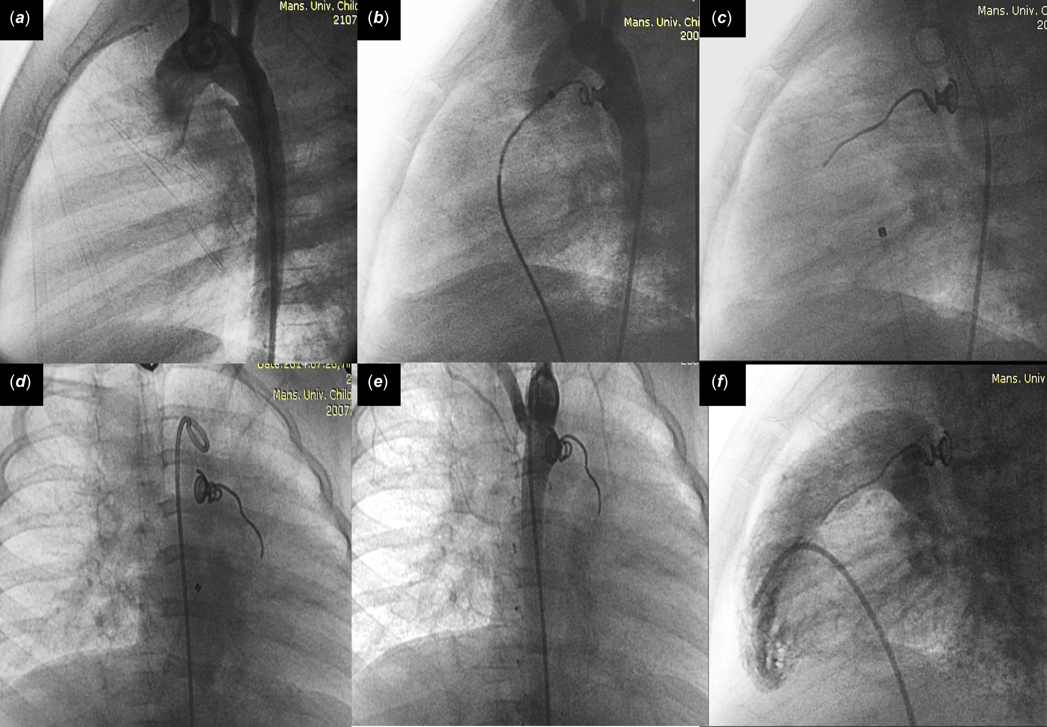
However, immediately following device release, unfolding of the coil loops on the pulmonary side was observed, resulting in coil straightening with extension across the pulmonary valve into the upper portion of the right ventricular outflow tract (Figure 1). Several attempts were made to reposition the straightened coil segment using balloon catheters to push the extended portion back into the pulmonary artery; however, these manoeuvres were unsuccessful in restoring the coil to its intended configuration. Given the risk of right ventricular outflow tract obstruction and the potential for pulmonary valve interference, the decision was made to retrieve the malformed device.
Fluoroscopic images of the abnormally straightened coil. (a) Aortogram in 90° straight lateral view showing the PDA. (b) Lateral view demonstrating the deployment of the PDA coil. (c, d) Lateral and anteroposterior views showing the straightened segment of the pulmonary coils after release. (e) Aortogram anteroposterior view showing no residual flow across the straightened coil, despite the unusual configuration of the coil. (f) The lateral view of the RVOT angiogram demonstrates the extent to which the tip of the straightened segment reaches.

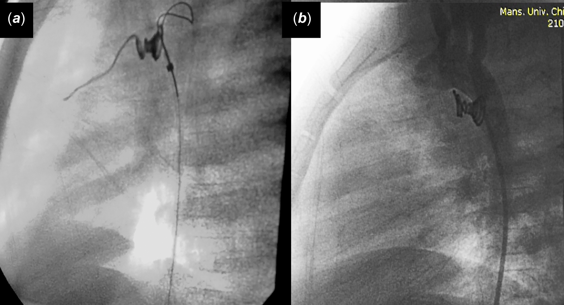
Prior to retrieval, an additional dose of heparin was administered, and the activated clotting time was measured to ensure adequate anticoagulation. The straightened coil was successfully retrieved using a snare catheter introduced from the aortic side. Following complete removal of the first device, a second Nit Occlud® PDA coil was deployed using the same technique. Post-deployment angiography confirmed complete occlusion of the patent ductus arteriosus with no residual flow and appropriate device positioning without complications (Figure 2).
Fluoroscopic images of (a) snaring of the deformed coil from the aortic side and (b) aortogram showing no residual flow across the second deployed coil with the expected usual morphology.

Discussion
The Nit-Occlud coil system has demonstrated high efficacy for transcatheter patent ductus arteriosus closure with minimal complications and limited reported mortality. The European Patent Ductus Arteriosus Registry retrospectively reported 1291 cases of transcatheter coil occlusion in 2001, reporting a success rate of 95% in patients with a patent ductus arteriosus minimal diameter of 2–6.2 mm and an adverse event rate of approximately 10%.Reference Magee, Huggon, Seed, Qureshi and Tynan2 Following the Federal Drug Administration approval in 2013,6 subsequent studies have confirmed these favourable outcomes. Moore et al. reported complete occlusion in 299 of 314 patients (96.8%) at one-year follow-up,Reference Moore, Greene and Palomares7 while Jung et al. documented a 98% complete occlusion rate at 12 months in 361 cases over 21 years, with residual shunts in only 2% and complications in 5% of cases.Reference Jung, Seol, Choi and Ha8
The Nit-Occlud coil is a nickel-titanium alloy-based, spiral-shaped device designed for small and medium patent ductus arteriosus.Reference Maksymenko, Kuzmenko and Dovhaliuk9 Its unique cone-in-cone configuration results from counter-directional winding of the proximal and distal segments. The tight distal windings (aortic side) prevent device migration into the pulmonary artery, while the more flexible proximal windings (pulmonary side) provide anchoring and adapt to individual ductal morphologies. The device is designed to assume its predetermined configuration at body temperature, and improper configuration may occur during room temperature testing.10 Moreover, the coil is repositionable and retrievable prior to release. In our case, we hypothesise that the coil straightening occurred due to excessive torsional stress at the device junction during the detachment process, preventing the proximal windings from achieving their intended configuration. This mechanical failure resulted in extension of the straightened segment across the pulmonary valve into the right ventricular outflow tract.
The management of this unusual complication required careful consideration of potential risks versus benefits. The decision to retrieve the malpositioned device was based on several theoretical complications associated with leaving the straightened coil in situ: (1) increased thromboembolism risk due to altered flow dynamics around the extended segment, (2) potential arrhythmogenesis from mechanical irritation within the right ventricular outflow tract, (3) risk of endothelial injury from the mobile device tip, potentially leading to endocarditis, and (4) possible late device embolisation. Given these considerations and the successful track record of coil retrieval procedures, device removal and replacement was deemed the safest approach.
Conclusions
To conclude, unexpected unfolding with straightening of the coil loops of patent ductus arteriosus occluder coils is possible. Hence, interventionists should have backup instruments in the catheterisation laboratory to remove the device using the proper snare and deploy another device, which is the most convenient and safest decision.
Financial support
This research received no specific grant from any funding agency, commercial, or not-for-profit sectors.
Competing interests
The authors declare no competing interests.
Ethical standard
This study was conducted in accordance with the 1964 Helsinki Declaration and its subsequent amendments or with comparable ethical standards.