Introduction
Space flight, in particular microgravity, imposes a wide range of physiological stresses on animals and their associated microbes. The adverse effects of microgravity have been well documented in animals and include bone loss, muscle atrophy and the dysregulation of the immune system (Sonnenfeld et al. Reference Sonnenfeld, Foster, Morton, Bailliard, Fowler, Hakenewerth, Bates and Miller1998; Lynch & Matin Reference Lynch and Matin2005; Crucian et al. Reference Crucian, Stowe, Pierson and Sams2008). In addition, there have been several studies regarding the impact of microgravity on microbial physiology (for review, see Horneck et al. Reference Horneck, Klaus and Mancinelli2010), such as increases in growth rates in liquid media (Taylor Reference Taylor1974; Ciferri et al. Reference Ciferri, Tiboni, Di Pasquale, Orlandoni and Marchesi1986; Klaus et al. Reference Klaus, Simske, Todd and Stodieck1997), higher resistance to antibiotics (Tixador et al. Reference Tixador, Richoilley, Gasset, Templier, Bes, Moatti and Lapchine1985), as well as elevated rates of conjugation, genetic transfer (Ciferri et al. Reference Ciferri, Tiboni, Di Pasquale, Orlandoni and Marchesi1986) and phage induction (Mattoni Reference Mattoni1968).
However, far less is known about the impact of the space environment on those microbes that comprise the healthy microbiome of animal hosts (McFall-Ngai et al. Reference McFall-Ngai, Nyholm and Castillo2010). In humans, the microbiome is comprised more than 1000 different microbial species that interact with host cells and are essential for normal host physiology, such as maintenance of various metabolic activities and the regulation of the host immune system (Rakoff-Nahoum et al. Reference Rakoff-Nahoum, Paglino, Eslami-Varzaneh, Edberg and Medzhitov2004; Mazmanian et al. Reference Mazmanian, Liu, Tzianabos and Kasper2005; Dethlefsen et al. Reference Dethlefsen, McFall-Ngai and Relman2007; Human Microbiome Consortium 2012). Although most interactions between hosts and microbes do not result in disease (Dethlefsen et al. Reference Dethlefsen, McFall-Ngai and Relman2007), much of what has been learned about host–microbe interactions in microgravity is derived from the study of pathogenic microbes (e.g. Nickerson et al. Reference Nickerson, Ott, Mister, Morrow, Burns-Keliher and Pierson2000; Chopra et al. Reference Chopra, Fadl, Sha, Chopra, Galindo and Chopra2006; Wilson et al. Reference Wilson2007; Rosenzweig & Chopra Reference Rosenzweig and Chopra2012; Vukanti et al. Reference Vukanti, Model and Leff2012; Kim et al. Reference Kim2013).
Several important studies have shown that microgravity can significantly impact the physiology of pathogenic microbes resulting in increases in virulence, resistance to environmental stress and increased survival in host macrophages (Nickerson et al. Reference Nickerson, Ott, Mister, Morrow, Burns-Keliher and Pierson2000, Reference Nickerson, Ott, Wilson, Ramamurthy and Pierson2004; Wilson et al. Reference Wilson, Ramamurthy, Porwollik, McClelland, Hammond, Allen, Ott, Pierson and Nickerson2002, Reference Wilson2007; Chopra et al. Reference Chopra, Fadl, Sha, Chopra, Galindo and Chopra2006; Crabbé et al. Reference Crabbé2011). Previous studies have shown that these changes in virulence are environment-dependent and, in some cases, can be attenuated through media supplementation. For example, in Salmonella typhimurium, the concentration of phosphate ion in the surrounding media was correlated to the pathogenesis-related acid stress response (Wilson et al. Reference Wilson2008) suggesting that it is possible to mitigate potential harmful effects of pathogens in space flight (Sarker et al. Reference Sarker, Ott, Barrila and Nickerson2010). These same studies have also determined that there are extensive changes in microbial gene expression both at the transcriptional and translational levels. One key finding is that microgravity alters the expression of the global regulator Hfq, an RNA-binding protein that binds to small regulatory RNA and message RNA (mRNA) molecules. These molecules foster the regulation of mRNA translation in response to environmental stress (e.g. acidic, osmotic, thermal), changes in metabolic concentrations (e.g. iron levels; Guisbert et al. Reference Guisbert, Rhodius, Ahuja, Witkin and Gross2007), as well as promote the virulence of several pathogens such as S. typhimurium (Wilson et al. Reference Wilson2007) and Pseudomonas aeruginosa (Crabbé et al. Reference Crabbé2011). Approximately half of all eubacteria have a homologue to Hfq (Valentin-Hansen et al. Reference Valentin-Hansen, Eriksen and Udesen2004), including several animal and plant symbionts (Ruby et al. Reference Ruby2005; Torres-Quesada et al. Reference Torres-Quesada, Oruezabal, Peregrina, Jofre, Lloret, Rivilla, Toro and Jimenez-Zurdo2010). In microgravity there is a down-regulation of hfq gene expression, which is thought to be involved in regulating virulence; however, the precise mechanisms are not known (Wilson et al. Reference Wilson2007). The hfq gene product has numerous regulatory functions that can differ significantly between species (for review Chao & Vogel Reference Chao and Vogel2010); however, the role of Hfq in beneficial animal–microbe interactions and the impact of space flight conditions on hfq expression in mutualistic bacteria remain unexplored.
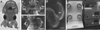
To address the role of Hfq in beneficial host–microbe interactions and to assess the impact of microgravity on this important regulator, we used the well-studied symbiosis between the bobtail squid Euprymna scolopes and the luminescent bacterium Vibrio fischeri as a model (for review Stabb & Visick Reference Stabb, Visick, Rosenberg, Delong, Stackebrandt, Lory and Thompson2013). V. fischeri colonizes a specialized organ in the centre of the host mantle cavity called the light organ (Fig. 1(A)). Once infected the bacteria trigger a series of developmental events that result in the morphogenesis of the host light organ over the first 4 days of colonization (McFall-Ngai & Ruby Reference McFall-Ngai and Ruby1991). Initially, in the juvenile squid there are fields of epithelial cells on either side of the light organ that form two appendage-like structures (Fig. 1(B)). These epithelial cells are covered in cilia that regularly beat to facilitate symbiosis-competent bacteria to aggregate, associate with the ciliated cells, and eventually enter pores found on the surface of the light organ (Montgomery & McFall-Ngai Reference Montgomery and McFall-Ngai1994; Nyholm et al. Reference Nyholm, Stabb, Ruby and McFall-Ngai2000; Altura et al. Reference Altura, Heath-Heckman, Gillette, Kremer, Krachler, Brennan, Ruby, Orth and McFall-Ngai2013). Persistent colonization of the light organ with V. fischeri for more than 12 h results in the onset of bacterial luminescence via quorum sensing (Lupp & Ruby Reference Lupp and Ruby2005), as well as an irreversible loss of the ciliated epithelial appendage (CEA) structures over a 96-h period (Fig. 1(C); Doino & McFall-Ngai Reference Doino and McFall-Ngai1995). An early event triggered by the bacteria is the onset of apoptotic cell death throughout the CEA (Fig. 1(D); Montgomery & McFall-Ngai Reference Montgomery and McFall-Ngai1994; Foster & McFall-Ngai Reference Foster and McFall-Ngai1998), which is triggered by the synergistic effects of two V. fischeri microbe-associated molecular pattern (MAMP) molecules lipopolysaccharide (LPS) and a peptidoglycan derivative known as tracheal cytotoxin (Foster et al. Reference Foster, Apicella and McFall-Ngai2000; Koropatnick et al. Reference Koropatnick, Engle, Apicella, Stabb, Goldman and McFall-Ngai2004). Typically, the cells undergoing cell death exhibit pycnotic (i.e., condensed) nuclei approximately 6 h after initial bacterial exposure. The number of pycnotic nuclei peaks 16 h after exposure to V. fischeri and slowly tapers off as the CEA structures begin to regress (Montgomery & McFall-Ngai Reference Montgomery and McFall-Ngai1994; Foster & McFall-Ngai Reference Foster and McFall-Ngai1998). The precise mechanism by which the CEA cells die and undergo regression is not completely understood; however, the regression occurs gradually over a 96-h period and is visible within 24 h after inoculation (Doino & McFall-Ngai Reference Doino and McFall-Ngai1995).
Overview of the symbiosis between the host squid Euprymna scolopes and luminescent bacterium Vibrio fischeri. (A) Hatchling squid with pronounced light organ in the centre of mantle cavity (arrow; bar=400 μm). (B) Micrograph of light organ in the absence of bacteria (i.e. aposymbiotic), depicting the pronounced ciliated epithelial appendages (CEA) extending from either side of the light organ (bar=200 μm). (C) Light organ exposed to symbiosis-competent V. fischeri for 96 h showing the complete loss of the CEA (bar=200 μm). (D) One half of the light organ stained with acridine orange depicting the pronounced pattern of dying pycnotic nuclei. The cell death event is triggered by exposure to V. fischeri and peaks 14 h after colonization under normal gravity conditions (bar=75 μm). (E) High-aspect-ratio vessel bioreactors that can simulate microgravity conditions.

The monospecificity of the Euprymna–Vibrio symbiosis in conjunction with the rapid developmental time line of bacteria-triggered events has been shown to be an effective model to examine the effects of microgravity on host–microbe interactions (Foster et al. Reference Foster, Kerney, Parrish, Khodadad and Ahrendt2011, Reference Foster, Khodadad, Ahrendt and Parrish2013). To simulate the microgravity environment we used high-aspect-ratio rotating wall vessel bioreactors (HARVs; Fig. 1(E)), which have been used for more than 20 years to mimic the low-shear environment of space flight (Wolf & Schwarz Reference Wolf and Schwarz1991; Schwarz et al. Reference Schwarz, Goodwin and Wolf1992; Nickerson et al. Reference Nickerson, Ott, Wilson, Ramamurthy and Pierson2004; Horneck et al. Reference Horneck, Klaus and Mancinelli2010). The HARV chambers simulate the microgravity environment by maintaining the fluids in constant suspension. These hydrodynamic forces within the HARV offset the effects of gravity; thus the cell culture, or squid, remains at constant terminal velocity. Previous studies have shown that the low-shear environment generated in the HARV chambers is strongly correlated to the experimental results obtained in actual space flight (Nickerson et al. Reference Nickerson, Ott, Wilson, Ramamurthy and Pierson2004; Wilson et al. Reference Wilson2007; Crabbé et al. Reference Crabbé2011) and has been used to assess the effects of the space environment on a wide range of physiological and genetic responses in both microbes (e.g. Nickerson et al. Reference Nickerson, Ott, Mister, Morrow, Burns-Keliher and Pierson2000; Wilson et al. Reference Wilson2008; Mauclaire & Egli Reference Mauclaire and Egli2010; Pacello et al. Reference Pacello, Rotilio and Battistoni2012; Rosenzweig & Chopra Reference Rosenzweig and Chopra2012; Vukanti et al. Reference Vukanti, Model and Leff2012; Arunasri et al. Reference Arunasri, Adil, Venu Charan, Suvro, Himabindu Reddy and Shivaji2013) and three-dimensional tissue cultures (e.g. Nickerson et al. Reference Nickerson, Ott, Wilson, Ramamurthy, LeBlanc, Honer zu Bentrup, Hammond and Pierson2003, Reference Nickerson, Ott, Wilson, Ramamurthy and Pierson2004, Reference Nickerson, Richter and Ott2007).
In this study, we assessed the impact of simulated microgravity on the expression of hfq in V. fischeri cultures. We also examined whether V. fischeri mutants defective in hfq were effective in colonizing and inducing normal light organ morphogenesis in the host squid. Both the juvenile squid and V. fischeri strains were co-incubated in the HARV chambers and the onset of apoptotic cell death and CEA regression, two of the most pronounced bacteria-induced phenotypes in light organ development, were assessed in the presence and absence of simulated microgravity. The resulting data provide evidence that the global regulating gene hfq is not required for host colonization but is required for normal light organ development. This study further confirms that mutualistic and pathogenic microbes often use similar mechanisms to interact with and colonize their animal hosts and that these symbiotic associations are susceptible to the effects of microgravity. By understanding the interactions of animals with their symbiotic microbiota in the space environment, we may be able to provide new insight into the mechanisms that maintain the balance between health and disease in animals.
Materials and methods
Host squid propagation
Specimen of adult E. scolopes were collected from O'ahu, Hawai'i and shipped to the Space Life Science Laboratory, Merritt Island, FL. The adult squid were maintained separately in 5-gallon aquaria at 23 °C on a 12 h light/dark cycle where they were mated bimonthly. The resulting egg clutches ranged in size from 150 to 300 eggs and were incubated in separate aquaria throughout their developmental period (∼21 days). Immediately after hatching, juvenile squid (2–3 mm in size) were transported to individual vials containing 5 ml of filtered sterilized seawater (FSW).
Bacterial strains, media and host colonization
The motile, symbiosis-competent strain V. fischeri ES114, which was originally isolated from an adult squid light organ (Boettcher & Ruby Reference Boettcher and Ruby1990), was used for all experiments and is referred to as wild-type. V. fischeri ES114 also served as the parent strain for the hfq mutant construction. The V. fischeri hfq mutant was kindly donated by K. Visick, Loyola University Chicago. The insertional hfq mutant was confirmed using standard PCR regularly throughout the study.
Prior to exposure to the host animals, both the wild-type and hfq mutant were grown in seawater tryptone broth (SWT) containing 0.5% tryptone, 0.3% yeast extract and 0.3% glycerol in 70% seawater (Boettcher & Ruby Reference Boettcher and Ruby1990) at 23 °C. Juvenile squid were then inoculated with V. fischeri strains at concentrations of 1×10⁵ cells ml−1 of FSW, replicating the approximate concentration of V. fischeri in the sand of the host squid habitat. The bacterial colonization of the host squid was monitored by measuring V. fischeri light production using a luminometer (GloMax 20/20, Promega Corp., Madison, WI). In all experiments, a subset of the juvenile squid was maintained without symbiosis-competent strains of V. fischeri, and these negative controls are referred as aposymbiotic animals.
Bacterial growth curves under simulated microgravity conditions
All V. fischeri cultures were grown in two rotary culture systems that contained eight 10 ml high-aspect-ratio vessels (HARVs, Synthecon, Houston, TX). The reactor vessels were positioned such that the axis of rotation was perpendicular to the direction of gravity, which simulated the low-shear microgravity conditions. For control purposes, HARVs were also positioned to replicate normal gravity conditions in which the axis of rotation was parallel to the gravity vector. The HARVs were inoculated with 1×10⁶ cells/ml−1 of SWT, incubated at 23 °C with a rotation rate of 20 rpm. The cellular density of the cultures were monitored spectrophotometrically (DU800, Beckman Coulter, Fullerton, CA) in triplicate every 2 h at a wavelength of 600 nm. Three technical replicates were completed for each growth curve.
RNA isolation and quantitative real-time PCR
To monitor the effects of microgravity on the expression of hfq in V. fischeri, cells were cultivated for 12, 24, 48, 72 and 96 h in the rotating HARV chambers in the microgravity and gravity positions. There were eight replicate cultures for each time point grown under both microgravity and gravity conditions. At each time point, the cultures were immediately placed in RNAlater to arrest cellular activity and stabilize the RNA. The RNAlater–cell mixtures were then centrifuged at 3000 g for 10 min to pellet the cells. Total RNA was isolated from each replicate culture using a modified RNAzolRT protocol (MRC, Cincinnati, OH). Briefly, each pellet was resuspended in 1 ml of RNAzolRT reagent and transferred to a PowerBiofilm bead tube (MoBio, Carlsbad, CA) and vortexed for 5 min. The samples were centrifuged at 12 000 g for 5 min at 23 °C and the resulting supernatants were transferred to a new tube where the RNA was precipitated with 75% (w/v) ethanol. The precipitated RNA was rinsed twice in 75% (w/v) ethanol to remove any additional RNAzolRT reagent. The RNA was then treated with Turbo DNA-free DNase (Ambion, Austin, TX) to remove residual DNA and eluted with an RNeasy MinElute Cleanup Kit (Qiagen, Valencia, CA). RNA concentrations were determined spectrophotometrically (Qubit 2.0 Fluorometer, Invitrogen, Carlsbad, CA) and normalized to 10 ng/μl−1 of RNAse-free water.
The relative copy number of hfq genes in V. fischeri cells cultivated under both gravity and simulated microgravity conditions were assessed with quantitative real-time PCR (qPCR). The expressed hfq genes were amplified and quantified using the Superscript III Platinum SYBR Green One-Step RT-qPCR kit (Invitrogen, Carlsbad, CA) with a Roche, Light Cycler 480-platform (Roche, Indianapolis, IN). PCR reactions were run in triplicate in a total volume of 25 μl per reaction. Each reaction contained a final concentration of 1×Superscript reaction master mix, 0.4 μgml−1 bovine serum albumin, 0.05 μM forward and reverse hfq primers and 50 ng RNA template. The primer set targeted the hfq gene found in the V. fischeri genome (Ruby et al. Reference Ruby2005) and included the forward (5′-CGTCGTGAAAGGATTCCAGT) and reverse (5′-ACGAGCCGGAACAACAGTAG) primers. The reaction conditions included initial 3 min of incubation at 42 °C for reverse transcription and then 5 min of incubation at 95 °C for polymerase activation. The running protocol was followed by 40 cycles of 95 °C for 15 s, 58 °C for 30 s and a single elongation step at 40 °C for 1 min. To assess the disassociation characteristics of the PCR products and to ensure that there were no primer dimers, a melting curve analysis was performed at 95 °C for 30 s and then at 65 °C for 30 s with continuous measurements at 95 °C. The results were compared to a calibration curve that was prepared from RNA extracted from 24 h V. fischeri cultures. RNA was serially diluted to 0.001, 0.01, 0.1, 1 and 10 ng/μl−1 generating a five-point standard curve for comparison. The copy number of the hfq genes was calculated, based on the calibration curve (i.e. it was normalized to ng of RNA) and average molecular mass of double-stranded DNA, as previously described (Fierer et al. Reference Fierer, Jackson, Vilgalys and Jackson2005).
Cell death determination and cellular regression
To visualize those cells of the CEAs undergoing apoptotic cell death, the juvenile squid were stained with the fluorescent marker acridine orange, which intercalates into the pycnotic (i.e. condensed) chromatin of the dying cells. Host squid were incubated in an equal mixture of 0.37 M MgCl2 and FSW to anesthetize the squid before dissection. The squid were then stained with acridine orange at a concentration of 5 ng/ml−1 of FSW for 5 min. The mantle and funnel of the juvenile were removed and the exposed light organ was then visualized with epifluorescence microscopy (Zeiss Axioplan Microscopy, Zeiss, Jena, Germany). Pycnotic nuclei throughout superficial ciliated epithelium were quantified and statistically compared using a parametric Student's t-test, as previously described (Foster et al. Reference Foster, Apicella and McFall-Ngai2000, Reference Foster, Khodadad, Ahrendt and Parrish2013). A minimum of eight animals were used for each treatment at each time point.
To measure the extent of cellular regression of the ciliated epithelium on the surface of the light organ, host squid were anesthetized, as previously described, and examined with microscopy. The lengths of the anterior CEAs were measured using the Zeiss Micro Imaging software (Carl Zeiss, Jena, Germany) and statistically compared between treatments using pairwise comparisons. The lengths were then normalized to the anterior appendage of hatchling squid observed at the start of each experiment (t=0 h). Results are reported as a per cent regression compared to the juvenile squid at t=0 h.
Results
Expression of hfq under simulated microgravity conditions
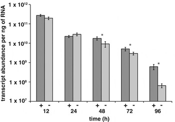
To examine the impact of simulated microgravity on hfq gene expression in the symbiont V. fischeri, quantitative real-time PCR (qPCR) was used. Cultures of V. fischeri were grown within the HARV bioreactors under simulated microgravity and gravity control conditions for 12, 24, 48, 72 and 96 h and RNA concentrations from each treatment were normalized (i.e. 50 ng per reaction). There was an overall decrease in hfq transcript abundance in both simulated microgravity and gravity treatments detected throughout the 96 h observation period (Fig. 2). However, significant differences (p<0.05) were observed between the V. fischeri parent and hfq mutant strain after 48 h incubation in simulated microgravity. The levels of hfq gene expression in cultures exposed to simulated microgravity continued to be significantly lower than normal gravity controls (p<0.05) throughout the 96 h experiment (Fig. 2).
Expression of gene hfq in V. fischeri cultures under gravity (+) and microgravity (−) conditions over time. Asterisk indicates significant difference between gravity and microgravity treatments with p<0.05.

Growth and luminescence of V. fischeri wild-type and hfq mutants under simulated microgravity conditions
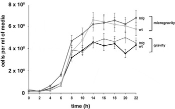
To address whether simulated microgravity impacts the growth of V. fischeri, motile cell cultures were incubated in HARV chambers and examined every 2 h spectrophotometrically. In addition to the wild-type strain of V. fischeri, an insertional mutant defective in hfq was also examined under identical growth conditions. There were few differences between wild-type and hfq mutant strains throughout the growth curves indicating that the lack of a functioning hfq gene did not impede normal growth (Fig. 3). However, differences were detected between simulated microgravity- and gravity-treated cells. Both strains had an altered growth rate under simulated microgravity conditions, beginning 10–14 h after initial inoculation. Although it was unclear whether the cultures were growing faster or experienced a shortened lag phase, the cultures reached cell densities that were higher than gravity-treated cells (Fig. 3).
Growth curve of wild-type V. fischeri (wt) and mutant strain defective in the gene hfq cultivated in HARV bioreactors under simulated microgravity and gravity conditions.

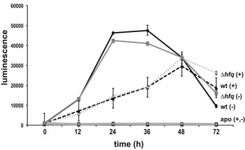
In addition to the growth rate, the ability of the strains to colonize the host squid light organ and luminesce in simulated microgravity was also examined. Once symbiosis-competent strains of V. fischeri colonize the light organ and reach sufficient cell densities, the cells begin to luminesce through quorum sensing approximately 12 h after initial colonization (Boettcher et al. Reference Boettcher, Ruby and McFall-Ngai1996; Lupp & Ruby Reference Lupp and Ruby2005). Results indicated that there was no delay in the onset of luminescence under simulated microgravity conditions; additionally, there was a pronounced increase in light production in symbiotic squid exposed to simulated microgravity (Fig. 4). Between 24 and 36 h there was an increase in the light levels of squid infected with the wild-type strain; however by 48 h, all the host animals showed comparable levels of luminescence, regardless of strain or environmental treatment. In aposymbiotic animals, no light production was observed under either simulated microgravity or gravity treatments throughout the 72-h observation period (Fig. 4).
Luminescence of host squid under microgravity (−) and gravity (+) conditions.
Squid colonized with V. fischeri wild-type (wt; black diamond) or mutant defective in hfq (grey square) under microgravity conditions were compared with those squid colonized with wild-type (black triangle) and hfq (grey circle) grown under 1×g gravity. Control animals maintained aposymbiotically (apo) showed no luminescence under microgravity (grey diamond) nor gravity (black square) controls.

Bacteria-induced phenotypes in host light organ under simulated microgravity conditions
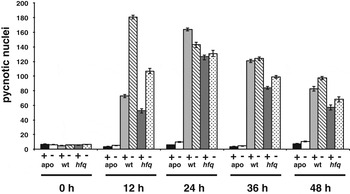
Although hfq mutants appear to colonize and initiate the symbiosis normally, there were differences observed in the onset of two key developmental phenotypes. Previous studies have shown that under simulated microgravity, the normal bacteria-induced apoptosis event that occurs along the surface epithelium of the light organ is accelerated and peaks several hours earlier than normal gravity conditions (Foster et al. Reference Foster, Khodadad, Ahrendt and Parrish2013; Fig. 5). In mutants defective in hfq, however, this simulated microgravity-induced phenotype was lost and the apoptosis event peaked at the same time point as wild-type (i.e. 24 h) under the normal gravity conditions. In addition, the hfq mutants also induced lower levels of apoptotic cell death under both simulated microgravity and gravity conditions compared with wild-type, although pycnotic nuclei counts were generally higher under simulated microgravity (Fig. 5).
Number of apoptotic pycnotic nuclei in host light organ under gravity (+) and microgravity (−) conditions over time. Host animals were either maintained without bacteria (apo) or inoculated with V. fischeri wild-type (wt) or mutants defective in the hfq gene (hfq).

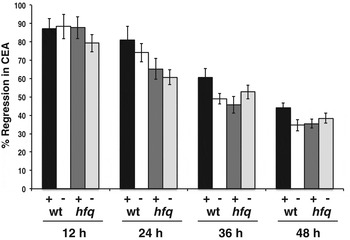
In addition to changes in the cell death event, there were also differences observed in the rates of cellular regression of the surface epithelium under simulated microgravity (Fig. 6). Previous results indicated that under simulated microgravity, the cells comprising the CEA structures on the surface of the light organ regress at a faster rate (Foster et al. Reference Foster, Khodadad, Ahrendt and Parrish2013; Fig. 6). Mutants defective in hfq showed accelerated cellular regression of the ciliated fields, which was comparable with wild-type cells under simulated gravity conditions (Fig. 6). This trend continued throughout the 48-h observation period.
Regression of the ciliated epithelial appendages (CEA) in the presence of V. fischeri wild-type (wt) and mutants defective in hfq (hfq) under 1×gravity (+) and microgravity conditions (−) over time.

Discussion
This study examined the role of the global regulator Hfq in the mutualistic association between the bobtail squid E. scolopes and its luminescent symbiont V. fischeri under normal gravity and simulated microgravity conditions. The results of this study indicate that: (1) hfq gene expression is down-regulated in symbiosis-competent V. fischeri cultures under simulated microgravity conditions; (2) the hfq gene in V. fischeri is not required for normal colonization of the host squid light organ; (3) loss of Hfq reduces the microgravity-induced phenotype of early-onset apoptosis in the CEAs; and (4) the synergistic effects of simulated microgravity and the loss of Hfq in V. fischeri accelerates normal regression of the CEA within the first 24 h.
The RNA-binding protein Hfq is an important global post-transcriptional regulator that is present in approximately half of all bacteria, including the symbiont V. fischeri (Ruby et al. Reference Ruby2005; Chao & Vogel Reference Chao and Vogel2010). Hfq is essential for pathogenesis in several microbes such as, but not limited to, Brucella abortus (Robertson & Roop Reference Robertson and Roop1999); entero-toxigenic Escherichia coli (Shakhnovich et al. Reference Shakhnovich, Davis and Waldor2009); P. aeruginosa (Sonnleitner et al. Reference Sonnleitner, Hagens, Rosenau, Wilhelm, Habel, Jager and Blasi2003); S. typhimurium (Sittka et al. Reference Sittka, Pfeiffer, Tedin and Vogel2007); Vibrio cholerae (Ding et al. Reference Ding, Davis and Waldor2004); and Yersinia pestis (Geng et al. Reference Geng2009). However, not all pathogens require Hfq and several strains show a wide range of virulence phenotypes in the absence of Hfq (for review, see Chao & Vogel Reference Chao and Vogel2010), with some strains such as the Gram-positive Listeria monocytogens (Christiansen et al. Reference Christiansen, Larsen, Ingmer, Sogaard-Andersen and Kallipolitis2004) and Staphylococcus aureus (Bohn et al. Reference Bohn, Rigoulay and Bouloc2007) having no Hfq phenotype.
Of the many microbes shown to possess an Hfq homologue, only two have been previously examined under a low-shear microgravity environment. In pathogenic strains of S. typhimurium and P. aeruginosa, there was a down-regulation of hfq expression under both actual and simulated microgravity conditions (Wilson et al. Reference Wilson, Ramamurthy, Porwollik, McClelland, Hammond, Allen, Ott, Pierson and Nickerson2002, Reference Wilson2007; Crabbé et al. Reference Crabbé2011). Previous studies have shown that as much as 20% of the S. typhimurium genome is directly or indirectly regulated by Hfq (Sittka et al. Reference Sittka, Lucchini, Papenfort, Sharma, Rolle, Binnewies, Hinton and Vogel2008) and that 32% of this Hfq regulon is differentially regulated during space flight (Wilson et al. Reference Wilson2007). Similar results are seen in P. aeruginosa with approximately 15% of all genes disrupted in hfq mutants (Sonnleitner et al. Reference Sonnleitner, Schuster, Sorger-Domenigg, Greenberg and Blasi2006) and 23% of genes differentially regulated in space (i.e., space flight stimulon) can be attributed to the Hfq regulon (Crabbé et al. Reference Crabbé2011). Although comparison of the two data sets showed a decrease in hfq expression in the two pathogens, only 16% of the down-regulated genes were shared between the two bacteria with no overlap in up-regulated genes, suggesting that the two pathogens have distinctive microgravity-induced stimulons (Crabbé et al. Reference Crabbé2011). These results may reflect the dynamic and differing roles that Hfq plays in various host-associated microbes. The down-regulation of hfq expression in the symbiotic V. fischeri in simulated microgravity conditions further confirms the role of Hfq as an important microgravity-induced regulator of microbial gene expression and that it is not limited to pathogens. However, future experiments that examine changes in the transcriptome of wild-type and hfq mutants will be required to fully assess the full role Hfq plays in the microgravity transcriptomic response of symbiotic V. fischeri, as well assess whether there is a core microgravity-induced stimulon response shared between mutualistic and pathogenic species.
Although Hfq appears to play an important role in the infection and virulence of a wide range of pathogens, the loss of Hfq function in V. fischeri did not impede normal colonization of the host light organ. The growth rates of V. fischeri with and without a functional hfq gene were comparable, suggesting that Hfq is not essential for V. fischeri cell growth. In addition, the onset of luminescence, a key indicator of light organ colonization, showed no statistical differences between the wild-type and hfq mutant, indicating that the loss of Hfq causes no initiation phenotype in the squid–vibrio symbiosis. A similar response has been seen in the nitrogen-fixing endosymbiont Sinorhizobium meliloti, which form a functional symbiosis with leguminous alfalfa (Torres-Quesada et al. Reference Torres-Quesada, Oruezabal, Peregrina, Jofre, Lloret, Rivilla, Toro and Jimenez-Zurdo2010). Mutant S. meliloti strains defective in Hfq are able to nodulate alfalfa roots at comparable levels to those found in the parent strain (Torres-Quesada et al. Reference Torres-Quesada, Oruezabal, Peregrina, Jofre, Lloret, Rivilla, Toro and Jimenez-Zurdo2010); however, numerous downstream defects were observed, such as the inability of the nodules to undergo normal levels of nitrogen fixation and long-term survival within acidic membrane compartments of the host (Barra-Bily et al. Reference Barra-Bily, Pandey, Trautwetter, Blanco and Walker2010; Gao et al. Reference Gao, Barnett, Long and Teplitski2010; Torres-Quesada et al. Reference Torres-Quesada, Oruezabal, Peregrina, Jofre, Lloret, Rivilla, Toro and Jimenez-Zurdo2010). Together, these results suggest that Hfq may not be critical for the initiation of beneficial host–microbe associations, but rather that Hfq may be important in the maturation and development of the symbioses.
Growth between the parent and the mutant strains were similar; however, there were differences observed under simulated microgravity and normal gravity treatments. Both V. fischeri strains grew to higher cell densities under simulated low-shear microgravity conditions mirroring what is found in other modelled and actual microgravity studies (Mattoni Reference Mattoni1968; Ciferri et al. Reference Ciferri, Tiboni, Di Pasquale, Orlandoni and Marchesi1986; Klaus et al. Reference Klaus, Simske, Todd and Stodieck1997; Foster et al. Reference Foster, Khodadad, Ahrendt and Parrish2013). The modest increase in cell densities of both V. fischeri wild-type and hfq mutant compared to other bacterial species may reflect the motile nature of V. fischeri in culture. V. fischeri is motile in its free-living state; however, once it colonizes the light organ it loses its flagella and becomes non-motile (Ruby & Asato Reference Ruby and Asato1993). Growth changes of bacterial cultures under natural and modelled microgravity conditions have been correlated to motility, as motile bacteria can often disrupt the quiescent fluid and nutrient environment of the culture thus changing the physiology and growth rates of the microbes (Benoit & Klaus Reference Benoit and Klaus2007).
The increase in luminescence observed in the host squid may also reflect the increased growth rate of V. fischeri strains under simulated microgravity conditions. Once the light organ has been colonized and V. fischeri cells reach a critical cell density (∼12 h), the cells utilize two quorum-sensing systems (e.g. ain and lux) to regulate luminescence (Lupp & Ruby Reference Lupp and Ruby2005). The V. fischeri cells continue to grow on nutrients provided by the host animal and reach maximum cell density approximately 24 h after initial inoculation (Graf & Ruby Reference Graf and Ruby1998). It is possible that under simulated microgravity conditions, the V. fischeri cells reached a higher cell density earlier than in the gravity controls and light production was subsequently elevated. By 48 h, the luminescence levels of both simulated microgravity and gravity controls were comparable. This decrease in light production during the second day of colonization may reflect the natural diel rhythm of the symbiosis (Boettcher et al. Reference Boettcher, Ruby and McFall-Ngai1996). At dawn, the host squid vents 95% of the bacterial populations and recultivates the bacterial population from the remaining 5% (Lee & Ruby Reference Lee and Ruby1994). It is possible that during the regrowth of the bacterial population within the light organ the effects of modelled microgravity were no longer present. Future work that examines the expression of the lux operon under normal and modelled microgravity throughout the diel cycle is required.
Although the loss of Hfq in V. fischeri had no impact on the initiation of the symbiosis under normal or reduced gravity conditions, Hfq did appear to be required for the full onset of apoptotic cell death in the host light organ CEAs. Levels of pycnotic nuclei were significantly attenuated in V. fischeri hfq mutants under both normal and simulated microgravity conditions. Previous studies have shown that simulated microgravity conditions accelerate the onset of V. fischeri cell death in the host light organ with a significant increase in the numbers of pycnotic nuclei (Foster et al. Reference Foster, Khodadad, Ahrendt and Parrish2013). The attenuated cell death response in hfq mutants under both gravity and simulated microgravity conditions may reflect an altered production of the microbe-associated molecular pattern (MAMP) molecule lipopolysaccharide (LPS). For example, examination of the transcriptome of several pathogens, such as P. aeruginosa, has shown that in hfq mutants there is a down-regulation of genes associated with cell wall, LPS and capsule production (Sonnleitner et al. Reference Sonnleitner, Schuster, Sorger-Domenigg, Greenberg and Blasi2006). Additionally, in S. typhimurium cultures exposed to simulated microgravity, there is a down-regulation of LPS O-antigen side chain synthesis and overall lower LPS production in microgravity (Wilson et al. Reference Wilson, Ramamurthy, Porwollik, McClelland, Hammond, Allen, Ott, Pierson and Nickerson2002; Rosenzweig & Chopra et al. Reference Rosenzweig and Chopra2012). Previous studies have also shown that microgravity can increase the sensitivity of the CEA cells to LPS-induced apoptosis (Foster et al. Reference Foster, Khodadad, Ahrendt and Parrish2013). Therefore, if levels of LPS are lower in V. fischeri hfq mutants, the increased sensitivity of CEA cells to LPS under microgravity conditions may play a factor in the attenuated cell death response seen in the host light organs. Further work is required to measure the levels of LPS in the symbiotic V. fischeri strains under normal and reduced gravity conditions.
Another important bacteria-induced development event that was altered under simulated microgravity conditions was the increased rate of regression of the CEA structures early on in light organ morphogenesis. Previous studies have shown that approximately 24 h post-inoculation of symbiotic V. fischeri, there is an irreversible loss of the CEA basement membrane, which provides structural support for the single layer of epithelial cells that comprise the CEA (Foster & McFall-Ngai Reference Foster and McFall-Ngai1998). It is thought that the loss of this structural membrane hastens the regression of the cells that comprise the CEA (Foster & McFall-Ngai Reference Foster and McFall-Ngai1998). The results of this study suggest that V. fischeri hfq mutants underwent a faster rate of regression during the first 24 h of the symbiosis compared with the wild-type strains. This regression occurred faster under simulated microgravity conditions; however, by 36 h, the level of CEA regression in both the V. fischeri wild-type and hfq mutant were not statistically different in simulated microgravity. These results may suggest that Hfq plays a role in regulating the bacterial signals that trigger the collapse of the basement membrane and the subsequent regression of the CEA structures. Hfq has been identified in regulating the expression of several quorum-sensing pathways (Hammer & Bassler Reference Hammer and Bassler2007) as well as several type III secretion systems that deliver effector molecules to the host cells (Sittka et al. Reference Sittka, Pfeiffer, Tedin and Vogel2007, Reference Sittka, Lucchini, Papenfort, Sharma, Rolle, Binnewies, Hinton and Vogel2008). The loss of Hfq in V. fischeri may disrupt the normal signal cascade required in the bacteria-induced light organ morphogenesis, thus accelerating the regression event. In addition, microgravity has also been shown to down-regulate the expression of genes associated with the extracellular matrix molecules, which are known to comprise the basement membrane structures (Zayzafoon et al. Reference Zayzafoon, Gathings and McDonald2004, Reference Zayzafoon, Meyers and McDonald2005). Furthermore, microgravity can alter and induce remodelling in the cytoskeleton in eukaryotic cells (Infanger et al. Reference Infanger2006). Together, the lack of Hfq and the effects of simulated microgravity may work synergistically to accelerate the regression of the CEA structures in symbiotic light organs.
In summary, the results of this study indicate that Hfq, an important post-transcriptional regulator of virulence in pathogenic microbes, is also an important molecule in symbiotic microbes that form associations with animal tissues. Although not essential for initiation of the symbiosis, Hfq does appear to play a critical role in two key developmental processes that restructure the host light organ tissues. The effects of simulated microgravity also appear to influence the expression of hfq in the symbiotic V. fischeri, suggesting that hfq may be an important component of the space flight stimulon in all host-associated microbes, not just pathogens. Further research is required to examine the influence of microgravity on the transcriptome of both the host squid and its symbiont in the presence and absence of Hfq. However, these results do indicate that the squid–vibrio symbiosis can serve as an important model to examine those molecular mechanisms required by both pathogens and mutualistic bacteria and whether the stress of the space flight environment can alter the regulation of these pathways, thus tipping the balance between health and disease in animal hosts.
Acknowledgements
The authors thank Karen Visick and Kati Geszvain for graciously providing the V. fischeri hfq mutant strain and Wayne Nicholson for the use of his HARV chambers. This work was supported by the NASA Florida Space Grant Consortium award UCF01-0000232913 and the Florida Space Institute Space Research Initiative awarded to JSF.