Introduction
This review describes the anatomical and physiological processes in sows that result in the passive transfer of immune constituents to the neonate through the ingestion of colostrum (colostrogenic immunity) or milk (lactogenic immunity), including both cell-mediated (CMI) and humoral immune components (Brambell, Reference Brambell1970; Saif, Reference Saif1990). The lengthy history of these concepts is worthy of note. In early Rome, the words immunitas and immunis meant ‘exempt from a social duty’, but there are also examples in which they were used to express exemption from, or resistance to, disease (Silverstein, Reference Silverstein and Silverstein1989). The widespread adoption of Edward Jenner's smallpox vaccine (1774) brought the idea of ‘immunization’ and ‘vaccination’ into the mainstream lexicon (Silverstein, Reference Silverstein and Silverstein1989). Passive immunity, the realization that the administration of immune serum could protect the naïve recipient, was brought to light in the work of Von Behring and Kitasato (Von Behring and Kitasato, Reference Von Behring and Kitasato1890a). Initially, they showed that mice (n = 8), intraperitoneally inoculated with blood from rabbits with antibodies against tetanus toxin, were able to survive exposure to the toxin. Later, they pioneered the use of equine antisera to treat human beings for tetanus and diphtheria (Von Behring and Kitasato, Reference Von Behring and Kitasato1890b). From there, it was a short step to the realization that passive immunity could be provided by a mother to her progeny. This idea was supported by clinical observations, and experiments showed that the disease resistance in offspring was related to the disease (or vaccination) history of the mother, as described below.
The recognition of maternal immunity
Otto Bollinger (Reference Bollinger1877) was the first to hypothesize the in utero transfer of maternal immunity. Initially, he had observed a case in which a newborn child from a smallpox-vaccinated mother was resistant to smallpox vaccine, i.e. the baby did not show the typical vesicular lesions associated with successful vaccination against smallpox. He then conducted a controlled clinical experiment and found that lambs from ewes (n = 700) vaccinated with sheep pox (Variola ovina) showed resistance to vaccination, i.e. the lambs from vaccinated mothers did not develop the vesicular lesions if vaccinated at 4–6 weeks of age.
Shortly thereafter, Albrecht E. Burckhardt (Reference Burckhardt1879), conducted a controlled clinical experiment and found that infants from mothers who had been successfully vaccinated against smallpox in late pregnancy did not develop the typical vesicular lesions associated with the smallpox vaccine when inoculated at 4–6 days of age. Burckhardt concurred with Bollinger that the mother's immunity against smallpox was transferred to the fetus in utero.
In Reference Chauveau1880, Auguste Chauveau (France) described a series of experiments that proved that lambs born from ewes inoculated during late gestation with a Bacillus anthracis preparation were resistant to the pathogen. Initially, Chauveau found that sheep repeatedly (seven or eight times) inoculated with a B. anthracis preparation at 15-day intervals stopped showing clinical signs post inoculation, e.g. malaise, swollen lymph nodes, ganglionic swelling, and/or fever. Chauveau concluded that exposure no longer produced clinical signs because the sheep had produced something in response to the initial inoculations that protected them from subsequent exposures. In the next phase of his studies, he repeatedly inoculated pregnant sheep and then inoculated their lambs. Because the lambs showed no clinical signs following their first exposure, Chauveau concluded that protection could be transferred from immune mothers to their offspring (Ladnyi, Reference Ladnyi1964).
In 1892, Paul Ehrlich proved that immunity against plant toxins (abrin and ricin) could be transferred from female mice to their offspring through mammary secretions. In one experiment, he ‘immunized’ 15 does against either abrin or ricin during pregnancy, then inoculated 22 pinkies from their litters to the same toxin. Because all pinkies survived, he hypothesized that passive immunity (‘passive immunität’) was transferred from the immune mother to their offspring through mammary secretions. To test this hypothesis, pinkies (n = 2) from immunized does were cross-fostered to a non-immunized doe and pinkies (n = 4) from non-immunized does were cross-fostered to immunized does (n = 4). None of the pinkies that nursed the non-immunized doe survived, but three of the four pinkies that suckled immunized does survived the inoculation. In addition, Ehrlich reported that the passive antibody could not be transferred from the male to the offspring. Bucks (n = 6) immunized with abrin were bred with non-immunized does (n = 6), then their pinkies were inoculated with the toxin. None of the pinkies survived and all showed the effects of the toxin, e.g. intestinal hemorrhage and tissue necrosis (Brieger and Ehrlich, Reference Brieger and Ehrlich1892).
A series of studies with ‘Bacillus diphtheriae’ (Corynebacterium diphtheriae) conducted by Theobald Smith between 1904 and 1909 provided further evidence and greater detail concerning the transfer of immunity from mother to offspring (Smith, Reference Smith1905; Reference Smith1907; Reference Smith1909). Working with C. diphtheriae culture fluid, Smith defined one unit of toxin as the dose able to kill one 250–280-g guinea pig after subcutaneous injection, and one antitoxin unit as the volume of serum able to completely neutralize one unit of toxin. In his experiments, an immune guinea pig was defined as an animal that survived a lethal dose of toxin.
Smith made four key observations:
(1) Immunity to diphtheria toxin in the mother is transmitted to her offspring (Smith, Reference Smith1905; Reference Smith1907). Female guinea pigs (n = 77) were administered one of 4 treatments: no treatment (n = 21), toxin (n = 4), antitoxin (n = 7), and a toxin–antitoxin mixture (n = 35) and then bred 2–3 months post-treatment. When their offspring weighed 250–280 g, they were subcutaneously injected with a lethal dose of toxin. All guinea pigs from mothers that received the toxin–antitoxin mixture survived the challenge and, over a range of challenge doses, showed greater resistance to the clinical effects of the toxin compared with other treatment groups.
(2) Immunity to diphtheria toxin in the father is not transmitted to his offspring (Smith, Reference Smith1907). Male guinea pigs (n = 6) were divided into two groups: treatment (n = 5) and control (n = 1). The guinea pigs in the treatment group were administered the toxin–antitoxin mixture and then bred with 11 untreated female guinea pigs. The control male guinea pig was bred with two untreated females. When the offspring of the female guinea pigs weighed 250–280 g, they were subcutaneously administered a lethal dose of diphtheria toxin. None of the offspring survived and Smith concluded that immunity could not be transmitted from the father to the offspring.
(3) Passive immunity does not persist in the offspring and is gradually lost over time (Smith, Reference Smith1907). Offspring from eight toxin–antitoxin-treated females (n = 18) and two untreated females (n = 2) were subcutaneously injected with different amounts of toxin (1.0–1.72 units) at different ages (32–119 days of age) and different body weights (251–650 g). The lethal dose in resistant guinea pigs gradually decreased by the time such that, by 3 months of age, guinea pigs from immunized females no longer showed any resistance to the toxin. Smith observed that maternally derived immunity did not persist; rather it gradually declined over time.
(4) Passive immunity is not transmitted from the first generation to the second generation (Smith, Reference Smith1907). Female guinea pigs (‘1st generation’) from immunized females were divided into two groups: positive control (n = 2) and untreated (n = 12). Guinea pigs in the positive control group were administered the toxin–antitoxin mixture and those in the untreated group were not. Thereafter, the offspring (‘second generation’) from both groups were administered a lethal dose of the toxin. None of the offspring from the untreated group survived, whereas offspring from the positive control group were resistant to the toxin. Therefore, Smith concluded that the passive immunity was not transmitted from the first generation to the second generation.
In 1912, Lemuel William Famulener conducted an experiment to establish whether maternal antibody was transmitted from mother to offspring via mammary secretions or in utero. Female goats (n = 5) were subcutaneously inoculated with sheep red blood cells (RBC) during late pregnancy. Kids farrowed from these does were either allowed to suckle their mother (treatment group) or fed with cow's milk (control group). Specific anti-sheep RBC antibody in serum, colostrum, and milk samples from the goats and kids was measured by complement fixation. The results showed that the goats inoculated with sheep RBCs during late pregnancy secreted high concentrations of anti-sheep RBC antibody in their colostrum and milk, and that the kids in the treatment group showed high antibody concentration in their blood whereas no anti-sheep RBC antibody was detected in the controls, i.e. no maternal antibody had been transferred to the kids in utero.
Although Famulener (Reference Famulener1912), working with goats, had concluded that the placenta was not permeable to maternal antibody, Ten broeck and Bauer (Reference Ten Broeck and Bauer1923) in a study of women (n = 6) known to be carriers of Clostridium tetani, showed that tetanus antitoxin was present in the mothers’ serum, placental cord blood, and children's serum. Despite the fact that they failed to account for the colostral contribution to serum antibody in the children, they concluded that ‘Since the antitoxin level in the mother's and child's blood is at approximately the same level it seems probably that the placenta is permeable to this antibody.’ McKhann and Chu (Reference McKhann and Chu1933) went on to show that antibodies recovered from human placenta showed neutralizing activity against diphtheria toxin, group A Streptococcus spp., poliovirus, and measles virus and, like Ten broeck and Baurer (Reference Ten Broeck and Bauer1923), concluded that neonatal immunity could have originated via transplacental transfer. Clarification was brought to the question of placental passive transfer of maternal antibody by Ladnyi (Reference Ladnyi1964). Doe rabbits immunized (n = 3) with tetanus toxoid and non-immunized controls (n = 3) were bred 15 days after vaccination. After birth, the kits from the immunized females were cross-fostered to the non-immunized females and vice versa. No antitoxin antibody was detected in the serum of the offspring born from non-immunized mothers nursing immunized females, but kits from immunized mothers fed by nonimmunized females had antitoxin antibody titers approximately equivalent to their mothers’. These results supported the conclusion that maternal antibody was transferred to fetal rabbits transplacentally.
Why are piglets born agammaglobulinemic?
In Reference Bauriedel, Hoerlein, Picken and Underkofler1954, Wallace Bauriedel et al., reported that maternal antibody could not be transferred from sow to the piglet in utero. Cesarean-derived, colostrum-deprived piglets (n = 12) from two sows were housed individually and fed purified casein and soybean protein diets. Analyses of serum samples collected from the piglets over time showed that gamma globulins appeared around 6 weeks of age, i.e. no maternal antibody was provided to the piglets transplacentally.
The work of Alvin Hoerlein (Reference Hoerlein1957) definitively established that maternal antibody was transferred to piglets via mammary secretions and not to fetal piglets in utero. A pregnant sow was subcutaneously inoculated with Brucella abortus and sheep RBC 14 days before delivery of her piglets by cesarean section. The piglets were then divided into control piglets (n = 5) that were left with the sow to receive colostrum and colostrum-deprived piglets (n = 4), which were isolated from other pigs and fed cow's milk. Subsequent testing showed that control piglets had antibodies against both B. abortus and sheep RBC, but colostrum-deprived piglets had no detectable antibodies against either.
The explanation for why piglets are born agammaglobulinemic lies in the structure of the placenta. An initial understanding of the nature of the placenta and its role in fetal development can be traced back to the ancient Greeks, i.e. the terms ‘placenta’, ‘chorion’, and ‘amnion’ were used in the treatise ‘De generatione animalium’ written by Aristotle in 340 B.C. Concerning the placenta, Aristotle wrote: ‘The vessels join on the uterus like the roots of plants and through them the embryo receives its nourishment’ (Longo and Reynolds, Reference Longo and Reynolds2009). A significant understanding of the mechanics of the maternal–fetal circulation within the placenta was not achieved until much later. In 1872, William Turner described the human placental structure and compared it to that of domestic animal species. In 1909, Otto Reference GrosserGrosser described the macroscopic and microscopic diversities of mammalian placental anatomy and proposed a system of classification based on placental structure. In 1961, Emmanuel Ciprian Amoroso modified Grosser's system and developed a system of classification based on the presence of tissue layers separating the maternal and fetal blood systems: maternal endothelium, maternal connective tissue, maternal (uterine) epithelium, fetal (chorionic) epithelium, fetal connective tissue, and fetal endothelium. The following mammalian placenta types are distinguishable by the presence of these different tissue layers:
(1) Epitheliochorial placentation (swine and equids) retains all six maternal and fetal tissue layers.
(2) Syndesmochorial placentation (ruminants) consists of five tissue layers, i.e. maternal endothelium, maternal connective tissue, chorionic epithelium, fetal connective tissue, and fetal endothelium. Thus, the maternal (uterine) epithelium is absent.
(3) Endotheliochorial placentation (most carnivores) separates maternal and fetal blood circulation by four tissue layers, including maternal endothelium, chorionic epithelium, fetal connective tissue, and fetal endothelium. Thus, the chorionic epithelium of the fetus is in direct contact with the endothelium of the maternal capillaries.
(4) Hemochorial placentation (human and most primates) is composed of three tissue layers (chorionic epithelium, fetal connective tissue, and fetal endothelium). This brings the maternal blood circulation into direct contact with the chorionic epithelium.
(5) Hemoendothelial placentation (most rodents and lagomorphs) is distinct in that maternal blood circulates in large sinus-like spaces in the trophoblast. Fetal capillaries projecting directly into the space are only separated from maternal blood by the fetal endothelial layer.
Both hemochorial and hemoendothelial placentation permit the transfer of maternal immunity to the fetal circulation (Leiser and Kaufmann, Reference Leiser and Kaufmann1994). In these species, progeny receives passive immunity both in utero and via mammary secretions after birth. The endotheliochorial placentation seen in most carnivores limits the transfer of maternal immunity to the fetus and the majority of passive immunity is transferred through mammary secretions, i.e. colostrum and milk, from the mother to their offspring after birth. The six-layered epitheliochorial placenta of swine and the five-layered syndesmochorial placenta of ruminants preclude any immune transfer through the placenta due to the number of layers constituting the placental barrier (Bertasoli et al., Reference Bertasoli, Santos, Paula, Barbosa, Silva and Jorge2015). Thus, the reason piglets are born agammaglobulinemic can be explained by the structure of the placenta.
Anatomy and physiology of lactation
Swine mammary gland anatomy
Anatomically, mammary glands in pigs are located in two parallel rows from the thoracic to the inguinal areas on the ventral body wall. Domestic pigs commonly have 14 mammary glands distributed in three areas of the body, i.e. four thoracic, eight abdominal, and two inguinal glands, but the total number of glands may vary between 12 and 18, depending on the genetic background of the individual (Labroue et al., Reference Labroue, Caugant, Ligonesche and Gaudré2001). Each mammary gland is usually composed of two complete gland systems, i.e. the gland has two separate teat canals that connect to one nipple (Martineau et al., Reference Martineau, Farmer, Peltoniemi, Zimmerman, Karriker, Ramirez, Schwartz and Stevenson2012).
The macroscopic and microscopic structures of the mammary gland differ between nulliparous and lactating sows. Mammary glands of nulliparous sows are composed of cell buds distributed among fat and connective tissue, but the tissues are displaced by glandular parenchyma in lactating sows. In lactating sows, mammary glands are composed of compound tubuloalveolar tissue-forming lobular structures. The lobules are lined by epithelial cells (lactocytes) that function in milk synthesis (Klopfenstein et al., Reference Klopfenstein, Farmer, Martineau, Straw, Zimmerman, D'Allaire and Taylor2006), and are surrounded by myoepithelial cells that function in milk ejection and vascularized connective tissue that contains lipid-depleted adipocytes and fibroblasts (McManaman and Neville, Reference McManaman and Neville2003). The secreting units are connected via a ductal system to an ostium found on the end of the teat.
Mammogenesis
Mammogenesis, the development of nulliparous mammary glands into functional, lactating mammary glands, begins with the onset of puberty. Over the course of puberty, mammary gland parenchymal tissue, i.e. alveoli, ducts, and associated connective tissues, increases by 51% and the extraparenchymal tissue, i.e. adipose tissues surrounding the parenchyma, decreases by 16% (Farmer et al., Reference Farmer, Fisette, Robert, Quesnel and Laforest2004). Ji et al. (Reference Ji, Hurley and Kim2006) reported that weight of swine mammary glands increased from 39.7 to 299.2 g per gland between 45 and 112 days of gestation, reaching the maximum mammary gland alveolar epithelial cell concentration by day 90.
Mammogenesis development during pregnancy is under the influence of specific reproductive hormones, e.g. relaxin, estrogen, and prolactin, particularly during the last one-third of gestation in the pregnant gilt. In swine, estrogens (estrone and estradiol 17-β) are initially produced by embryonic blastocysts beginning at 10–15 days of pregnancy (Spencer and Bazer, Reference Spencer and Bazer2004). At 17–19 days of gestation, fetal placental tissue becomes the primary source of estrogens (Ainsworth and Ryan, Reference Ainsworth and Ryan1966; Bazer and Thatcher, Reference Bazer and Thatcher1977; Knight, Reference Knight1994; Farmer, Reference Farmer2001). However, porcine myometrium and endometrium serve as supplemental sources of estradiol 17-β beginning at 14–16 days of gestation (Franczak and Kotwica, Reference Franczak and Kotwica2008). Relaxin is initially produced by theca interna cells of preovulation follicles during early pregnancy. Once the corpora lutea develop (10 days after ovulation), they become the major source of relaxin for the remainder of the pregnancy (Bagnell et al., Reference Bagnell, Zhang, Ohleth, Connor, Downey, Tsang and Ainsworth1993). Prolactin, secreted by the anterior pituitary, is essential for both mammogenesis and lactogenesis (Neville et al., Reference Neville, McFadden and Forsyth2002). Prolactin secretion during pregnancy is controlled by dopamine released by the hypothalamus. Kraeling et al. (Reference Kraeling, Barb and Rampacek1992) reported that prolactin concentration does not vary significantly during the first 90 days of gestation (P < 0.05) but increases markedly around 110 days of gestation, as discussed in the subsection ‘Prolactin’.
Estrogen
The effect of estrogens on mammogenesis is well described in swine. Sørensen et al. (Reference Sørensen, Sejrsen and Purup2002) studied mammary development in gilts in 10-day increments from birth to day 112 of pregnancy. The rate of mammary tissue development and the DNA accumulation increased with age, but was highest after 90 days of age. The shift in mammary gland development coincided with the upward shift in estrogen production at 90 days of age. Sørensen et al. (Reference Sørensen, Sejrsen and Purup2002) found that mammogenesis in pregnant gilts was slow during the first two trimesters of pregnancy. In the third trimester, under the influence of increasing concentrations of estrogen and progesterone and a low concentration of prolactin, the mammary glands underwent major histological remodeling, as adipose tissue and stroma were displaced by lobuloalveolar tissue and milk secretory units were formed (Kensinger et al., Reference Kensinger, Collier and Bazer1986a; Ji et al., Reference Ji, Hurley and Kim2006).
Kensinger et al. (Reference Kensinger, Collier and Bazer1986b) created sows with different numbers of conceptuses: pseudopregnant sows (no conceptuses) were created by the administration of estradiol-valerate on days 11–15 of the estrous cycle, 4–7 conceptuses by ligation of one oviduct, and 8–11 conceptuses by normal mating. The number of viable fetuses the sow carried was correlated with the level of serum estrogen-sulfate, which in turn reflected the wet weight, dry weight, and mammary DNA content of her mammary glands (Kensinger et al., Reference Kensinger, Collier, Bazer and Kraeling1986c).
Relaxin
Relaxin plays a role in mammary gland parenchymal growth during the last trimester of gestation. To demonstrate the effects of relaxin on mammogenesis, Hurley et al. (Reference Hurley, Doane, O'Day-Bowman, Winn, Mojonnier and Sherwood1991) designed an experiment consisting of 38 gilts allocated to three different groups: ovariectomized gilts that received progesterone to maintain pregnancy, but no relaxin; ovariectomized gilts that received both progesterone and relaxin, and control gilts. Mammary parenchymal tissue of the gilts was measured and compared between groups on day 100 of gestation. The results showed that ovariectomized gilts that did not receive relaxin had less mammary parenchymal tissue than controls (P < 0.05), whereas no difference was detected in the mammary parenchymal tissue between ovariectomized gilts that received progesterone and relaxin and controls (P > 0.05).
Prolactin
The effect of prolactin on mammogenesis was reported by Farmer et al. (Reference Farmer, Sorensen and Petitclerc2000). Bromocriptine (10 mg) was orally administered three times daily to pregnant gilts between 70 and 110 days of gestation to inhibit prolactin production. Treated gilts and control gilts were sacrificed at 110 days of gestation. Parenchymal and extraparenchymal tissues were collected from both groups and mammary DNA, RNA, dry matter, protein and fat content were measured. No difference was detected between the two groups in the weight of extraparenchymal tissue (P ≥ 0.1), but significant decreases in the weight of parenchymal tissue, total DNA, and RNA and significant increases in fat and dry matter were observed in gilts with inhibited prolactin production (P < 0.01). Farmer et al. (Reference Farmer, Sorensen and Petitclerc2000) concluded that prolactin is essential for both normal mammary gland development during the last trimester of gestation and promoting lactogenesis during late gestation and after parturition (discussed in the subsection ‘Lactogenesis’).
Lactogenesis
Lactogenesis is the process by which the components of colostrum and milk are created and then secreted. Phase I of lactogenesis, beginning between 90 and 105 days of gestation, marks the synthesis of milk components (lactose, casein, and lipids) in mammary gland alveoli (Hartmann et al., Reference Hartmann, Smith, Thompson, Wakeford and Arthur1997). Phase II, overlapping with Phase I but initiated at farrowing, involves milk synthesis and secretion initiated by progesterone withdrawal after lysis of the corpora lutea.
Phase II can be described in terms of colostral, ascending, plateau, and descending phases. The colostral phase marks the beginning of lactation immediately post farrowing (Hartmann et al., Reference Hartmann, Atwood, Cox, Daly, Wilde, Peaker and Knight1995). The total yield of colostrum per day is <50% of early lactation milk production, but colostrum contains more protein, less fat, and less sugar than milk (Dorland, Reference Dorland1900). In litters of 8–12 piglets, Hartmann et al. (Reference Hartmann, Whitely and Willcox1984) found that 2.5–5.0 (average of 3.5) kg of colostrum was produced in the first 24 h after parturition (Hartmann et al., Reference Hartmann, Whitely and Willcox1984). The ascending phase occurs 24–48 h after parturition and is characterized by the switch from colostrum to milk (Klobasa et al., Reference Klobasa, Werhahn and Butler1987). During the ascending phase, the frequency and volume of milk secretion increase, with maximum milk production achieved about 10 days after the switch from colostrum to milk. The plateau phase, the period when a lactating sow maintains the maximum level of milk production, can last up to 28 days (King et al., Reference King, Mullan, Dunshea and Dove1997). The descending phase is the period of declining milk production. In modern production systems, sows usually do not reach the descending phase, because piglets are weaned prior to 28 days of age (Martineau et al., Reference Martineau, Farmer, Peltoniemi, Zimmerman, Karriker, Ramirez, Schwartz and Stevenson2012).
Similar to mammogenesis, lactogenesis is under the influence of hormones, e.g. prolactin, oxytocin, and growth hormone. Prolactin is responsible for maintaining milk synthesis and oxytocin stimulates the contraction of myoepithelial cells surrounding the alveolar lumen to force milk through the mammary gland ductal system to the teats (Ellendorf et al., Reference Ellendorff, Forsling and Poulain1982). Growth hormone plays a role in the regulation of lactogenesis by attracting nutrients necessary for the synthesis of milk components (Neville et al., Reference Neville, McFadden and Forsyth2002).
Prolactin
The secretion of prolactin and growth hormone from the anterior pituitary and oxytocin from the posterior pituitary are stimulated by a neuroendocrine milk ejection reflex (Tucker, Reference Tucker, Conn and Freeman2000; Farmer, Reference Farmer2001). Specifically, the physical stimulation of the piglets’ suckling on the nipples sends a signal to the hypothalamus to reduce the release of dopamine into the circulation, which in turn promotes the release of prolactin by the anterior pituitary (Freeman et al., Reference Freeman, Kanyicska, Lerant and Nagy2000). Other hormones, e.g. thyrotropin-releasing hormone, oxytocin, and vasopressin, are other possible stimulators of the prolactin-releasing reflex in pigs (Maeda and Frohman, Reference Maeda and Frohman1978; Kendall et al., Reference Kendall, Richards and Li-Chen1983; Dubreuil et al., Reference Dubreuil, Couture, Pelletier, Petitclerc, Delorme, Lapierre, Gaudreau, Morisset and Brazeau1990).
Research supports the crucial role of prolactin in lactogenesis. Kopinski et al. (Reference Kopinski, Blaney, Downing, McVeigh and Murray2007) fed ergot to pregnant sows to inhibit prolactin secretion and, thereby, evaluated the effect of prolactin on lactogenesis. Pregnant sows were fed diets containing 0% (negative control), 0.3, 0.6, 1.2, or 1.5% w/w of sorghum ergot sclerotia beginning 6–10 days before farrowing, then removed the toxin after farrowing. Sows fed 1.5% w/w ergot had lower plasma prolactin concentration compared with the controls and produced no milk. Sows fed 0.6 or 1.2% ergot showed a lesser effect on plasma prolactin reduction and milk production.
Farmer et al., (Reference Farmer, Robert and Rushen1998) evaluated the role of prolactin in the initiation and maintenance of lactation by feeding a diet containing a dopamine agonist (bromocriptine) immediately pre-farrowing or for specific periods of lactation. Gilts were assigned to six groups, i.e. control (n = 6), bromocriptine-contaminated feed (10 mg per 2.2 kg feed sow−1 day−1) from 110 days of gestation until farrowing (n = 7), from 1 to 6 days post-partum (n = 6), from 7 to 13 days post-partum (n = 7), from 14 to 20 days post-partum, and from 21 to 27 days post-partum. Regardless of the stage of lactation, gilts fed bromocriptine showed significantly lower serum prolactin concentration and lower production of mammary secretions compared with the controls (P < 0.01) during the week of treatment.
Oxytocin
During lactation, piglets stimulate the neuroendocrine reflex by physically massaging the udder, thereby causing a release of oxytocin (Algers et al., Reference Algers, Rojanasthien and Uvnäs-Moberg1990). Oxytocin stimulates the contraction of myoepithelial cells surrounding the alveoli, which results in the removal of milk from the mammary gland ductal system (Fraser, Reference Fraser1980; Hartmann and Holmes, Reference Hartmann and Holmes1989). This stimulus-reflex response is fast, but highly transient, i.e. the increase in oxytocin concentration occurs 30 s before milk ejection, but oxytocin is cleared from circulation in 10–20 s (Fraser, Reference Fraser1980; Ellendorf et al., Reference Ellendorff, Forsling and Poulain1982).
Growth hormone
The effect of growth hormone on lactation in swine has been demonstrated by exogenous hormone administration experiments. Initially, Harkins et al. (Reference Harkins, Boyd and Bauman1989) compared milk yield in lactating sows (n = 8) administered recombinant growth hormone (porcine somatotropin (pST)) at the rate of 8.22 mg day−1 on lactation days 12–29 versus untreated control sows (n = 8). Significant differences in milk composition (milk fat, milk protein, lactose, and solids) were not detected, but sows that received pST produced more milk when compared with controls (P < 0.05). Conversely, Cromwell et al. (Reference Cromwell, Stahly, Edgerton, Monegue, Burnell, Schenck and Schricker1992) compared milk production in three groups: controls (n = 84), sows (n = 42) that received pST (60 mg day−1) from day 108 of gestation to day 24 post-farrowing, and sows (n = 42) that received pST on days 3, 10, 17, and 24 post-farrowing. In this study, it was found that sows receiving pST had a higher concentration of serum somatotropin within 4 h after injection compared with controls (p < 0.01), but no difference in milk yield was detected among the three groups. King et al. (Reference King, Campbell, Smits, Morley, Ronnfeldt, Butler and Dunshea2000) hypothesized that the effect of pST in lactating sows is related to demand. That is, there may be an interaction between exogenous pST and litter size such that greater milk yield is only observed in the presence of both pST and large litters with bigger piglets.
Immunological functions of mammary secretions
Cellular components in swine mammary secretion
Cellular components in swine mammary secretions were first described by Evans et al., in 1982. Colostrum and milk were collected from multiparous, large white sows (n = 3) following intravenous injection with oxytocin to stimulate milk letdown. Cells harvested from the samples were counted in a Neubauer hemocytometer and cell viability assessed using a trypan blue exclusion procedure. Total cell yield was highest in colostrum (1 × 10⁷ cells ml−1) and then decreased by day post-partum. The population of cells in colostrum consisted of 71.7% neutrophils, 26.4% lymphocytes, 1.3% macrophages, 0.4% epithelial cells, and 0.2% eosinophils. The number and proportion of each cell type in mammary secretions varied by day post-partum, but tended to decrease until 20 days post-partum. Later estimates of the number and proportion of cell types in mammary secretions vary widely (Evans et al., Reference Evans, Newby, Stokes and Bourne1982; Lee et al., Reference Lee, McCauley and Hartman1983; Schollenberger et al., Reference Schollenberger, Degorski, Frymus and Schollenberger1986; Hurley and Grieve, Reference Hurley and Grieve1988; Magnusson et al., Reference Magnusson, Rodriguez-Martinez and Einarsson1991; Wuryastuti et al., Reference Wuryastuti, Stowe, Bull and Miller1993; Magnusson, Reference Magnusson1999). Magnusson et al. (Reference Magnusson, Rodriguez-Martinez and Einarsson1991) suggested that this variation in cell numbers and types reflects both the developmental stage of the mammary gland and the physiologic and immunologic conditions of the sow.
Lymphocytes
Le Jan (Reference Le Jan1994) evaluated cells in sow milk and found that 10–25% were lymphocytes, among which 70–90% were CD4+ and CD8+ T lymphocytes. In colostrum, Le Jan (Reference Le Jan1994) reported 57% as CD4+ or CD8+ presenting cells. Notably, this work did not identify ‘double negative’ (dual CD4-/CD8-) T lymphocytes, which will have led to inaccuracies in the reported proportions of CD4+ and CD8+ T lymphocytes.
T lymphocytes in mammary secretions are a selected population of cells that migrated from circulation (Le Jan, Reference Le Jan1996). CD4+ or CD8+ T lymphocytes in mammary secretions are known to differ from circulating T lymphocytes in specific responses, e.g. they proliferate in response to exposure to ovalbumin (Evans et al., Reference Evans, Newby, Stokes and Bourne1982), and do not express interleukin-2 (IL-2) receptors, except when stimulated with mitogenic substances (Le Jan, Reference Le Jan1994). Large questions remain, however. For example, characteristics and functions of γ/δ T lymphocytes in swine mammary secretions are largely under-researched.
Research in human beings suggests that B lymphocytes in mammary secretions may have been plasmablast or plasma cells that were transported across epithelial cells of the mammary gland alveoli (Bush and Beer, Reference Bush and Beer1979). Like T lymphocytes, B lymphocytes in mammary secretions differ from their counterparts in the circulatory system. Only 2% of B lymphocytes in mammary secretions contain complement receptors versus 31% of B lymphocytes in circulation (Schollenberger et al., Reference Schollenberger, Degorski, Frymus and Schollenberger1986). Overall, the function of B lymphocytes in mammary secretions and their contribution to passive immunity in the piglet is unclear.
Phagocytic cells
Phagocytic cells in swine mammary secretions include both neutrophils and macrophages, with neutrophils constituting the largest proportion, i.e. 72% in colostrum and 32–55% in milk (Evans et al., Reference Evans, Newby, Stokes and Bourne1982). Under light and electron microscopy, Lee et al. (Reference Lee, McCauley and Hartman1983) found that neutrophils and macrophages recovered from mammary secretions had phagocytic vacuoles containing lipid, casein, and cellular debris.
Schollenberger et al. (Reference Schollenberger, Degorski, Frymus and Schollenberger1986) confirmed that neutrophils and macrophages in sow mammary secretions were capable of phagocytosis, but found they were not capable of inactivating Escherichia coli. Schollenberger et al. (Reference Schollenberger, Degorski, Frymus and Schollenberger1986) estimated the killing index of phagocytic cells in circulation at between 0.14 and 0.25, i.e. they effectively inactivated phagocytized E. coli. In contrast, the intracellular killing index of neutrophils and macrophages in sow mammary secretion was >1, i.e. phagocytized E. coli multiplied because the cells were unable to kill them. Evans et al. (Reference Evans, Newby, Stokes and Bourne1982) compared neutrophils and macrophages in sow mammary secretion versus alveolar macrophages and blood neutrophils in terms of their ability to phagocytosis yeast cells. The intracellular killing index of mammary secretion neutrophils was not significantly different than alveolar macrophages and blood neutrophils, but the activity of mammary secretion macrophages was significantly lower than the other phagocytic cells (P < 0.1).
Cumulatively, these studies suggested that phagocytic cells in swine mammary secretion are capable of phagocytosis, but may lack some intracellular functions. These reports are consistent with reports that neutrophils recovered from human mammary secretions showed a reduction in functionality, e.g. motility, phagocytosis, respiratory burst, and PMN (polymorphonuclear) cell-mediated cytotoxicity, compared with serum-derived cells (Kohl and Loo, Reference Kohl and Loo1980; Nair et al., Reference Nair, Schwartz, Slade, Johnson, Quebbeman and Beer1985; Schollenberger et al., Reference Schollenberger, Degorski, Frymus and Schollenberger1986; Özkaragöz et al., Reference Özkaragöz, Rudloff, Rajaraman, Mushtaha, Schmalstieg and Goldman1988).
Epithelial cells
In swine, mammary alveolar epithelial cells serve an important role in transferring antibody from the parenchyma into the alveolae (Hurley and Theil, Reference Hurley and Theil2011), as will be fully described in the next section. In other respects, our understanding of the function of epithelial cells in swine mammary secretions and their role in neonatal immunity is incomplete and primarily descriptive. In mammary secretions, sloughed epithelial cells constitute ~2.5% of cells in colostrum and 35–80% of cells in milk (Boutinaud and Jammes, Reference Boutinaud and Jammes2002). Lee et al. (Reference Lee, McCauley and Hartman1983) reported that the number of epithelial cells increased from the day one through day 28 of lactation. The phenotype, composition, and perhaps the functionality of epithelial cells change over the course of lactation. When comparing epithelial cells in colostrum to those in milk, Le Jan (Reference Le Jan1993) found that colostral cells were smaller (9–15 versus 15–40 µm), expressed less cytoplasmic secretory component and no membrane secretory component, contained little or no intracellular immunoglobulin A (IgA), and were unable to replicate under in vitro conditions. Just 10% of the epithelial cells in colostrum versus 66% of the epithelial cells in milk contained secretory component (J chain) and intracytoplasmic dimeric IgA. Le Jan (Reference Le Jan1996) hypothesized that, similar to epithelial cells in the mammary secretions of other species, swine mammary secretion epithelial cells might be able to produce cytokines and function as antigen-presenting cells. For example, epithelial cells in bovine mammary secretions secrete some innate immune factors, including lactoferrin, β-defensin, and lipopolysaccharide-binding protein (Sanchez et al., Reference Sanchez, Calvo and Brock1992; Miles, Reference Miles2004; Swanson et al., Reference Swanson, Gorodetsky, Good, Davis, Musgrave, Stelwagen, Farr and Molenaar2004). Milk fat globule secreted by epithelial cells in bovine mammary secretions contains membrane components with antimicrobial activity, e.g. xanthine oxidase, and sphingolipids (Sprong et al., Reference Sprong, Hulstein and Van der Meer2001; Hancock et al., Reference Hancock, Salisbury, Ovejero-Boglione, Cherry, Hoare, Eisenthal and Harrison2002).
Antibodies in swine mammary secretions
Antibody isotypes were first identified in human plasma in Reference Tiselius1937 (Tiselius) and milk in Reference Hanson and Berggård1962 (Hanson and Berggård). Thereafter, Porter (Reference Porter1969) identified antibody isotypes in swine milk as 79.7% IgG, 14.1% IgA, and 6.2% IgM. Curtis and Bourne (Reference Curtis and Bourne1971) reported that the immunoglobulin composition of swine mammary secretions differed between colostrum and milk. Colostrum contained an average of 61.8 mg ml−1 IgG1, 40.3 mg ml−1 IgG2, 9.7 mg ml−1 IgA, and 3.2 mg ml−1 IgM. In contrast, milk (at 72 h of lactation) contained an average of 1.9 mg ml−1 IgG1, 1.3 mg ml−1 IgG2, 3.4 mg ml−1 IgA, and 1.2 mg ml−1 of IgM.
Bourne and Curtis (Reference Bourne and Curtis1973) identified the sources of IgG, IgA, and IgM in sow mammary secretion by injecting sows (n = 18) intravenously with 125iodine-labeled IgG, IgA, and IgM 7 days prior to farrowing, and then collecting mammary secretion samples and measuring the proportion of serum-derived immunoglobulin. In colostrum, 100% of IgG, 24–54% of IgA, and 70–100% of IgM came from serum. In contrast, 20–38% of IgG, 12–27% of IgA, and 4–32% of IgM in milk came from serum. By default, the remainder of immunoglobulins is produced locally, i.e. within the mammary parenchyma.
Thus, an antibody in mammary secretions is derived from two distinct sources: serum antibody produced by plasma cells in lymphoid follicles reaches the mammary glands via the circulatory system and antibody (IgG, S-IgA, S-IgM) produced by plasma cells located in the mammary tissue (Brantzaeg, 1981). Studies in mice showed that mammary tissue plasma cells originate as B lymphocytes that have been stimulated by exposure to antigen. These cells mature into plasma cells in mucosal lymphoid follicles, then re-enter the circulatory system and migrate to the mammary parenchyma. The migrating cells bind to ‘mucosal addressin cell-adhesion molecule 1’ (MADCAM1) presented by cells in the mammary parenchyma via adhesive molecule-like α4, β7-integrin on the plasma cell membrane (Brandtzaeg, Reference Brandtzaeg2010).
Transport of antibody into mammary secretions
Regardless of the source, immunoglobulins in mammary gland tissue are transcytosed into the mammary gland alveolae via antibody isotype-dependent mechanisms, one for IgG and one for secretory IgA (S-IgA) and IgM (S-IgM).
Roopenian and Akilesh (Reference Roopenian and Akilesh2007) reported that IgG transport in human beings is mediated by the binding of IgG heavy chain and the neonatal Fc receptor (FcRn) on the basolateral surface of mammary epithelial cells. Specifically, the Fc portion of IgG binds with high affinity to FcRn at pH <6.5. The receptor-bound IgG is then endocytosed and transported to the mammary gland alveolae (apical end of the epithelial cell). At the physiological pH of mammary secretions (7.2–7.8), the FcRn receptor loses its affinity for IgG and releases the molecule into the mammary alveolae.
Studies in mammal species showed that the selective transport of IgG from serum results in the concentration of IgG in mammary secretions during late pregnancy, i.e. correlates to a period that coincides with high progesterone and low estrogen levels (Chamley et al., Reference Chamley, Buckmaster, Cerini, Cumming, Goding, Obst, Williams and Winfield1973; Killian et al., Reference Killian, Garverick and Day1973; Hammer and Mossmann, Reference Hammer and Mossmann1978; Watson, Reference Watson1980; Porter et al., Reference Porter, Coley and Gani1988; Butler, Reference Butler1998; Schnulle and Hurley, Reference Schnulle and Hurley2003). On the day of parturition, the concentration of IgG in colostrum is three times that of the sow's serum. For this reason, piglet serum IgG levels during the colostral period are often higher than those found in the dam's serum (Simpson-Morgan and Smeaton, Reference Simpson-Morgan and Smeaton1972; Newby et al., Reference Newby, Stokes and Bourne1982; Porter et al., Reference Porter, Coley and Gani1988). Likewise, this explains why sows seronegative for certain pathogens may have neonates that are seropositive for the same pathogen.
Studies in mice showed that the transepithelial transport of S-IgA and S-IgM is mediated by the polymeric immunoglobulin receptor (pIgR) on mammary epithelial cells. pIgR binds the J-chain peptide of polymeric immunoglobulins (dimeric IgA and pentameric IgM), then the pIgR-bound polymeric immunoglobulin is internalized by endocytosis and transported from the basolateral surface to the apical surface of the epithelial cells. At the apical surface, the pIgR is cleaved by proteolytic enzymes and the antibody is released into the mammary alveolae. The pIgR secretory component remains bound to the immunoglobulin J-chain (Otten and van Egmond, Reference Otten and van Egmond2004; Hurley and Thiel, Reference Hurley and Theil2011). pIgR mediates the transport of both IgM and IgA in mammary secretions, for which reason the free secretory component can be detected in both colostrum and milk (Le Jan, Reference Le Jan1993; Kumura et al., Reference Kumura, Sone, Shimazaki and Kobayashi2000).
In particular, the transport of S-IgA, and therefore its concentration in mammary secretions, mirrors the expression of pIgR during lactation (De Groot et al., Reference De Groot, Kuik-Romeijn, Lee and De Boer2000). Although the hormonal regulation of the expression of pIgR on sow mammary epithelial cells has not been described, in vitro work by Rosato et al. (Reference Rosato, Jammes, Belair, Puissant, Kraehenbuhl and Djiane1995), using mammary tissue explants from pregnant or lactating rabbits, suggested that the expression of pIgR was stimulated by an increase in prolactin and a decline in progesterone and estradiol concentrations during lactation.
Acquisition of maternal immunity by the neonatal piglet
The immature systemic and mucosal adaptive immune systems of newborn piglets are characterized by limited peripheral lymphoid cells, undeveloped lymphoid tissues, and the absence of effector and memory T-lymphocytes (reviewed by Sinkora and Butler, Reference Sinkora and Butler2009; Talker et al., Reference Talker, Käser, Reutner, Sedlak, Mair, Koinig, Graage, Viehmann, Klingler, Ladinig and Ritzmann2013). To partially fill this lack, lymphocytes ingested by suckling piglets are able to cross the duodenal and jejunal epithelium by intercellular migration for at least 1 week after birth (Tuboly et al., Reference Tuboly, Bernath, Glavits and Medveczky1988; Williams, Reference Williams1993; Bianchi et al., Reference Bianchi, Scholten, Moonen-Leusen and Boersma1999). For reasons that are not yet clear, only maternally-derived cells are able to cross (Tuboly et al., Reference Tuboly, Bernath, Glavits and Medveczky1988; Williams, Reference Williams1993), i.e., cross-fostered piglets cannot absorb cells from a substitute dam (Bandrick et al., Reference Bandrick, Pieters, Pijoan, Baidoo and Molitor2011). Thereafter, these cells migrate to mesenteric lymph nodes and a variety of other tissues, including liver, lung, spleen, and the lamina propria and submucosal spaces of the duodenum and jejunum (Tuboly et al., Reference Tuboly, Bernath, Glavits and Medveczky1988; Williams, Reference Williams1993; Tuboly and Bernath, Reference Tuboly and Bernath2002). Williams (Reference Williams1993) demonstrated that colostral mononuclear leukocytes could be detected in liver, lung, spleen, and mesenteric lymph nodes 24–48 h after suckling.
These cells retain functionality within the neonate. Maternally-derived immune cells from human babies displayed weak cytotoxicity against Herpes simplex virus-infected target cells (Kohl and Loo, Reference Kohl and Loo1980). In piglets that received colostral cells, peripheral blood mononuclear T-lymphocytes showed a significantly higher response to mitogens, e.g., phytohemagglutinin and concavalin A, compared to controls and peripheral B-lymphocytes which showed significant responses to pokeweed mitogen (Williams, Reference Williams1993). In an investigation of 1-week-old piglets, Bianchi et al. (Reference Bianchi, Scholten, Moonen-Leusen and Boersma1999) postulated that IgG secretory cells detected in spleen, peripheral lymph nodes, and bone marrow were the progeny of B cells originally provided in colostrum. There is limited published research on the role of colostral cells in protecting piglets against pathogens. However, it has been shown that the transfer of peripheral blood mononuclear cells from adult pigs to neonates delayed the onset of transmissible gastroenteritis virus (TGEV) (Cepica and Derbyshire, Reference Cepica and Derbyshire1984).
Piglets are born agammaglobulinemic and do not become fully immunologically competent until about 4 weeks of age (Blecha, Reference Blecha2001; Salmon et al., Reference Salmon, Berri, Gerdts and Meurens2009; Langel et al., Reference Langel, Paim, Lager, Vlasova and Saif2016). Transmission of swine maternal antibody to neonates was first demonstrated by Speer et al., (Reference Speer, Brown, Quinn and Catron1959). Sows intravenously immunized with E. coli were shown to produce mammary secretions with antibodies against the pathogen. Thereafter, colostrum collected from the immunized sows was fed to piglets born to E. coli-naïve sows. Testing of blood samples from the piglets showed that E. coli-specific antibody was detectable in piglet serum within 12 h after colostrum administration.
Until they become immunocompetent, the antibodies provided in colostrum and milk play a crucial role in the protection of the piglets against infectious disease. Ingested maternal immunoglobins remain intact within the neonatal digestive tract because the proteolytic activity in the digestive tract is low and is further reduced by ‘sow colostrum trypsin inhibitor (SCTI)’ (Jensen, Reference Jensen1978). Trypsin is able to cleave immunoglobulins, but SCTI irreversibly binds trypsin, thereby protecting proteins in mammary secretions (Kunitz, Reference Kunitz1947; Chamberlain et al., Reference Chamberlain, Perry and Jones1965).
Transcytosis of maternal immunoglobulins across neonatal intestinal epithelial cells is initiated by one of two mechanisms: non-selective endocytosis or specific receptor-mediated binding of the antibody (Leary and Lecce, Reference Leary and Lecce1979; Bush and Staley, Reference Bush and Staley1980; Pácha, Reference Pácha2000). Non-selective endocytosis relies on the fact that the neonatal small intestine is lined with mucosal epithelial cells temporarily able to absorb intact macromolecules (Hurley and Thiel, Reference Hurley and Theil2011).
Immunoglobulins that transcytose the enterocytes are released in the lamina propria, diffuse to the intestinal lymphatic system, and enter the circulatory system (Murata and Namioka, Reference Murata and Namioka1977; Moog, Reference Moog1979). Colostral IgG, a reflection of the sow's antibody profile, appears in the neonatal circulatory system within 48 h after birth (Watson, Reference Watson1980; Porter et al., Reference Porter, Coley and Gani1988; Wilson et al., Reference Wilson, Stokes and Bourne1989). In addition, absorbed maternal IgM, IgA, and IgG can undergo transudation through the epithelial cells to the intestinal mucosal surfaces of the piglet where it plays a major role in protecting neonates against enteric pathogens, e.g. porcine epidemic diarrhea virus (PEDV), TGEV, and rotavirus (Saif et al., Reference Saif, Bohl and Gupta1972; Bohl et al., Reference Bohl, Gupta, McCloskey and Saif1972a, Reference Bohl, Gupta, Olquin and Saifb; Bradley et al., Reference Bradley, Bourne and Brown1976; Ward et al., Reference Ward, Rich and Besser1996; Saif, Reference Saif1999; Poonsuk et al., Reference Poonsuk, Giménez-Lirola, Zhang, Arruda, Chen, da Silva Carrion, Magtoto, Pineyro, Sarmento, Wang, Sun, Madson, Johnson, Yoon and Zimmerman2016a, Reference Poonsuk, Zhang, Chen, Gonzalez, da Silva Carrion, Sun, Ji, Wang, Main, Zimmerman and Giménez-Lirolab).
This neonatal ability to absorb maternal antibody exists for a very limited period of time. According to Murata and Namioka (Reference Murata and Namioka1977), pinocytosis of colostral immunoglobulins disappeared by 2 h after birth in the duodenum, 48 h in the jejunum, and 72 h in the ileum (‘gut closure’). In addition, the mucosal epithelium is completely replaced by intestinal epithelial cells incapable of transcytosis within 9 days after birth (Smith and Jarvis, Reference Smith and Jarvis1978; Moog, Reference Moog1979). Although these ‘new’ cells can pinocytose macromolecules, they cannot complete the transfer of immunoglobulins to the lamina propria (Pácha, Reference Pácha2000; Rooke and Bland, Reference Rooke and Bland2002).
After gut closure occurs, the majority of macromolecules in the intestinal tract are degraded by digestive enzymes, but S-IgA and S-IgM are stabilized by binding to secretory component (Brandtzaeg, Reference Brandtzaeg1981). Some of these intact antibodies are transcytosed via antibody-specific FcRn and pIgR receptors on intestinal epithelial cells (Pácha, Reference Pácha2000); a process stimulated by EGF (epithelial growth factor) and TGF-β (transforming growth factor β) in colostrum (Jensen et al., Reference Jensen, Elnif, Burrin and Sangild2001; Danielsen et al., Reference Danielsen, Thymann, Jensen, Jensen, Sangild and Bendixen2006).
Maternal immunity and swine health: past, present, and future applications
The timeline of scientific discoveries that led to the recognition of maternal immunity was reviewed in the section ‘The recognition of maternal immunity’. Directly pertaining to swine, Friedberger and Frohner (1908) were the first to document the role of maternal antibody in piglet health, i.e. piglet susceptibility to erysipelas was inversely related to the level of maternal immunity. Subsequently, McArthur (Reference McArthur1919) found that 70% of 1-day-old piglets born from ‘hog cholera’ (classical swine fever virus, CSFV) immune sows were resistant to CSFV infection.
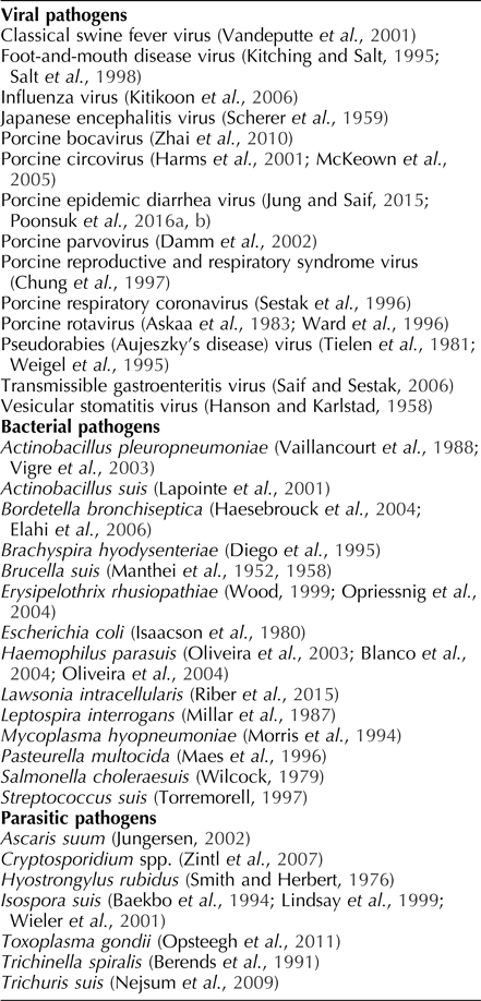
Overall, however, the relevance of passive immunity to piglet health was slow to gain recognition in swine medicine. Despite the seminal work on classical swine fever virus (De Schweinitz and Dorset, Reference De Schweinitz and Dorset1903; Dorset et al., Reference Dorset, Bolton and McBryde1904) and the creation of a substantial body of work on bacterial infections in swine (see Merchant, Reference Merchant1940), little work was done on porcine maternal immunity until after World War II (see Dunne, Reference Dunne and Dunne1958). After WWII, the role of maternal immunity in neonatal protection was demonstrated against a wide range of pathogens of swine (Table 1). As management and production practices improved over this period, the natural question was, How can breeding herd immunity be manipulated so as to optimize maternal immunity in neonates?
Swine pathogens against which maternal immunity can provide protection to piglets

There are essentially two methods for managing immunity in the host: intentional exposure to the agent or vaccination. Intentional exposure is based on choosing the time, place, and route by which the host will be exposed to material presumed to be contaminated with the agent. This approach was used in smallpox (variola virus) control as early as 1000 A.D. (Parrino and Graham, Reference Parrino and Graham2006). ‘Feedback’, infecting sows by exposing them to biological material (tissues, manure) contaminated with the pathogen of interest is a form of intentional exposure that has been widely used in swine production. A search of the literature suggested that Bay et al. (Reference Bay, Doyle and Hutchinigs1953) was the first to document this process. Using an inoculum prepared from macerated gastrointestinal tracts recovered from TGEV-infected pigs, pregnant sows were exposed orally (n = 10), intramuscularly (n = 4), subcutaneously (n = 2), or served as uninoculated controls (n = 10). The piglets (n = 200) from these sows were then orally exposed to the same inoculum at 3 to 7 days of age. Piglet survivability by the group was 50 of 70 (71%) from orally exposed sows, 14 of 27 (52%) from intramuscularly exposed sows, 0 of 23 from subcutaneously exposed sows, and 12 of 80 (15%) from uninoculated control sows. Feedback has significant drawbacks, including non-uniform and uncontrolled pathogen exposure, the unintended transmission of adventitious agents likewise present in the inoculum, and the perpetuation of virulent viruses in the herd (Chase and Lunney, Reference Chase, Lunney, Zimmerman, Karriker, Ramirez, Schwartz and Stevenson2012; Saif et al., Reference Saif, Pensaert, Sestak, Yeo, Jung, Zimmerman, Karriker, Ramirez, Schwartz and Stevenson2012; Clement et al., Reference Clement, Singrey, Lawson, Okda, Nelson, Diel, Nelson and Christopher-Hennings2016).
Based on earlier work with rinderpest (Edington, Reference Edington1897), USDA scientists developed a method to control hog cholera based on intentional exposure of swine to CSFV and simultaneous administration of anti-CSFV antibody (Dorset et al., Reference Dorset, McBryde, Niles and Dorset1908). Abuse of this method, i.e. preparation and sales of inadequate or substandard products by commercial entities, led to the Virus-Serum-Toxin Act of 1913 and the establishment of specific criteria for the production of vaccines (Skidmore, Reference Skidmore and Crawford1927; Conner, Reference Conner1952). Even so, the simultaneous method continued in use until effective killed (McBryde and Cole, Reference McBryde and Cole1936) and live-attenuated CSFV vaccines (Baker, Reference Baker1946; Koprowski et al., Reference Koprowski, James and Cox1946) became available.
The advent of vaccines, particularly those produced under the guidelines of the Virus-Serum-Toxin Act of 1913, addressed many of the pitfalls of intentional exposure by providing assurance that the inoculum was pure, safe, potent, and efficacious. Vaccination of sow herds has been used to stimulate maternal immunity and to protect piglets against various pathogens, e.g. porcine parvovirus (Antonis et al., Reference Antonis, Bruschke, Rueda, Maranga, Casal, Vela, Luuk, Belt, Weerdmeester, Carrondo and Langeveld2006), porcine circoviruses (Chae, Reference Chae2012), porcine reproductive and respiratory syndrome virus (Meng, Reference Meng2000), pseudorabies (Aujeszky's disease) virus (Mettenleiter et al., Reference Mettenleiter, Klupp, Weiland and Visser1994), Leptospira spp. (Fish and Kingscote, Reference Fish and Kingscote1973), and Mycoplasma hyopneumoniae (Ruiz et al., Reference Ruiz, Utrera and Pijoan2003). In general, vaccines that stimulate immunity in the dam will improve maternal immunity in the neonate, but there are exceptions. Bohl et al. (Reference Bohl, Gupta, Olquin and Saif1972b) showed that the route of immunization affected the antibody isotypes present in mammary secretions. In particular, sows orally inoculated or naturally infected with virulent TGEV developed higher levels of neutralizing antibodies and higher S-IgA responses in mammary secretions than sows intramuscularly injected with TGEV. In particular, piglets nursing orally inoculated or naturally infected sows received higher levels of TGEV-specific IgA and survived exposure to TGEV at a higher rate. In 1975, Reference Bohl and SaifBohl and Saif discovered that TGEV-specific IgA in milk reflected ‘productive’ intestinal replication of the virus, whereas parenteral antigenic exposure stimulated TGEV-specific IgG. As reviewed in the subsection ‘Antibodies in swine mammary secretions’, enteric infections stimulate plasmacytes to mature into plasma cells, which then migrate to the mammary parenchyma where they produce the IgA that is incorporated into mammary secretions. For these reasons and until vaccines capable of stimulating pathogen-specific S-IgA are available, feedback will be needed to provide adequate levels of maternal immunity against certain enteric pathogens in piglets, e.g. TGEV and PEDV.
These concepts are clear and well-established: Maternal immunity is transferred from sows to their piglets via mammary secretions. This process provides protection against infectious agents during the lactation period and until the immune system of the piglet is more fully developed. Maternal immunity can be manipulated by intentional exposure or vaccination to enhance herd health and productivity. The challenge to implementing and optimizing this approach in the field is that quantitative methods for measuring lactogenic immunity have not been fully developed and, in many cases, the levels of maternal immunity required to protect piglets against specific agents are not well-characterized. The issue is compounded by the fact that pre-farrow sow serum antibody levels do not directly reflect the antibody levels she will provide in colostrum or milk. Antibodies can be directly measured in colostrum and milk, but at that point it is too late to stimulate maternal immunity. Therefore, methods to measure immunity in gestating animals and to predict the level of lactogenic immunity they will provide their piglets is an area in need of investigation.