Introduction
Biases in face emotion processing have been suggested to represent an important neurobiological marker for depression (Fu et al. Reference Fu, Williams, Cleare, Brammer, Walsh, Kim, Andrew, Pich, Williams, Reed, Mitterschiffthaler, Suckling and Bullmore2004, Reference Fu, Williams, Brammer, Suckling, Kim, Cleare, Walsh, Mitterschiffthaler, Andrew, Pich and Bullmore2007; Harmer et al. Reference Harmer, O'Sullivan, Favaron, Massey-Chase, Ayres, Reinecke, Goodwin and Cowen2009) and antidepressant administration has been shown to modulate neural response to emotional faces (e.g. Del-Ben et al. Reference Del-Ben, Deakin, McKie, Delvai, Williams, Elliott, Dolan and Anderson2005; Anderson et al. Reference Anderson, Del-Ben, McKie, Richardson, Williams, Elliott and Deakin2007). Evidence also indicates that negatively biased face emotion processing is associated with vulnerability to depressive relapse in addition to occurring in the depressed state (Gur et al. Reference Gur, Erwin, Gur, Zwil, Heimberg and Kraemer1992; Persad & Polivy, Reference Persad and Polivy1993; Bradley et al. Reference Bradley, Mogg, Millar, Bonham Carter, Fergusson, Jenkins and Parr1997; Suslow et al. Reference Suslow, Junghanns and Arolt2001; Bhagwagar et al. Reference Bhagwagar, Cowen, Goodwin and Harmer2004; Surguladze et al. Reference Surguladze, Young, Senior, Brebion, Travis and Phillips2004; Joormann & Gotlib, Reference Joormann and Gotlib2006, Reference Joormann and Gotlib2007). Personality factors, especially neuroticism (a tendency to experience negative emotional states such as anxiety, anger, guilt and depressed mood), are known to play a key role in vulnerability to depression (Kendler et al. Reference Kendler, Gatz, Gardner and Pedersen2006). Recent research has suggested that rumination may be the key variable mediating the link between neuroticism and depression (Roberts et al. Reference Roberts, Gilboa and Gotlib1998; Roelofs et al. Reference Roelofs, Huibers, Peeters, Arntz and van Os2008). Rumination is the tendency to prolonged, repetitive thinking upon one's own thoughts, feelings and problems (Nolen-Hoeksema, Reference Nolen-Hoeksema1991) and is associated with both vulnerability to depression (e.g. Nolen-Hoeksema & Morrow, Reference Nolen-Hoeksema and Morrow1991) and negative cognitive biases including attentional bias towards sad faces and more negative ratings of facial affect (Joormann & Gotlib, Reference Joormann and Gotlib2006; Raes et al. Reference Raes, Hermans and Williams2006). No studies have yet addressed whether rumination influences perception of facial emotions in non-depressed individuals but high neuroticism is associated with impaired sensitivity to happy facial expression in healthy volunteers (Chan et al. Reference Chan, Goodwin and Harmer2007).
Brain imaging studies of response to facial affect in depressed patients have shown altered patterns of brain response (Sheline et al. Reference Sheline, Barch, Donnelly, Ollinger, Snyder and Mintun2001; Fu et al. Reference Fu, Williams, Cleare, Brammer, Walsh, Kim, Andrew, Pich, Williams, Reed, Mitterschiffthaler, Suckling and Bullmore2004, Reference Fu, Williams, Brammer, Suckling, Kim, Cleare, Walsh, Mitterschiffthaler, Andrew, Pich and Bullmore2007, Reference Fu, Mourao-Miranda, Costafreda, Khanna, Marquand, Williams and Brammer2008; Lawrence et al. Reference Lawrence, Williams, Surguladze, Giampietro, Brammer, Andrew, Frangou, Ecker and Phillips2004; Gotlib et al. Reference Gotlib, Sivers, Gabrieli, Whitfield-Gabrieli, Goldin, Minor and Canli2005; Keedwell et al. Reference Keedwell, Andrew, Williams, Brammer and Phillips2005; Surguladze et al. Reference Surguladze, Brammer, Keedwell, Giampietro, Young, Travis, Williams and Phillips2005; Dannlowski et al. Reference Dannlowski, Ohrmann, Bauer, Deckert, Hohoff, Kugel, Arolt, Heindel, Kersting, Baune and Suslow2008; Lee et al. Reference Lee, Seok, Lee, Cho, Yoon, Lee, Chae, Choi and Ham2008). Typically these studies report enhanced responses to negative faces in limbic regions and decreased responses in cortical regions. These changes have been suggested as neurobiological markers for depression (Fu et al. Reference Fu, Williams, Cleare, Brammer, Walsh, Kim, Andrew, Pich, Williams, Reed, Mitterschiffthaler, Suckling and Bullmore2004). However, there remain significant inconsistencies in the literature that may be attributable to methodological factors, including variation in the characteristics of the depressed patients such as severity of depression, medication and hospitalization. To avoid such confounds, Bhagwagar & Cowen (Reference Bhagwagar and Cowen2008) advocate studying unmedicated remitted patients to identify trait abnormalities and vulnerabilities, and Joormann & Gotlib (Reference Joormann and Gotlib2007) reported that behavioural biases towards sad faces persist after recovery from depression. Using positron emission tomography (PET), Neumeister et al. (Reference Neumeister, Drevets, Belfer, Luckenbaugh, Henry, Bonne, Herscovitchs, Goldman and Charney2006) found that remitted patients had exaggerated responses to sad faces in the amygdala, and blunted responses in the ventral striatum, also suggesting that a key brain response (amygdala hyperactivation to negative faces) persists after clinical recovery. By contrast, Norbury et al. (Reference Norbury, Selvaraj, Taylor, Harmer and Cowen2010), in a functional magnetic resonance imaging (fMRI) study, reported enhanced responses in remitted patients compared to controls in prefrontal and striatal regions for fearful faces, but no change in the amygdala.
One possible explanation for these inconsistencies is that personality variables play a crucial intervening role. Canli et al. (Reference Canli, Zhao, Desmond, Kang, Gross and Gabrieli2001; also Canli, Reference Canli2008) highlight the potential impact of individual differences on neural responses, particularly to emotional stimuli. Several brain imaging studies have found personality to influence neural response to emotional stimuli (e.g. Canli et al. Reference Canli, Sivers, Whitfield, Gotlib and Gabrieli2002; Haas et al. Reference Haas, Omura, Constable and Canli2007, Reference Haas, Constable and Canli2008; Chan et al. Reference Chan, Norbury, Goodwin and Harmer2009). Most of these studies have focused on neuroticism as a personality trait conferring risk for depression and reported increased limbic and/or medial prefrontal responses to negative emotions associated with high levels of neuroticism. Less is known about rumination, although Siegle et al. (Reference Siegle, Steinhauer, Thase, Stenger and Carter2002) showed that, in depressed participants, rumination correlated with prolonged amygdala responses to negative words. In another study trait rumination predicted the extent of amygdala and prefrontal activation when healthy participants deliberately increased and decreased their levels of negative affect (Ray et al. Reference Ray, Ochsner, Cooper, Robertson, Gabrieli and Gross2005). Recently, rumination was shown to mediate the relationship between neuroticism and depressive symptoms (Roelofs et al. Reference Roelofs, Huibers, Peeters, Arntz and van Os2008) and it has been suggested that a ruminative response style may be a cognitive manifestation of neuroticism.
In this study we investigated the differential neuronal responses to facial affect between remitted subjects with a past history of depression and controls, and the extent to which these interacted with the personality trait of rumination. We used a region of interest (ROI) approach that focused on specific areas associated with face emotion processing reported in the literature, including limbic, striatal and prefrontal regions. We predicted that scores on the Ruminative Response Scale (RRS; Nolen-Hoeksema, Reference Nolen-Hoeksema1991) would correlate with increased neural response to negative facial emotions and that this would result in increased neural responses in the remitted group compared to the controls.
Method
Participants
Diagnosis was established using the Structured Clinical Interview for DSM-IV (SCID-I; First et al. Reference First, Spitzer, Gibbon and Williams2002). Participants were selected from a larger cohort recruited to the New Molecules in Mood disorders (NewMood) study. Recruitment was conducted using a website, local advertisement, general practitioner (GP) surgeries and word of mouth. All participants completed a comprehensive battery of interviews, questionnaires and neuropsychological testing. Sixty-seven participants who met the following criteria were included in this next stage. All participants gave informed consent and the study was approved by the local National Health Service (NHS) research ethics committee.
All participants were right-handed, had no contraindications to MRI scanning, reported normal or corrected to normal (with contact lenses) vision and from inside the scanner were comfortably able to read back text presented on a test slide projected onto the screen. Participants had suffered no significant medical or neurological conditions and had no history of head trauma.
Control participants had no personal or immediate family history of any emotional or psychiatric problems. Remitted depressed participants met DSM-IV criteria for at least one past major depressive episode. They did not meet DSM-IV criteria for a current major depressive episode or any other current psychiatric disorders and had no history of bipolar disorder, psychosis or obsessive–compulsive disorder. At the time of scanning all participants were required to have a Montgomery–Asberg Depression Rating Scale (MADRS; Montgomery & Asberg, Reference Montgomery and Asberg1979) score <13 and a Clinical Anxiety Scale (CAS, seven items adapted from the Hamilton Anxiety Rating Scale; Hamilton, Reference Hamilton1959) score <8.
Assessments
Participants completed the 22-item RRS and the 240-item NEO Personality Inventory – Revised (NEO PI-R; McCrae & John, Reference McCrae and John1992), which includes a neuroticism subscale. Factor analyses of the RRS have identified factors termed brooding and reflection (Treynor et al. Reference Treynor, Gonzalez and Nolen-Hoeksema2003) and some researchers have suggested that whereas brooding predicts future depression, reflection may be a more positive facet of rumination (Treynor et al. Reference Treynor, Gonzalez and Nolen-Hoeksema2003; Joormann et al. Reference Joormann, Dkane and Gotlib2006). However, this has not been shown consistently (Joormann, Reference Joormann2006; Miranda & Nolen-Hoeksema, Reference Miranda and Nolen-Hoeksema2007) and recent research by Roelofs et al. (Reference Roelofs, Huibers, Peeters, Arntz and van Os2008) found that both brooding and reflection were significant positive mediators between neuroticism and depressive symptoms. On this basis, we used the total RRS score in the analysis.
Procedure
Participants completed a blocked design implicit emotional faces task. Faces were taken from a standardized series (Ekman & Friesen, Reference Ekman and Friesen1976). Morphing software was used to create faces with varying intensities of emotion from neutral (0%) to 100%; faces at 80% intensity were used.
Three emotions (happy, sad and fear) were used, and also neutral. Each block consisted of six faces, each of a different actor, three male and three female. Each face was displayed for 3000 ms, with an interstimulus interval of 500 ms, giving each block a total length of 21 s. Three blocks of each emotion were presented in a pseudo-random order, interspaced with 10 neutral blocks. For all participants, except eight of the controls, there were also three rest blocks of 21 s each where a fixation cross was displayed. Total task length was therefore either 6 min and 39 s or 7 min and 42 s.
Participants were asked to press a button to indicate the sex of each face. They were not given any instructions regarding the emotions expressed. The task was back-projected onto a screen visible to the participant using two mirrors attached to the head coil. Responses were given using a fibre-optic button box, using the right hand.
Scanning protocol
Scanning was conducted using a Philips Intera 1.5-T MRI scanner. Data were acquired with T2*-weighted, gradient echo, echo planar imaging. Repetition time (TR) was 2.1 s with an echo time (TE) of 40 ms. Each volume consisted of 29 contiguous axial slices with a slice thickness of 4.5 mm, a slice gap of 0.5 mm and an in-plane resolution 3.5 mm×3.5 mm. A T1-weighted structural scan was acquired for each participant to be used in the normalization stage of the spatial preprocessing. This image was also examined by a consultant radiologist to exclude any structural abnormality.
Analysis
fMRI data were analysed using Statistical Parametric Mapping (SPM5; The Welcome Department of Cognitive Neurology, UK). The functional images were realigned using the first image as a reference. Each participant's T1-weighted structural image was then co-registered to the resulting mean functional image from the realignment process. This co-registered structural image was segmented and the resulting grey matter image was normalized by a non-linear transformation into a standard stereotactic space. This transformation was then applied to each functional image and finally the functional images were spatially smoothed using a Gaussian kernel filter of 10 mm to allow for intersubject differences.
In this work we focused on three key subtractions, happy-neutral, sad-neutral and fear-neutral, allowing us to identify the neural substrates unique to processing each of these three emotions. The group analysis takes a region of interest (ROI) approach based on areas previously identified in the emotional face processing literature as being altered in depression. The ROIs identified were the amygdala, hippocampus, parahippocampal gyrus, globus pallidus, thalamus, caudate nucleus, putamen, insula, fusiform gyrus, prefrontal cortex (PFC) Brodmann area (BA) 9/44/45, orbitofrontal cortex BA 11/47 and cingulate gyrus BA 23/24/25/32.
For the ROI analysis we used small volume corrections (SVCs) with masks developed using the Wake Forest University (WFU) PickAtlas (Tzourio-Mazoyer et al. Reference Tzourio-Mazoyer, Landeau, Papathanassiou, Crivello, Etard, Delcroix, Mazoyer and Joliot2002; Maldjian et al. Reference Maldjian, Laurienti, Kraft and Burdette2003, 2008) in the above ROIs. A small volume corrected threshold of family-wise error (FWE) <0.05 was used for each ROI with a minimum cluster extent threshold of 5 voxels.
Group comparisons were carried out using two-sample t tests and correlational analyses were conducted by performing separate one-sample t tests with variables of interest for correlations entered as covariates.
Results
Participant characteristics
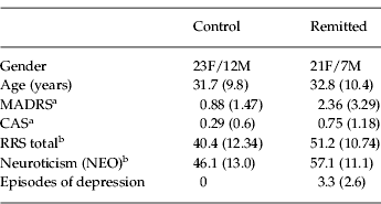
Table 1 shows that participants were well matched for age and gender. Two control participants (both female) and two remitted participants (one male, one female) were excluded from the analyses because of either excessive movement in the scanner (movement that was beyond the boundaries of 1 voxel: 3.5 mm/3°) or excessive magnetic susceptibility drop-out in their functional images. Therefore, the analysis was conducted on 35 control and 28 remitted depressed participants.
Participant characteristics
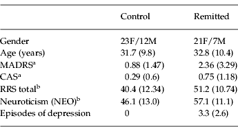
F, Female; M, male; MADRS, Montgomery–Asberg Depression Rating Scale; CAS, Clinical Anxiety Scale; RRS, Ruminative Responses Scale.
Values given as mean (standard deviation).
a Significant group difference, p<0.05.
b Significant group difference, p<0.0005.
Clinical rating scales showed that MADRS, CAS and RRS scores differed between the two groups. Although the MADRS and CAS scores were very low and well within the healthy range in both groups, the scores in the remitted group were significantly higher on both scales. Remitted participants also rated themselves as significantly more ruminative (t 61=3.66, p<0.001) and more neurotic (t 61=3.54, p<0.001) than the control participants. Three remitted participants were currently taking antidepressant medication.
Behavioural and questionnaire analysis
All participants correctly identified the sex of over 80% of the faces, indicating adequate attention to the task. There were no significant differences in accuracy between the groups or any of the emotions.
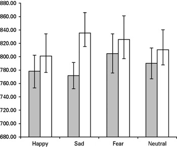
A repeated-measures ANOVA of the reaction time data showed that there was a significant main effect of emotion (F 192,3=2.772, p=0.043). Post-hoc tests showed that this was due to longer reaction times to fearful than happy faces (p=0.005). There were no other significant differences between the emotions. There was no main effect of group (F 64,1=0.715, p=0.401). There was a trend towards a significant interaction between emotion and group (F 192,3=2.602, p=0.053). This was due to a trend towards a significant group difference in reaction time to sad faces, with the remitted participants taking longer to respond than the controls (t 64=1.790, p=0.078 two-tailed). There were no significant correlations between accuracy or reaction time data and RRS scores. The reaction time data are illustrated in Fig. 1.
Reaction times to identify the gender of emotional faces, by group and emotion. Error bars ±1 standard error. □, Remitted;
 , control.

Neuroticism and RRS score correlated significantly in all participants (r=0.53, n=63, p<0.001). In remitted participants, there was a significant positive correlation between RRS score and number of past episodes of depression (r=0.58, n=28, p=0.001). However, there was no correlation between neuroticism and number of past episodes (r=0.033, n=28, p=0.87).
fMRI analysis
Group comparisons
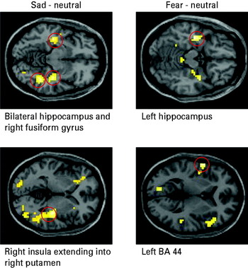
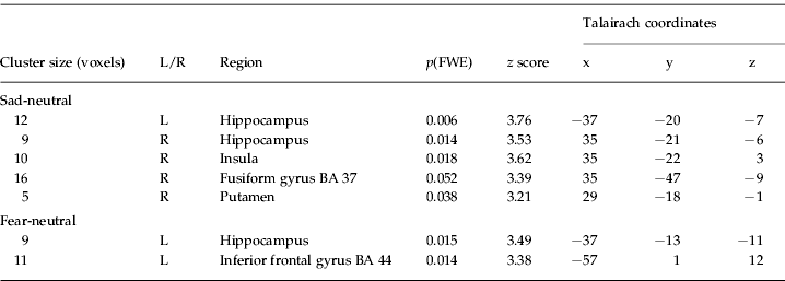
Group comparisons are detailed in Table 2 and illustrated in Fig. 2. ROIs with a significantly greater blood oxygen level-dependent (BOLD) signal in either the control or the remitted group were identified for each emotion contrast. In response to sad faces, remitted participants showed significantly reduced activity in the right insula, right fusiform gyrus, right putamen and bilateral hippocampus. In response to fearful faces remitted participants showed significantly reduced activity in the left hippocampus and left BA 44. No significant differences in any ROIs were found for happy faces. When the group comparisons were re-examined covarying for RRS score, the significant clusters remained significant.
Regions of attenuated activity in remitted depressed participants in response to negative emotions, thresholded at p<0.01 uncorrected for display purposes.

Group comparisons. All regions shown are less active in remitted participants than controls
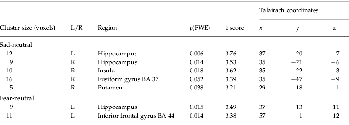
L, Left; R, right; FWE, family-wise error; BA, Brodmann area.
To investigate the possibility that the attenuated response to negative facial emotions in the remitted group was due to overactivity to neutral faces resulting in a ceiling effect, we examined the neutral compared with rest data. We did not find that remitted patients had greater responses to neutral faces compared to rest (data not shown), suggesting that this is not the explanation for our findings. We also used the main effect of neutral-rest, at p<0.05 uncorrected, as a mask in examining group differences for the individual emotions. We found that the group differences in the hippocampus and fusiform gyrus occurred in regions active in the neutral-rest main effect. This further supports the notion that, in these regions at least, the attenuated response in the remitted group cannot be explained by baseline hyperactivity.
Correlations with RRS scores
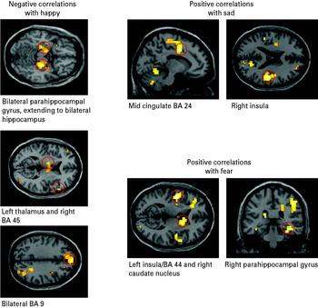
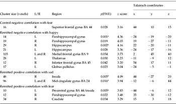
To investigate the relationship between past depression history, rumination and response to emotional faces we performed a correlation analysis separately in control and remitted participants. Table 3 and Fig. 3 give illustrations and full details and coordinates of peak activity. For control participants, a significant correlation with RRS score was found only in the right superior frontal gyrus (negative correlation with response to fear). However, for the remitted participants, a very different and more extensive pattern of correlations was found. Negative correlations with RRS score for happy faces were found in the bilateral hippocampus, parahippocampal gyrus, thalamus and medial frontal gyrus (BA 9), right inferior frontal gyrus (BA 45) and left globus pallidus. Positive correlations with RRS score were found in the insula and mid-cingulate (BA 24) for sadness and bilateral inferior frontal gyrus (BA 44, extending to the insula on the left) and right caudate nucleus and parahippocampal gyrus for fear. When the whole group was considered together, the only significant correlation found was a positive relationship with RRS in the fear-neutral contrast, in the right BA 44 [15 voxels, z=3.29, p(FWE)=0.018]. Given that three independent correlations (for happy, sad and fear) were performed for each group, a Bonferroni correction of 1/3 should strictly be applied to these correlations. Using this approach, only correlations with RRS in the remitted patients remain significant: the left parahippocampal and right hippocampal correlations with RRS for happy, the right insula and mid-cingulate correlations for sad and the left precentral/insula correlation for fear.
Regions where activity in response to emotional faces correlates with Ruminative Responses Scale (RRS) score in remitted depressed participants, thresholded at p<0.01 uncorrected for display purposes.

Regions where BOLD signal to emotion-neutral correlates with RRS score significantly using small volume correction (SVC) in the regions of interest (ROIs)

BOLD, Blood oxygen level-dependent; RRS, Ruminative Responses Scale; L, left; R, right; FWE, family-wise error; BA, Brodmann area.
a Regions survive Bonferroni correction for performing three independent correlations.
As reported above, RRS scores correlated significantly with number of previous depressive episodes in the remitted group. It was therefore possible that this past history was the crucial variable rather than RRS. To assess this possibility, we also ran correlational analyses in the remitted group with number of previous episodes as a crucial variable. A similar but less extensive pattern of positive correlations was seen in response to sad and fearful faces. In the fear-neutral contrast, significant negative correlations were also found in some ROIs, including the fusiform gyrus and thalamus, similar to the group effects. In addition, although we have argued above that rumination may mediate the effects of neuroticism on vulnerability to depression, it is also possible that our results are related directly to neuroticism. We therefore performed a correlational analysis using neuroticism score, revealing no significant response in our ROIs.
Discussion
The aim of this study was to investigate the relationship between rumination, past depression and neural response to emotional stimuli. Both past history of depression and underlying personality traits confer risk of future depressive episodes. This study allowed us to compare the effects of these different risk factors on a hypothesized neurobiological marker for depression. We have demonstrated decreased neural responses to negative facial emotions in remitted depressed participants, compared to never depressed participants. We also found that self-reported rumination correlated positively with neural response to negative facial emotions (sadness and fear) and negatively with neural response to positive facial emotion (happiness) in remitted depressed participants only.
Averaging across groups, the neural effects of emotional compared to neutral faces were broadly consistent with the literature (e.g. Breiter et al. Reference Breiter, Etcoff, Whalen, Kennedy, Rauch, Buckner, Strauss, Hyman and Rosen1996). In particular, we found that the amygdala was specifically involved in the neural response to fear but not sadness or happiness, as many (Adolphs et al. Reference Adolphs, Tranel, Damasio and Damasio1994; Morris et al. Reference Morris, Frith, Perrett, Rowland, Young, Calder and Dolan1996; Phan et al. Reference Phan, Wager, Taylor and Liberzon2002), but not all (Sander et al. Reference Sander, Grafman and Zalla2003; Fitzgerald et al. Reference Fitzgerald, Angstadt, Jelsone, Nathan and Phan2006), previous studies have suggested.
Comparing remitted patients with controls, behavioural data revealed that the remitted participants tended to be slower to identify the sex of a sad face. This could be interpreted as indicating that expressions of sadness were more distracting to the remitted participants. This is consistent with Joormann & Gotlib (Reference Joormann and Gotlib2007) who found the attention of remitted participants to be more diverted by sad faces using a dot-probe task. Behaviourally, therefore, our results suggest that attentional biases towards negative faces observed in depression persist after recovery.
However, the imaging results are not consistent with the theory that the effects observed in depression persist after remission. Remitted depressed participants demonstrated significantly attenuated neural response to sad faces in the hippocampus, insula, fusiform gyrus and putamen and to fearful faces in the hippocampus and bilateral inferior frontal gyrus. These results were not attributable to hyperactivity in the neutral faces condition.
This is the opposite pattern to that typically observed in currently depressed patients, who are characterized by limbic hyperactivation to negative faces. In currently depressed patients, Fu et al. (Reference Fu, Williams, Cleare, Brammer, Walsh, Kim, Andrew, Pich, Williams, Reed, Mitterschiffthaler, Suckling and Bullmore2004) found increased responses to sad faces in areas including the hippocampus extending to the amygdala and parahippocampal gyrus, ventral striatum (putamen/globus pallidus), insula, caudate nucleus, dorsal cingulate gyrus, and inferior parietal cortex. These responses, in similar areas to those less active in our remitted group in response to sad and fearful faces, attenuated with antidepressant treatment, which suggests that limbic and striatal hyperactivity may be state dependent and not persistent. A similar state-dependent effect on amygdala activity in response to sad faces in bipolar depression was demonstrated by Almeida et al. (Reference Almeida, Versace, Hassel, Kupfer and Phillips2010). However, previous studies have found mixed results in fully remitted unipolar depressed groups. Neumeister et al. (Reference Neumeister, Drevets, Belfer, Luckenbaugh, Henry, Bonne, Herscovitchs, Goldman and Charney2006) used PET and found blunted striatal responses to sadness in remitted subjects compared to controls, consistent with our results. However, their study reported enhanced amygdala responses to sadness and no differences for fear, results that are not consistent with our findings. By contrast, Norbury et al. (Reference Norbury, Selvaraj, Taylor, Harmer and Cowen2010) reported enhanced responses to fearful faces in prefrontal and striatal areas, which is inconsistent with our results. However, they reported no differences between remitted patients and controls in terms of amygdala response, similar to our findings. There are no obvious features of the remitted groups in these three studies that could explain the discrepancies. A possible explanation for the inconsistencies is that different tasks were used. Although Neumeister et al. (Reference Neumeister, Drevets, Belfer, Luckenbaugh, Henry, Bonne, Herscovitchs, Goldman and Charney2006) used a similar task to ours, Norbury et al. (Reference Norbury, Selvaraj, Taylor, Harmer and Cowen2010) used an emotion-matching task that explicitly required attention to emotional information and decision making on the basis of that emotion.
Another possible explanation for the different patterns of results observed in the different studies is that, in the relatively small subject groups typical of imaging studies, individual differences in personality variables have an important impact (Canli et al. Reference Canli, Zhao, Desmond, Kang, Gross and Gabrieli2001; Canli, Reference Canli2008), To address this possibility explicitly, we considered whether the neural responses to facial affect in the control and remitted groups were significantly correlated with rumination, a personality trait that is known to confer risk for depression. There were very few regions exhibiting a correlation between RRS score and response to facial emotion in the control group, suggesting that rumination is not a personality factor associated with differential emotional processing in normal subjects. This can be contrasted with studies suggesting that the trait of neuroticism is associated with abnormal face processing in never-depressed volunteers. However, it should be noted that these studies specifically recruited groups of subjects with high and low neuroticism scores rather than using a correlational approach in a representative population sample (Chan et al. Reference Chan, Norbury, Goodwin and Harmer2009). Consistent with our RRS findings, we did not find that neuroticism influenced emotional processing in this group of normal controls. Our findings therefore do not support the idea that rumination in normal subjects, without vulnerability to depression, is associated with negative emotional biases in a face emotion task. However, in subjects with a demonstrated vulnerability to depression, the degree of rumination is associated with neuronal responses to emotional processing in several ROIs. Within the remitted group, more ruminative participants had enhanced BOLD responses to sad and fearful faces, and attenuated responses to happy faces. We suggest that this effect of rumination is an interaction with a past history of depression and not simply a reflection of needing a particular ‘threshold’ of rumination, given the lack of significant effects if the whole group is considered together. In addition, the range of rumination scores was similar in both groups even though the means were significantly different.
Paradoxically, therefore, it would seem that past depression and rumination have opposite effects on response to emotional faces. Those areas that were less active in remitted depressives than controls in response to negative expressions were also areas in which responses to these expressions were positively correlated with ruminative tendency in the remitted patients. Although the group comparisons showed the opposite pattern of brain response to negative faces to that observed in previous studies of currently depressed subjects, the correlations with RRS score in remitted participants were in the direction expected based on cognitive biases described in depression previously (e.g. Fu et al. Reference Fu, Williams, Cleare, Brammer, Walsh, Kim, Andrew, Pich, Williams, Reed, Mitterschiffthaler, Suckling and Bullmore2004, Reference Fu, Williams, Brammer, Suckling, Kim, Cleare, Walsh, Mitterschiffthaler, Andrew, Pich and Bullmore2007; Surguladze et al. Reference Surguladze, Brammer, Keedwell, Giampietro, Young, Travis, Williams and Phillips2005).
The regions in which activity correlated with rumination have been implicated in mechanisms of memory and emotion and overlap with areas showing state-dependent changes in PET studies of resting blood flow and metabolism and fMRI studies of treatment response in depression (Mayberg et al. Reference Mayberg, Liotti, Brannan, McGinnis, Mahurin, Jerabek, Silva, Tekell, Martin, Lancaster and Fox1999; Fu et al. Reference Fu, Williams, Cleare, Brammer, Walsh, Kim, Andrew, Pich, Williams, Reed, Mitterschiffthaler, Suckling and Bullmore2004, Reference Fu, Williams, Brammer, Suckling, Kim, Cleare, Walsh, Mitterschiffthaler, Andrew, Pich and Bullmore2007; Surguladze et al. Reference Surguladze, Brammer, Keedwell, Giampietro, Young, Travis, Williams and Phillips2005). Along with the insula, activations in the hippocampus showed the most consistent and focal group differences and correlations with rumination. This is in keeping with the idea that the hippocampus may play a key role in rumination and that the ability to limit this process underlies resilience and recovery from depression (Deakin, Reference Deakin1991, Reference Deakin1993; Graeff et al. Reference Graeff, Guimaraes, DeAndrade and Deakin1996). Much preclinical evidence implicates hippocampal 5-hydroxytryptamine (5-HT)1A receptors as a key site of action of selective serotonin reuptake inhibitors (Drevets et al. Reference Drevets, Frank, Price, Kupfer, Holt, Greer, Huang, Gautier and Mathis1999). In humans, Mayberg et al. (Reference Mayberg, Brannan, Tekell, Silva, Mahurin, McGinnis and Jerabek2000) showed that recovery on fluoxetine was associated with focal bilateral suppression of hippocampal metabolism. Monk et al. (Reference Monk, Klein, Telzer, Schroth, Mannuzza, Moulton, Guardino, Masten, Clure-Tone, Fromm, Blair, Pine and Ernst2008) found that amygdala and nucleus accumbens overactivity to fear and attenuation to happiness were present in children with a family history of, and therefore a high risk of developing, depression. The clusters of neural response to happy faces, which we found to correlate negatively with remitted RRS scores in the hippocampus, also extended into the amygdala.
Our proposal to resolve the apparent paradox that rumination and past depression have opposite influences on neural responses to emotion is that the group differences demonstrate a mechanism for maintaining remission, whereas correlations with rumination indicate increased vulnerability to future depressive episodes. In a PET study, Liotti et al. (Reference Liotti, Mayberg, McGinnis, Brannan and Jerabek2002) found that increases in regional cerebral blood flow (rCBF) in subgenual cingulate during experimentally induced sadness were least in remitted depressives compared with controls and currently depressed patients. They hypothesized that remission from depression involves sustained suppression of activity in this area, even in the face of transient sadness. Although we did not observe any group differences in BA 25, we suggest that, to successfully sustain remission, other areas that are important to emotion processing and in which we saw reduced BOLD signals in remitted participants must also be suppressed in response to negative emotional stimuli.
Rumination in the remitted group could be an intervening trait that reduces the efficiency of this suppression of neural response to emotional stimuli. Although, overall, the remitted group showed reduced neuronal response to negative faces, correlational analysis suggests that this was most marked in the least ruminative subjects and least marked in the most ruminative. The more ruminative subjects may be most vulnerable to relapse. Rumination has been associated with the likelihood of future depressive symptoms developing (Nolen-Hoeksema & Morrow, Reference Nolen-Hoeksema and Morrow1991; Just & Alloy, Reference Just and Alloy1997; Spasojevic & Alloy, Reference Spasojevic and Alloy2001). In our own participants, we found a significant positive correlation between RRS score and the number of previous episodes of depression. Behaviourally, Bouhuys et al. (Reference Bouhuys, Geerts and Gordijn1999) showed that greater perception of negative emotion in ambiguous faces in remission predicts relapse. We hypothesize that rumination reduces the tendency of remitted patients to suppress instinctive responses to negative emotional material and thus renders them more vulnerable to relapse. Thus, remission from depression may be maintained by sustained suppression of neural responses to emotional material that are exaggerated in depression. By contrast, vulnerability to relapse associated with rumination is related to a relative failure of this suppression.
To further support our assertion that differences seen here between the control and remitted participants represent suppression of neural responses to emotional material that are exaggerated in depression, the inclusion of a currently depressed group would be beneficial. The assertions made regarding the link between rumination and vulnerability to depression are based on correlational data. A longitudinal design, assessing whether relapse was associated with reduced suppression of neural responses to negative emotional material, would lend significant strength to this hypothesis. Explicit group comparisons of high and low ruminators (both control and remitted) would also be valuable; the distribution of RRS scores did not allow such a comparison in the present study.
Despite these limitations, our study suggests that a crucial interplay between past depression history and a personality trait conferring depression vulnerability determines neuronal responses to emotional faces. This generates testable hypotheses for future research and may be of clinical importance in predicting vulnerability to relapse following remission of symptoms.
Acknowledgements
This study was funded by the NewMood EU Integrated Programme LSHM-CT-2004-503474. E. T. was supported by a Medical Research Council (MRC) Studentship. For all their assistance, we thank the staff of the Wellcome Trust Clinical Research Facility, Manchester, where this research was carried out. We also thank Dr D. Chase for her useful comments on earlier drafts of this manuscript, K. Lloyd-Williams, Dr K. Lythe, Dr I. Koychev and C. Holliday for their hard work and assistance with recruitment and data acquisition, and the participants for taking part in the research.
Declaration of Interest
None.