Introduction
The genus of lichen-forming fungi Circinaria Link, comprising c. 40 species, is the second largest in the family Megasporaceae, after Aspicilia A. Massal. (Wijayawardene et al. Reference Wijayawardene, Hyde, Al-Ani, Tedersoo, Haelewaters, Rajeshkumar, Zhao, Aptroot, Leontyev and Saxena2020; Paukov et al. Reference Paukov, Davydov, Nordin, Owe-Larsson, Sohrabi and Teptina2021).The genus shows a wide variety of growth forms, including fruticose, crustose and umbilicate species. Whereas crustose species of the genus have long been included within the genus Aspicilia A. Massal., fruticose species, usually vagrant, have been included in the genera Agrestia J. W. Thomson, Sphaerothallia Eversm. or Chlorangium Link (Sohrabi et al. Reference Sohrabi, Ahti and Litterski2013a). Circinaria species have received great attention in recent years, especially the vagrant taxa (Nordin et al. Reference Nordin, Savić and Tibell2010; Sohrabi et al. Reference Sohrabi, Ahti and Litterski2013a; McCune & Di Meglio Reference McCune and Di2021), and several new species have been described from Iran (Sohrabi et al. Reference Sohrabi, Ahti and Litterski2013a), China (Ismayil et al. Reference Ismayil, Wen and Abbas2015, Reference Ismayil, Abbas and Guo2019), Greece (Sohrabi et al. Reference Sohrabi, Leavitt, Obermayer and Mayrhofer2023), Italy (Nascimbene et al. Reference Nascimbene, Nimis, Klüßendorf and Thüs2023) and the USA (Owe-Larsson et al. Reference Owe-Larsson, Nordin, Tibell and Sohrabi2011; McCune & Di Meglio Reference McCune and Di2021). Crustose species of the genus are primarily epilithic, and have grey to olive-brown thalli, with or without a grey prothallus. Recent molecular studies have greatly improved our understanding of the infrageneric relationships and taxonomic value of the morphological characters of the genus (Nordin et al. Reference Nordin, Savić and Tibell2010; Sohrabi et al. Reference Sohrabi, Stenroos, Myllys, Søchting, Ahti and Hyvönen2013a; McCune & di Meglio Reference McCune and Di2021).
Persepolis, capital of the Achaemenid Empire (c. 550–330 BC), located in south-western Iran and surrounded by the southern Zagros Mountains of the Iranian plateau, is one of 27 UNESCO World Heritage Sites in Iran (https://whc.unesco.org/en/list/). The monumental stone of its construction, including those displaying bas-reliefs of incalculable artistic and archaeological value (Mousavi Reference Mousavi2012), is colonized by a large number of lichen-forming fungi, including several species of the genus Circinaria. Species of this genus also occur on stone surfaces in other UNESCO World Heritage Sites in Iran such as Pasargadae (Sohrabi et al. Reference Sohrabi, Favero-Longo, Pérez-Ortega, Ascaso, Haghighat, Talebian, Fadaei and de los Ríos2017), which is part of the Sassanid Archaeological Landscape in Fars Province, and Bistone in Kermanshah and Sousha in Khuzestan Province.
During a recent lichenological survey of Persepolis, an apparently new species of Circinaria colonizing the bas-reliefs and stone walls was found. In this study, we describe the new taxon, Circinaria persepolitana, based on morphological observations and molecular analyses of representative specimens. Given the high abundance of C. persepolitana at the historic site of Persepolis and the need to avoid biodeterioration of this emblematic site for its conservation, we also analyzed the interactions established by the lichen thalli of this species with the lithic substratum and their capacity to deteriorate the colonized stone surface.
During our research, we also conducted a phylogenetic analysis of several species belonging to the Circinaria genus, including C. persepolitana, C. mansourii (Sohrabi) Sohrabi and Aspicilia reptans (Looman) Wetmore. Our aim was to re-evaluate and determine the status of these species. Our analysis revealed that the species we were investigating is closely related to C. mansourii and A. reptans. Based on our findings, we make the new combination Circinaria reptans and propose a correction to its nomenclature.
Material and Methods
Sampling sites
We collected two types of samples from two locations during our research. One was directly obtained from bare rock suitable as a lichen substratum while the other included rock colonized by lichens. The locations were at Kuh-e Rahmat but c. 5 km apart. The first location was at the Naghsh-e-Rajab limestone outcrop (29.9661°N, 52.8867°E) and the second was within the historic site or palace complex (29.9358°N, 52.8919°E).
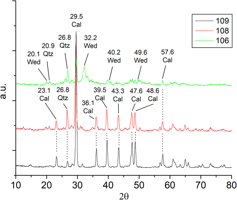
To compare the mineral composition of rocks, we collected small stone pieces (1 cm2) from the historic area with permission from the general office of Persepolis. These were analyzed by X-ray diffraction (XRD). We collected samples identified as no. 109 (where the holotype of the new species was also recorded) from the Naghsh-e-Rajab limestone outcrop, located outside the historic site's limits. Additionally, we gathered substratum samples (no. 106) in powder form from the Hall of a Hundred Columns and sample no. 108, comprising small stone pieces measuring c. 1 cm2, from the Unfinished Gate.
To compare the DNA of lichen samples found at the historic site with the type specimens (collected at the Naghsh-e-Rajab limestone outcrops), we sampled small lichen fragments, c. 1 mm2 in size, from significant areas of the Persepolis site, including the Apadana Palace, Hadish Palace, Tachara Palace, Hall of a Hundred Columns, and the Unfinished Gate.
X-ray diffraction (XRD)
Samples 109, 108 and 106 were ground to powder using an agate mortar and then analyzed using an x-ray diffractometer (Philips PW 1730, The Netherlands). The analysis involved Cu K-alpha radiation and a scan range of 3–35°26, with a scan step of 0–02°26 and 1 s per step. Mineral identification was aided by connecting a graphical terminal to the JCPDS—International Center for Diffraction Data ‘Powder Diffraction File’ (PDF) database.
Morphology and anatomy
The present study is based mainly on material collected by the first author (MS) and managed according to Obermayer (Reference Obermayer, Kranner, Beckett and Varma2002); specimens were later deposited in the ICH herbarium at the Iranian Research Organization for Science and Technology (IROST) in Tehran. Field photographs were taken with a Canon PowerShot SX60 HS camera.
Morphological observations of specimens were performed with a Wild M7 stereomicroscope (Wild Heerbrugg, Switzerland). Thallus measurements were made at ×25 magnification and rounded to the nearest 0.1 mm. Anatomical features (Ryan et al. Reference Ryan, Bungartz, Nash, Nash, Ryan, Gries and Bungartz2002; Owe-Larsson et al. Reference Owe-Larsson, Nordin, Tibell, Nash, Gries and Bungartz2007) were measured in hand-cut sections and observed on material mounted in water under a PZO DIC microscope (Warsaw, Poland). Measurements were taken at ×1000 magnification using an oil immersion lens and a Wild vernier micrometer (scale of 1 μm), to an accuracy of 0.5 μm for microscopic structures and 5 μm for the anatomical layers of thalli, according to the formula (min–) x̄ – SD – x̄ + SD (–max) (n), where x̄ is the mean, SD is the standard deviation and n is the number of measurements.
Chemical analyses
Spot tests, UV and TLC
Spot tests with 10% potassium hydroxide (K), 5.25% sodium hypochlorite (C) and Lugol's iodine (I) following Nylander (Reference Nylander1866), and an ethanol solution of 5% para-phenylenediamine (PD) following Orange et al. (Reference Orange, James and White2010) were conducted to examine the chemical characteristics of the cortex and medulla. Reagent C was also applied after pretreatment with K on the same thallus fragment (KC). Long-wave ultraviolet light (350 nm) was used to observe lichen thalli. Thin-layer chromatography was performed following Orange et al. (Reference Orange, James and White2010), using solvent systems A, B and C.
Liquid chromatography and tandem mass spectrometry (LC-MS/MS) analysis
The metabolic profile was analyzed following the protocol described by Norouzi et al. (Reference Norouzi, Azizi, Gholami, Sohrabi and Boustie2020). In brief, the compounds were separated using a Waters Alliance e2695 separation module (Milford, MA, USA) equipped with an Atlantis T3 C18 column (2.1 mm × 100 mm, 3 μm; Milford, MA, USA). The temperature of the column was set at 30 ℃. The extract was dissolved in methanol, followed by filtration (PTFE membrane filters, 0.45 μm; Simplepure, China). A 20-min gradient elution programme was used on 10 μl of the sample with the following conditions: 95% A (water + 0.1% formic acid, v/v), gradually reduced to 5% A within 20 min and followed by 95% B (acetonitrile) for 5 min. The eluent flow rate was set at 0.25 ml min−1.
The MS/MS analysis was conducted on a Waters Micromass Quattro micro API mass spectrometer (Milford, MA, USA) in negative ionization mode. The following conditions were adopted: source temperature 120 ℃, dissociation temperature 300 ℃, capillary voltage 3.5 kV, cone voltage 30 V, collision energy 30 eV. N2 was used for both nebulizing and gas drying. Data acquisition, processing and visualization were performed using MassLynx v. 4.1 and MZmine v. 2.37. Finally, the MS/MS spectra database of lichen substances (LDB), provided by Olivier-Jimenez et al. (Reference Olivier-Jimenez, Chollet-Krugler, Rondeau, Beniddir, Ferron, Delhaye, Allard, Wolfender, Sipman and Lücking2019) and hosted by the GNPS public spectral library (Wang et al. Reference Wang, Carver, Phelan, Sanchez, Garg, Peng, Nguyen, Watrous, Kapono and Luzzatto-Knaan2016), was used to confirm the identity of most of the detected compounds.
DNA extraction, amplification and sequencing
DNA extraction was performed on a total of seven specimens of the new putative taxon, including two recently collected herbarium specimens (of which Sohrabi 32109 was chosen as the holotype), as well as five thalli collected directly from surfaces of historical monuments. In addition, we also carried out DNA extraction of three specimens of Circinaria mansourii. Areoles were excised from thalli with a razor blade, placed in microcentrifuge tubes and stored at −80 °C for 1 h after which they were pulverized using a Qiagen TissueLyser II and glass beads. DNA was extracted using E.Z.N.A.® Forensic DNA Kit (Omega Bio-Tek), following the manufacturer's instructions. The fungal nrITS region was amplified using the primers ITS1-F (White et al. Reference White, Bruns, Lee, Taylor, Innis, Gelfand, Sninsky and White1990) and ITS4_KYO2 (Toju et al. Reference Toju, Tanabe, Yamamoto and Sato2012). Amplification reactions were prepared for a 15 μl final volume containing 7.5 μl of MyTaq™ Red Mix (Bioline) which includes the MyTaq™ DNA polymerase, 0.5 μl of each of the primers at 10 μM, 5.5 μl of H2O, and 1 μl of template. PCR conditions for the amplification of the nrITS were as follows: 5 min at 95 °C; 10 cycles of 30 s at 95 °C, 30 s at 66 °C, 1 min 30 s at 72 °C; 34 cycles of 30 s at 95 °C, 30 s at 56 °C, 1 min 30 s at 72 °C; 10 min at 72 °C. Seven sequences of the nrITS region of the putative new species were obtained.
Alignment and phylogenetic analysis
For this study, a total of 10 new ITS sequences were generated and 36 additional sequences were downloaded from GenBank. A summary, including material and locality information, names of collectors and where specimens were deposited, is given in Table 1. Species nomenclature follows Nordin et al. (Reference Nordin, Savić and Tibell2010), Owe-Larsson et al. (Reference Owe-Larsson, Nordin, Tibell and Sohrabi2011), Sohrabi et al. (Reference Sohrabi, Stenroos, Myllys, Søchting, Ahti and Hyvönen2013a, Reference Sohrabi, Leavitt, Rico, Halıcı, Shrestha and Stenroosb) and Paukov et al. (Reference Paukov, Nordin, Tibell, Frolov and Vondrák2017).
Voucher information and GenBank Accession numbers for ITS sequences of Circinaria specimens used in this study. New sequences are indicated in bold.

ITS sequences were aligned with BioEdit v. 7.1.3.0 (Hall Reference Hall1999) and MAFFT v. 7 (https://mafft.cbrc.jp/alignment/server/) using the progressive G-INS-1 method (Katoh et al. Reference Katoh, Rozewicki and Yamada2019) and later corrected by hand. The optimal substitution model was inferred using the ModelTest-NG algorithm (Darriba et al. Reference Darriba, Posada, Kozlov, Stamatakis, Morel and Flouri2020) implemented in raxmlGUI v. 2.0 software (Kozlov et al. Reference Kozlov, Darriba, Flouri, Morel and Stamatakis2019; Edler et al. Reference Edler, Klein, Antonelli and Silvestro2021); the general time reversible model (GTR + G) was selected. Bayesian inference with the Markov chain Monte Carlo (BMCMC) method (Larget & Simon Reference Larget and Simon1999) was performed using BEAST v. 2.6.6 (Bouckaert et al. Reference Bouckaert, Vaughan, Barido-Sottani, Duchêne, Fourment, Gavryushkina, Heled, Jones, Kühnert and de Maio2019). Two independent runs of BEAST were made with a chain length of 107, and every 1000th generation was recorded. Tree files from two independent runs were combined in LogCombiner v. 2.6.6, a part of the BEAST 2 package (http://beast2.cs.auckland.ac.nz/). A maximum clade credibility tree with mean node heights was inferred with a 50% burn-in fraction and a posterior probability of 0.5. Tree files were visualized with FigTree v. 1.4.2 (http://tree.bio.ed.ac.uk/software/figtree/). The most likely tree and 1000 rapid bootstrap pseudoreplicates were calculated using RAxML v. 8.0.26 (Stamatakis Reference Stamatakis2014) with raxmlGUI v. 2.0 software (Edler et al. Reference Edler, Klein, Antonelli and Silvestro2021), applying the GTRGAMMA model of substitution. Since both phylogenetic inference methods returned identical tree topologies, the final topology shown in Fig. 1 corresponds to that recovered using maximum likelihood (ML) inference. Bootstrap support values and BMCMC posterior probability were noted on the best-scoring tree. Aspicilia goettweigensis (Zahlbr.) Hue, a representative of the large monophyletic genus Aspicilia s. str. after Nordin et al. (Reference Nordin, Savić and Tibell2010) within the family Megasporaceae, was selected as outgroup for phylogenetic reconstruction.
Maximum likelihood (ML) phylogeny of selected Circinaria ITS sequences. The reliability of each branch was tested by ML and Bayesian methods. Values at tree nodes indicate ML bootstrap percentages (left) and Bayesian inference with the Markov chain Monte Carlo (BMCMC) posterior probabilities (right). Thicker branches indicate ML bootstrap values ≥ 70% or BMCMC posterior probability ≥ 0.95. Accession numbers and voucher information are provided in Table 1. Aspicilia goettweigensis was used as an outgroup. Branch lengths represent the estimated number of substitutions per site assuming the respective models of substitution. The type specimen is marked with an asterisk.

Scanning electron microscopy study
Rock fragments (sample no. 106) colonized by Circinaria persepolitana were prepared following the procedure developed by Wierzchos & Ascaso (Reference Wierzchos and Ascaso1994) for the observation of the lichen-lithic substratum interface by scanning electron microscopy using the back-scattered electron mode (SEM-BSE). Lichen-colonized rock fragments were fixed in glutaraldehyde and osmium tetroxide solutions, dehydrated in a graded ethanol series, and embedded in LR-White resin. Blocks of resin-embedded rock samples were finely polished, carbon coated, and observed using an FEI INSPECT scanning electron microscope (Oregon, USA) at the Museo Nacional de Ciencias Naturales (CSIC). Microprobe analyses were performed using an Oxford Instruments INCA X-Ray Energy Dispersive Spectrometer (EDS) microanalytical system (Oxford, UK) during SEM observation. To obtain high-resolution images of crystal morphology, small fragments of rocks harbouring the lichen thallus were coated with gold (10 nm) and analyzed with a TESCAN Mira3 FESEM (Brno, Czechia). The microscope was operated at an acceleration voltage of 0.2 to 1 kV using the Beam Deceleration Mode.
Results and Discussion
The topology of the phylogenetic tree recovered by ML analysis is shown in Fig. 1. The supported relationships among the species agree with previously published phylogenetic reconstructions of the group (Owe-Larsson et al. Reference Owe-Larsson, Nordin, Tibell and Sohrabi2011; Sohrabi et al. Reference Sohrabi, Stenroos, Myllys, Søchting, Ahti and Hyvönen2013a, Reference Sohrabi, Leavitt, Rico, Halıcı, Shrestha and Stenroosb). The Bayesian tree returned an identical topology to that obtained by maximum likelihood using RAxML.
The recovered phylogram based on the analyses of nrITS sequences showed low support for most of the relationships within the genus Circinaria (Fig. 1). A well-supported basal clade (BS = 95, PP = 1) included the species Circinaria contorta (Hoffm.) A. Nordin et al., C. hoffmanniana (S. Ekman & Fröberg ex R. Sant.) A. Nordin and C. serenensis (Cl. Roux & M. Bertrand) A. Nordin. The rest of the relationships among species were not supported, with the exception of the clade containing C. cerebroides (Mereschk.) Sohrabi, C. rostamii Sohrabi and C. lacunosa (Mereschk.) Sohrabi (95/1), the clade containing C. hispida (Mereschk.) A. Nordin et al. and C. rogeri (Sohrabi) Sohrabi (85/1), and the clade containing the new species, C. ochracea Thüs & Nascimbene, C. mansourii and Aspicilia reptans (Looman) Wetmore (100/1). The specimens of C. persepolitana formed a well-supported clade (95/1) sister to the clade formed by C. ochracea, C. mansourii and Aspicilia reptans (99/1). A high level of molecular diversity was found in C. persepolitana compared to other species in the group (Fig. 1).
Taxonomy
Circinaria persepolitana Sohrabi & Pérez-Ort. sp. nov.
MycoBank No.: MB 847202
A species of Circinaria with an areolate crustose thallus. Areoles rimose or squamuliform to verrucose, or bullate-areolate in the centre, hollow and cracking into popcorn-like tufts, and angular or elongated in the margins, with diverging and branching prothallus-like extensions in the grey rim. On calcareous rocks (limestone). The only known lichen substance found in this species is α-alectoronic acid. By its bullate hollow areoles, the new species resembles Aspicilia goettweigensis. The latter species differs from Circinaria persepolitana by containing the stictic acid complex.
Type: Iran, Fars, Marvdasht, Naghsh-e-Rajab limestone outcrops, northern slope, in the quarry of stone monuments of Persepolis, 1635 m, on grey limestone, GPS: 29.9661°N, 52.8867°E, 15 June 2019, M. Sohrabi 32109 (ICH—holotype; IRAN, hb. M. Sohrabi—isotypes). GenBank Accession: OR365438.
Circinaria persepolitana (A–F) and its distribution in south-western Iran (G & H). A, projections of bullate-areolate surfaces in the central part of the thallus. B, projections of angular to irregular areoles. C & D, papillate areoles, somewhat verrucose, hollow and cracking into popcorn-like tufts, more or less squamuliform areolate. E, cross-section of the green photobiont algal layer, and the white and grey mycobiont layer. F, chlorococcoid algal cell (Trebouxia-type). G, location of the Persepolis UNESCO World Heritage Site and distribution of C. persepolitana in the area, with the holotype locality indicated with a triangle. H, distribution map of C. persepolitana in the historic site of Persepolis. Scales: A–D = 1 mm; E = 200 μm; F = 10 μm. In colour online.

Circinaria persepolitana on historic rock surfaces. A, diffuse thallus margin with some areoles peeled off. B, verrucose thallus. C, hollow squamuliform areolate thallus, with visible white medulla. D & E, thallus with rimose-areolate to bullate-areolate, dispersed or rarely overlapping areoles, verrucose to squamuliform (slightly vertically elevated areoles). F, the outer part of the thallus with grey prothallus-like extensions. Scales = 1 cm. In colour online.

Thallus crustose, (1–)2–10(–12) cm diam., (0.2–)0.5–1(–3) mm thick. Areoles rimose or squamuliform (slightly vertically elevated areoles) to verrucose or bullate-areolate, angular to irregular, flat to ±convex, sometimes rounded, (0.2–)0.5–1.5(–2.5) mm diam., contiguous or dispersed, rarely overlapping. Central areoles somewhat papillate, in older parts verrucose, hollow and cracking into popcorn-like tufts, more or less squamuliform areolate. Thallus edge usually formed by elongated areoles different from the central areoles. Prothallus usually distinct, branching, sparsely developed along the thallus edge, more or less fimbriate, forming a white-grey margin, 0.1–0.4(–1) mm wide. Surface usually greyish olive-brown, sometimes partly olive-brown, ochre, grey-brown, white-grey, dull, slightly pruinose. Pseudocyphellae not seen. Epinecral upper layer hyaline, consisting of dead cells, without crystals, (5–)7–9(–11) μm (n = 15). Cortex uneven, paraplectenchymatous, (10–)30–45(–60) μm thick (n = 20), uppermost part ±brown or rarely olive-brown, with cells (5–)7–9(–12) μm diam. (n = 15), (5–)10–22(–40) μm thick (n = 18), covered with crystals. Medulla white, containing crystals of calcium oxalate c. 1–3 μm diam., I−; algal layer discontinuous. Photobiont chlorococcoid (Trebouxia-type), cells spherical to subspherical, (10–)15–25(–30) μm diam. (n = 30).
Apothecia and pycnidia not observed.
Chemistry
Spot tests: cortex and medulla I–, K−, P−, C−. TLC: no lichen substances detected. LC-MS: α-alectoronic acid, [M-H]- m/zb: 543.99, chemical formula: C28H32O9 [M-H+CH3OH], calculated mass: 543.223, Rt (min)a: 17.17, MS2 ions, m/z (relative intensity)c: 528, 516, 513, 497, 487, 471, 358, 256, 146. In addition, seven unidentified lichen substances were detected; however, further investigation with more material is needed.
Etymology
The species is named after Persepolis, capital of the Persian Achaemenid Empire from the reign of Darius I (the Great, r. 522–486 BC) until its destruction in 330 BC and a UNESCO World Heritage Site in Iran, where the type collection was obtained.
Ecology, habitat and distribution
This species typically grows on large limestone boulders in various locations on Kuh-e Rahmat (Mountain of Mercy) in Fars Province, south-western Iran, where several archaeological sites can be found. It is also distributed in Naghsh-e Rajab, an archaeological site located just west of the historic city of Istakhr and c. 5 km north of the Persepolis site. The species can be found in the northern sector of the Persepolis site, including areas such as the walls of the Gate of All Nations (Xerxes Gate), Apadana Palace, Throne Hall (Hall of a Hundred Columns), Treasury, Council Hall, Palace of Artaxerxes III, Imperial Stables, Tripylon (Three-gated Hall), Tachara (Palace of Darius), Hadish (Palace of Xerxes), and the Palace of Artaxerxes I. Additionally, it can be found on calcareous rocks, both on vertical and horizontal surfaces exposed to the sun, in mountainous regions near the UNESCO World Heritage Site of Persepolis (Fig. 2 G & H). Circinaria persepolitana is often a predominant species (e.g. in the type locality) in suitable habitats, usually with co-dominant species such as Acarospora cervina A. Massal., A. laqueata Stizenb., Calogaya biatorina (A. Massal.) Arup et al., C. decipiens (Arnold) Arup et al., Circinaria scabridula (H. Magn.) Sohrabi, Lobothallia radiosa (Hoffm.) Hafellner s. lat. and Protoparmeliopsis usbekica (Poelt) S. Y. Kondr.
Remarks
Circinaria persepolitana is a well-delimited species easily distinguished from other saxicolous species of Circinaria by its convex to squamuliform areolate thallus, thin angular to elongated marginal areoles, the grey prothallus-like extensions, branched at the margin, fimbriate or forming a grey rim, and by its exceptional lichen chemistry (α-alectoronic acid by LC-MS analysis) and negative reactions to K, C, I, P and UV in both medulla and cortex. Circinaria persepolitana is also characterized by areoles that become hollow and crack into popcorn-like tufts of tissue or upright squamules with the white exposed medulla similar to Aspicilia goettweigensis. The species differs from Circinaria contorta, a common species of the genus in the region, by having a different thallus morphology. Circinaria contorta has generally scattered, rounded, ultimately ±convex to squamuliform areoles and contains aspicilin. The thalli of Circinaria contorta are usually fertile with crateriform apothecia and pruinose discs.
In the phylogenetic tree, Circinaria persepolitana is subdivided into two supported subclades. Specimens from both subclades differ by 13–18 nucleotides in nrITS, but inhabit the same ecotopes and there are no obvious morphological differences in specimens forming both subclades. We therefore interpret them as two intraspecific lineages within C. persepolitana. However, we do not exclude the possibility that future studies may show the coexistence of two cryptic species.
Circinaria persepolitana is phylogenetically close to two terricolous species that grow in arid regions of Eurasia and North America, Aspicilia reptans and C. mansourii. Both species differ from C. persepolitana by their centrally warted and peripherally lobed thalli, which grow on soil and plant detritus, the presence of rhizomorphs on the lower surface of the thalli, and by containing aspicilin as a secondary metabolite. Circinaria mansourii, like C. persepolitana, has long prothallus-like extensions, which are attached to the tips of lobes. Both A. reptans and C. mansourii lack a saxicolous habit and have convex to squamuliform areoles and α-alectoronic acid. The recently described Circinaria ochracea (Nascimbene et al. Reference Nascimbene, Nimis, Klüßendorf and Thüs2023) belongs to the same branch of the phylogenetic tree as C. persepolitana, C. mansourii and Aspicilia reptans but differs ecologically by growing in a water-splash zone and it has a uniform crustose thallus with flat ochraceous areoles. The new species is also similar to Circinaria crespiana (V. J. Rico) Sohrabi & V. J. Rico, a Mediterranean species known from Spain and Italy, grows among mosses, on siliceous rocks and on soil. It has large squamulose thalli attached to the substratum with rhizomorph-like extensions (Sanders & Rico Reference Sanders and Rico1992), and large, appressed apothecia up to 3 mm in diameter.
Note
Due to restrictions in collecting any kind of material from UNESCO World Heritage Sites, the holotype specimen and specimen Sohrabi 32099 were collected from calcareous rocks (limestone) outside Persepolis, from Naghsh-e-Rajab, c. 5 km north of the historic site. X-ray diffraction (XRD) analyses conducted on samples 106, 108 and 109 (Fig. 4), revealed that the rock composition in the locality where the type specimen was collected (Naghsh-e Rajab, sample 109) and that of the historical substratum (Persepolis, sample 108) exhibited a remarkable degree of similarity, displaying an analogous pattern of calcite and subordinate quartz and clay minerals.
X-ray diffraction analysis of rock samples indicating the substratum of Circinaria persepolitana in three distinct locations. Sample 106 corresponds to the lichen-lithic substratum (with thallus of C. persepolitana) found in the Hall of a Hundred Columns. Sample 108 corresponds to the substratum without a lichen thallus, sampled from the Unfinished Gate. Sample 109 corresponds to the substratum of the holotype specimen (without lichen thallus) located in Naghsh-e-Rajab limestone outcrops. Qtz = quartz peak; Wed = weddellite peak (a mineral form of calcium oxalate); Cal = calcite peak; a.u. = arbitrary scale; 2θ (theta angle) = the angle of diffraction, measured in degrees. In colour online.

Additional specimens examined
Circinaria contorta. Iran: Mazandaran: Nour, Roian, Koop Village, open forest area surrounding the village, 36.333778°N, 51.859027°E, 1500 m, saxicolous, on calcareous rock, 14 x 2002, Mehdi Mofid 3074 (hb. M. Sohrabi).
Circinaria mansourii. Iran: Golestan: Golestan National Park, Mirzabaylou towards Almeh valley, 37.35°N, 56.2°E, 1300 m, 2008, Sohrabi 15077 & Ghobad-Nejhad (hb. M. Sohrabi-MS016192—isotypes).—Russia: Astrakhanskaya Oblast: Akhtubinskiy District, Bogdinsko-Baskunchakskiy, ‘Bolshoye Bogdo’ mountain, 48.133889°N, 46.836389°E, 2018, A. G. Paukov 3049; ibid., 2019, A. G. Paukov 3285, 3503 (UFU).
Nomenclatural notes
In their work on the Aspicilia reptans group in western North America, McCune & Di Meglio (Reference McCune and Di2021) proposed the synonymization of A. reptans and Circinaria mansourii. Study of additional material of C. mansourii from the Astrakhan oblast in Russia shows that it has morphological and anatomical features that merit keeping C. mansourii as a separate taxon. It differs from A. reptans in having many ‘prothalline spicate tips’ and an algal layer similar to ‘type 2’ (McCune & Di Meglio Reference McCune and Di2021) with vertical projections 30–40 μm wide towards the cortex, connected at their bases to form a more or less continuous, but sometimes fragmented, algal layer. Rhizomorphs are abundant in the specimens studied and they are moderately fertile with 1–3-spored asci and ascospores 22–25 × 15–20 μm. Conidia are similar to those reported for A. reptans, 6–9 μm. Furthermore, in the phylogenetic tree, C. mansourii forms a statistically supported sister group with Aspicilia reptans.
McCune & Di Meglio (Reference McCune and Di2021) made no taxonomic rearrangements in the genus Aspicilia; however, according to our phylogenetic reconstruction, A. reptans is placed within Circinaria. Therefore, we propose a new combination here:
Circinaria reptans (Looman) Sohrabi, Owe-Larsson & Paukov comb. nov.
MycoBank No.: MB 847207
Lecanora reptans Looman, Bryologist 65, 301 (1962).—Aspicilia reptans (Looman) Wetmore, Mycotaxon 23, 243 (1985); type: Canada, Saskatchewan, Webb, near Swift Current, on soil in eroded grassland, elevation 792 m a.s.l., 12 April 1959, Looman 596114 (WIS—holotype).
New records
Circinaria mansourii has previously been recorded from the Iranian provinces of Golestan and East Azerbaijan (Lumbsch et al. Reference Lumbsch, Ahti, Altermann, de Paz G, Aptroot, Arup, Bárcenas Peña, Bawingan, Benatti and Betancourt2011). It is found on soil and dead plant debris, often on dead tufts of Poa bulbosa L. in arid communities. The study of lichens in the Bogdinsko-Baskunchakskiy Nature Reserve revealed the presence of the species in the north of the Caspian Region, in Astrakhanskaya Oblast, Russia (Loktionov et al. Reference Loktionov, Voloboeva and Nurmuhambetova2016). Here it grows in herbaceous communities almost exclusively on the basal parts of dead Poa bulbosa.
Weathering capacity
Areoles of Circinaria persepolitana (sample 106) were observed closely attached to the rock surface, which appeared markedly disintegrated (white arrows in Fig. 5A). Mycobiont hyphae formed rounded cavities in the attachment area (black arrows in Fig. 5A and white arrow in Fig. 5B) or penetrated, generating fissures within the stone (arrow in Fig. 5C). As a result of these interactions with the colonized rock, and the associated biogeochemical and biogeophysical alterations, mineral fragments accumulate within the lichen thallus structure (Fig. 5D (arrows) & 5E). These mineral fragments from the rocky substratum (black asterisk in Fig. 5E, EDS spectra in 5F) appeared intermixed with calcium-rich crystals (white asterisk in Fig. 5E, EDS spectra in 5G, SEM-SE images in 5H & I). The XRD analysis of the thallus-rock interface of C. persepolitana sample 106 (Fig. 4) confirmed the presence of weddellite (CaC2O4⋅2H2O), the dihydrate calcium oxalate.
Scanning electron microscopy of the Circinaria persepolitana-lithic substratum interface using the back-scattered electron mode (SEM-BSE). A–E, Circinaria persepolitana-lithic substratum interface with beige limestone. A, lichen-rock interface showing penetration of mycobiont hyphae within the lithic substratum (black arrows) and disintegration of stone surface (white arrows). B, detail of the rounded cavities formed by dissolution under C. persepolitana thalli (white arrow). C, detail of fissures in the lithic substratum induced by the penetration of mycobiont hyphae (black arrow). D, detail of the lower part of the thallus harbouring mineral fragments retrieved from the stone surface (white arrows). E, detail of the accumulation of calcium oxalate crystals (white asterisk) within the medulla together with mineral fragments (black asterisk). F, EDS spectra of limestone fragments (black asterisk in E). G, EDS spectra of calcium-rich crystals (white asterisk in E). H & I, SEM-BSE images of calcium oxalate crystals from the thalli. Scales: A = 100 μm; B–D = 50 μm; H = 10 μm; I = 2 μm.

The space between C. persepolitana areoles was frequently occupied by fungal growths associated with numerous mineral fragments of different sizes (Fig. 6A), which also induced rock surface disintegration (Fig. 6A & B). In these communities dominated by fungi (Fig. 6C), colonies of cyanobacteria were also observed (asterisk in Fig. 6B) as well as heterotrophic bacterial colonies (arrows in Fig. 6B–D).
Scanning electron microscopy using the back-scattered electron mode (SEM-BSE) showing images of areas in between Circinaria persepolitana areoles. A, area extensively colonized by fungal hyphae. B, area showing association of cyanobacteria (asterisk) and heterotrophic bacteria colonies at the surface of the fungal growth (white arrows). C, detail of free-living fungi and heterotrophic bacteria colonies (white arrows) in the proximity of the stone surface. D, detail of the presence of several heterotrophic bacterial colonies (white arrows) amongst fungal hyphae. Scales: A = 100 μm; B & D = 50 μm; C = 40 μm.

Final Remarks
The identification of biodeteriogenic micro-organisms in historical monuments is essential for protecting the cultural heritage today and transferring it to future generations. In this regard, molecular biology techniques make it possible to identify with certainty the micro-organisms that colonize monumental and historic stone surfaces. Among them, phylogenetic analyses can play a key role in the identification and taxonomic classification of these organisms, but they have hardly been used in the context of cultural heritage management.
The new species Circinaria persepolitana, which has great potential for contributing to the biodeterioration of bas reliefs in Persepolis (Fig. 7), has been described here. This biodeteriogenic potential has been attributed because of its capacity to induce biogeophysical and biogeochemical actions on the lithic substratum. In fact, lichens are important biodeteriogen agents, especially in cultural heritage assets made with carbonate rocks (Ascaso et al. Reference Ascaso, Wierzchos, Souza-Egipsy, de los Ríos and Rodrigues2002; de los Ríos et al. Reference de los Ríos, Cámara, García, Rico, Galván and Ascaso2009). In addition, the establishment of C. persepolitana thalli facilitated the colonization of heterotrophic bacteria and cyanobacteria, which also can be involved in biodeterioration processes (de los Ríos et al. Reference de los Ríos, Wierzchos and Ascaso2002, Reference de los Ríos, Galván and Ascaso2004; Nir et al. Reference Nir, Barak, Kramarsky-Winter, Kushmaro and de los Ríos2022). Mineral dissolution and disintegration of stone surfaces are frequent phenomena associated with the establishment and development of lichen thalli on limestone from archaeological sites, threatening its conservation (Sohrabi et al. Reference Sohrabi, Favero-Longo, Pérez-Ortega, Ascaso, Haghighat, Talebian, Fadaei and de los Ríos2017; Matteucci et al. Reference Matteucci, Scarcella, Croveri, Marengo, Borghi, Benelli, Hamdan and Favero-Longo2019; Nir et al. Reference Nir, Barak, Kramarsky-Winter, Kushmaro and de los Ríos2022). These biodeterioration processes can be particularly harmful in areas with bas reliefs because the sculptural artwork may disappear. Hence, the survey of Circinaria persepolitana and long-term monitoring of new establishments in different areas of Persepolis is essential for preserving these valuable bas reliefs.
Colonization of Circinaria persepolitana on historic rock reliefs of Persepolis in south-western Iran. A & B, Achaemenid warriors and cuneiform reliefs in the northern wall of the Hadish Palace. C, a lotus flower, with visible colonization. D, colonization of old Persian cuneiform. E & F, detail from the outer Achaemenid warriors from Persepolis. In colour online.

Conversely, the description of C. persepolitana confirms the valuable contribution of archaeological areas to lichen diversity in man-made habitats. Archaeological areas in Latium (central Italy) have been shown to host a rich lichen flora, and most of the saxicolous species reported for Latium occur solely on heritage surfaces (Nimis et al. Reference Nimis, Monte and Tretiach1987). Although C. persepolitana has also been observed in natural mountainous areas surrounding Persepolis, its presence in the anthropized plain, largely characterized by an agricultural landscape, seems rather limited to the monumental area. To explain the lichen richness of the archaeological sites in Latium, a relationship with the provenance of different lithic materials from heterogeneous regions of the Roman Empire has been hypothesized (Nimis et al. Reference Nimis, Monte and Tretiach1987). The observation of C. persepolitana in the site of Kuh-e-Rahmat, where the building stones used in Persepolis were quarried (Torabi-Kaveh et al. Reference Torabi-Kaveh, Heidari, Mohseni and Ménendez2019), similarly suggests a possible human-driven origin for the observed colonization in the monumental site. The current absence of apothecia and pycnidia, however, appears somehow in contrast to the widespread colonization observed through the UNESCO site, and suggests some limits to a potential further expansion of the species in the area. The abundant production and release of spores was shown to be a common trait of species displaying the highest frequencies on heritage surfaces, at least in the decades following cleaning interventions (Morando et al. Reference Morando, Matteucci, Nascimbene, Borghi, Piervittori and Favero-Longo2019). By contrast, the spread but limited number of large thalli of C. persepolitana seem to testify to a very old colonization event and/or transport to the site. Similarly, Pyrenodesmia erodens (Tretiach et al.) Søchting et al. had been observed as a deteriogenous lichen species on monuments, but no apothecia were found on the heritage surfaces and its description became possible only with fertile specimens from a natural outcrop (Tretiach et al. Reference Tretiach, Pinna and Grube2003). To our knowledge, several black fungal taxa have been isolated and described from monumental stone surfaces (Sert et al. Reference Sert, Sümbül and Sterflinger2007, Reference Sert, Sümbül and Sterflinger2011; Labuda et al. Reference Labuda, Eliáš, Sert and Sterflinger2008; Sert & Sterflinger Reference Sert and Sterflinger2010), but there are no descriptions of new species directly from UNESCO World Heritage monuments.
Despite the threat to stone conservation posed by lichen colonization, this study suggests the need to address strategies that combine the safeguarding of heritage surfaces with caring for the biological value of this species. Indeed, lichen diversity may represent an additional element and a potential object of valorization in the context of the archaeological site (Seaward Reference Seaward, St Clair LL and Seaward2004). Although the removal of this species seems generally advisable to limit its deteriogenous impact on the stone surfaces in Persepolis (see above), limits to the expansion of its colonization (related to the exclusive occurrence of sterile thalli) suggest the possibility of a local conservation of some thalli, such as on surfaces without reliefs or other decorations, and therefore allowing visitors to see them. Conserving the habitat and substrate of new species and preserving historic surfaces while reducing biodeterioration pose the greatest challenge for environmentalists and restorers. We must establish new regulations to manage the overlapping natural habitats and UNESCO historic sites.
Acknowledgements
MS would like to express his thanks to the Center for International Research and Collaboration in Tehran for grant no. 874. The work of AP is supported by RSCF grant 23-44-00070. SPO was supported by the grant RYC-2014-16784 from the Spanish Ministry of Science, Innovation and Universities. Particular thanks are due to Prof. P. L. Nimis at the University of Trieste (Italy) for his encouragement, useful comments, and suggestions during his personal visit to the historic site of Persepolis. Additional financial support for this study was provided by the Iranian Research Organization for Science and Technology (IROST) through grant no. 1402-023586 and Comunidad de Madrid through TOP-HERITAGE S2018/NMT-4372.
Author ORCIDs
Mohammad Sohrabi, 0000-0003-4864-3905; Alexander Paukov, 0000-0001-6689-7189; Sergio Pérez-Ortega, 0000-0002-5411-3698; Sergio Enrico Favero-Longo, 0000-0001-7129-5975.