Introduction
Nasopharyngeal carcinoma (NPC) is derived from the nasopharyngeal epithelium, and particularly from the fossa of Rosenmüller. Nasopharyngeal carcinoma development is thought to be a multi-step process involving many genetic changes in oncogenes and tumour suppressor genes. Gene abnormalities in cell cycle regulators that function in the transition from the G1 to S phase have been associated with tumourigenesis and the malignant potential of a tumour.Reference Baba, Tsukuda, Mochimatsu, Furukawa, Kagata and Satake1 Radiation therapy is the primary treatment for NPC, due to its sensitivity to irradiation and close proximity to the skull base. The addition of systemic chemotherapy may improve outcomes.Reference Al-Sarraf, LeBlanc, Giri, Fu, Cooper and Vuong2
The 14-3-3 sigma gene was originally characterised as a human mammary epithelial marker (also termed ‘HME1’) in normal mammary epithelium.Reference Prasad, Valverius, McDuffie and Cooper3 The 14-3-3 proteins are involved in many biological processes, and it has been suggested that they play a regulatory role in many cellular processes, including apoptotic cell death, mitogenic signal transduction and cell cycle control.Reference Yaffe4 In addition, Hermeking et al. demonstrated that the 14-3-3 proteins control the G2-M checkpoint of the cell cycle (subject to regulation by the p53 protein), in response to DNA damage.Reference Hermeking, Lengauer, Polyak, He, Zhang and Thiagalingam5
The 14-3-3 sigma protein is known to play a role in cell cycle arrest by sequestering cyclin-dependent kinase 1 cyclin B1 complexes, as well as cyclin-dependent kinases 2 and 4,Reference Chan, Hermeking, Lengauer, Kinzler and Vogelstein6, Reference Laronga, Yang, Neal and Lee7 hence its definition as a cyclin-dependent kinase inhibitor. Moreover, in primary human epidermal keratinocytes, down-regulation of 14-3-3 sigma results in evasion of senescence. Taken together, these findings suggest that the functional inactivation of 14-3-3 sigma may be associated with carcinogenesis. Diminished expression of 14-3-3 sigma in human cancers (including gastric cancer, hepatocellular carcinoma, breast cancer, lung cancer and ovarian cancer) has been reported.Reference Iwata, Yamamoto, Sasaki, Itoh, Suzuki and Kikuchi8–Reference Kaneuchi, Sasaki, Tanaka, Shiina, Verma and Ebina10In contrast, over-expression of 14-3-3 sigma has been reported in cervical, pancreatic and colorectal cancers.Reference Perathoner, Pirkebner, Brandacher, Spizzo, Stadlmann and Obrist11–Reference Sano, Shimooka, Weixa, Segawa, Jian and Motegi13 Moreover, both normal and malignant lymphoid cells commonly express the 14-3-3 sigma gene.Reference Bhatia, Siraj, Hussain, Bu and Gutierrez14 Thus, the association between 14-3-3 sigma expression and cancer should be evaluated separately for each individual cancer type.
The present study aimed to identify any association between the expression of these factors and carcinogenesis, via concurrent immunohistochemical analysis of the 14-3-3 sigma, cyclin-dependent kinase 2 and 4, and Epstein-Barr nuclear antigen 1 proteins and the p16 gene, within NPC and normal nasopharyngeal tissue.
Materials and methods
Patients and specimens
The study population consisted of 30 nasopharyngeal carcinoma (NPC) patients (25 men and 5 women), with a mean age of 52.5 years (range, 32–78 years). The control group comprised 10 patients with a nasopharyngeal mass later confirmed not to be NPC (7 men and 3 women), with a mean age of 35.7 years (range, 19–53 years). The study design avoided using the NPC patients as their own histological controls (as done in other studies) because, given the small size of the nasopharynx, normal-appearing contralateral nasopharyngeal tissue from NPC patients may actually harbour occult, microscopic, submucosally spread tumour cells.Reference Hwang, Huang, Chen, Chien, Hsu and Li15, Reference Tsao, Liu, Wang, Yuen, Leung and Yuen16
All primary tumour specimens were obtained at the time of initial biopsy in our hospital's otolaryngology department clinic. Fresh biopsy tissue blocks were cut into two pieces, of which one was used for pathological examination and the other was stored at −80°C for later use.
Specimens for immunoblot assay were obtained from a further 6 NPC patients and 4 non-NPC patients, rather than the 30 study patients and 10 control patients described above, as the volume of fresh biopsy material obtained from the latter two groups was insufficient for both reverse transcriptase polymerase chain reaction and immunoblot assay.
The study protocol was reviewed and approved by the local hospital Institutional Review Board.
Extraction of RNA and reverse transcription polymerase chain reaction
Total RNA (i.e. messenger RNA (mRNA), ribosomal RNA, and transfer RNA) was isolated from the biopsy tissue using TRIzol (Life Technologies, Gaithersburg, Maryland, USA), purified by phenolchloroform extraction, and quantified by spectrophotometry with absorbance at 260 nm. Two micrograms of the RNA thus extracted was annealed with 1 µg of oligo (dT) 15 primers (Promega, Madison, Wisconsin, USA) and converted to copy DNA by reverse transcription with Moloney murine leukaemia virus reverse transcriptase (Promega), RNasin and deoxynucleoside triphosphates mixture.
The primers used were as follows: p16 protein, forward 5′-ATGGAGCCTTCGGCTGACT -3′, reverse 5′-CATCATCATGACCTGGATCG -3′; 14-3-3 sigma, forward 5′- AGAGCGAAACCTGCTCTCAG-3′, reverse 5′- CTCCTTGATGAGGTGGCTGT-3′; cyclin-dependent kinase 2, forward 5′-CATTCCTCTTCCCCTCATCA-3′, reverse 5′- CAGGGACTCCAAAAGCTCTG-3′; cyclin-dependent kinase 4, forward 5′-GAAACTCTGAAGCCGACCAG-3′, reverse 5′- AGGCAGAGATTCGCTTGTGT-3′; Epstein–Barr nuclear antigen 1, forward 5′-TCATCATCATCCGGGTCTCC-3′, reverse 5′-CCTACAGGGTGGAAAAATGGC-3′; where A = adenine, T = thymine, G = guanine and C = cytosine.
Polymerase chain reaction products were electrophoresed on 1.5 per cent agarose gels to confirm their size. Messenger RNA levels were assessed by densitometry (Kodak Digital Science 1D; Eastman Kodak, Rochester, New York, USA).Reference Moore, Shen, Gao, Busuttil and Coito17, Reference Ko, Su, Huang, Chen and Hwang18 All gels containing polymerase chain reaction amplicons were scanned using the Kodak Image System and evaluated electronically using Kodak Digital Science 1D software, which compared the grey scales of the scanned amplicons to glyceraldehyde 3-phosphate dehydrogenase positive controls.Reference Mustafa, Klonisch, Hombach-Klonisch, Kehlen, Schmutzler and Koehrle19 Relative mRNA concentrations were determined. The same nasopharyngeal carcinoma tissues were used as external positive controls in all polymerase chain reaction examinations. A glyceraldehyde 3-phosphate dehydrogenase copy DNA probe was used as an internal positive control.Reference Ko, Su, Huang, Chen and Hwang18
Nasopharyngeal carcinoma cell culture
The TW02, TW04 and Hone-1 NPC cell lines, derived from the primary nasopharyngeal tumours of untreated NPC patients, were used for functional assays.Reference Lin, Chan, Chen, Huang, Wu and Hsu20–Reference Glaser, Zhang, Yao, Zhu, Wang and Li22 All cell culture related reagents were purchased from Gibco-BRL (Grand Island, New York, USA). The TW02 and TW04 cells were grown in Dulbecco's modified Eagle medium, while the Hone-1 cells were grown in Roswell Park Memorial Institute medium containing 10 per cent fetal blood serum and 100 U/ml penicillin and streptomycin (Gibco-BRL).
Immunoblot analysis
Immunoblotting was performed according to standard procedures. For protein extraction from NPC tissues and cell lines, samples were frozen and homogenised in radio-immunoprecipitation assay buffer (50 mM Tris-HCl, pH 7.5, 150 mM NaCl, 1 per cent Nonidet-P40, 0.5 per cent Na-deoxycholate and 0.1 per cent sodium dodecyl sulphate). The protein concentration in each sample was estimated by Bio-Rad Protein Assay (Bio-Rad, Hercules, California, USA). Equal amounts of protein (50 µg) were separated using 10 per cent sodium dodecyl sulphate polyacrylamide gel electrophoresis, and transferred to polyvinylidene difluoride membranes. Polyclonal antibodies against cyclin-dependent kinases 2 and 4, 14-3-3 sigma, Epstein–Barr nuclear antigen 1, p16 protein and β-actin (monoclonal; Santa Cruz Biotechnology, Santa Cruz, California, USA) were incubated with the membranes at room temperature for 1 hour. Adherent immunoglobulin G antibodies were detected by incubation with secondary antibodies conjugated to horseradish peroxidase (Bio/Can Scientific, Mississauga, Ontario, Canada) and developed using Western Lightning reagent.
Immunohistochemistry
Formalin-fixed, paraffin-embedded NPC tissue specimens from the 30 NPC patients were retrieved for immunohistochemical analysis of 14-3-3 sigma, cyclin-dependent kinases 2 and 4, p16, and Epstein–Barr nuclear antigen 1 proteins. Monoclonal antibodies for each of these 5 proteins were diluted 1:100 in phosphate-buffered saline according to the manufacturers' instructions. Tissue sections were deparaffinised with xylene, treated with 3 per cent hydrogen peroxide for 10 minutes to inhibit endogenous peroxidase activity, and then microwaved in 10 mM citrate buffer at pH 6.0 to unmask the epitopes. After antigen retrieval, the sections were incubated with diluted primary antibodies overnight at 4°C, followed by washing with phosphate-buffered saline. A polymer conjugate containing horseradish peroxidase and fragment antigen binding region was then applied to the sections for 30 minutes. After extensive washing, the sections were incubated with peroxidase substrate diaminobenzidine for 5 minutes. Sections were then counterstained with Gill's haematoxylin and mounted with mounting medium.
All sections were interpreted by a pathologist who was blinded to the clinical data. Immunoreactivity was graded as ++ (strong staining, >50 per cent of cells), + (moderate staining, 10–50 per cent of cells) and − (no staining, <10 per cent of cells).
Statistical analysis
All analyses were performed using the SPSS statistical software program (version 13.0 for Windows, SPSS Inc, Chicago, Illinois, USA). Analysis of variance and calculation of correlation coefficients were used to compare results for 14-3-3 sigma, cyclin-dependent kinases 2 and 4, p16, and Epstein–Barr nuclear antigen 1, as regards the NPC group, the normal control group and clinicopathological parameters. Survival rates were calculated using the Kaplan–Meier method utilising a log-rank test. A p value of less than 0.05 was considered to indicate statistical significance. All statistical tests used a two-tailed p value.
Results
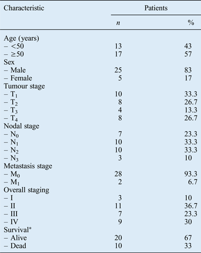
Thirty patients with nasopharyngeal carcinoma (NPC) and 10 patients with a non-NPC nasopharyngeal mass were included in this study. All NPC patients underwent examinations of the head and neck before treatment, along with staging examinations including whole body bone scanning, abdominal ultrasonography, computed tomography and magnetic resonance imaging (Table I). The American Joint Committee on Cancer 2010 tumour–node–metastasis staging system was used for clinical staging.Reference Edge, Byrd, Compton, Fritz, Greene and Trotti23
Nasopharyngeal carcinoma patients' demographic data

*At time of writing. T = tumour; N = node; M = metastasis
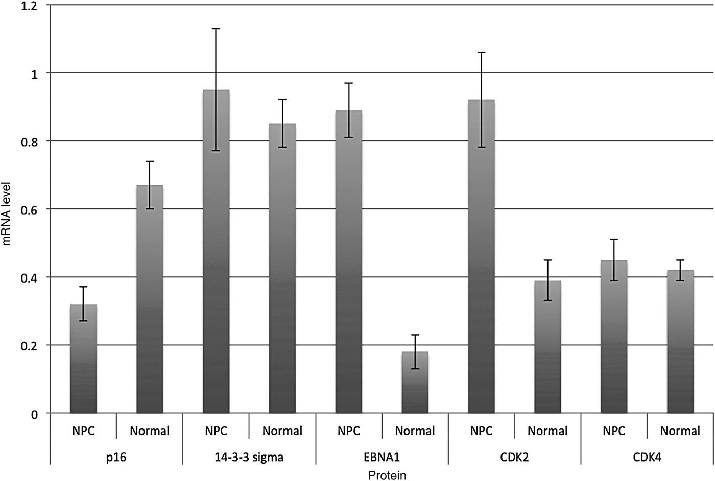
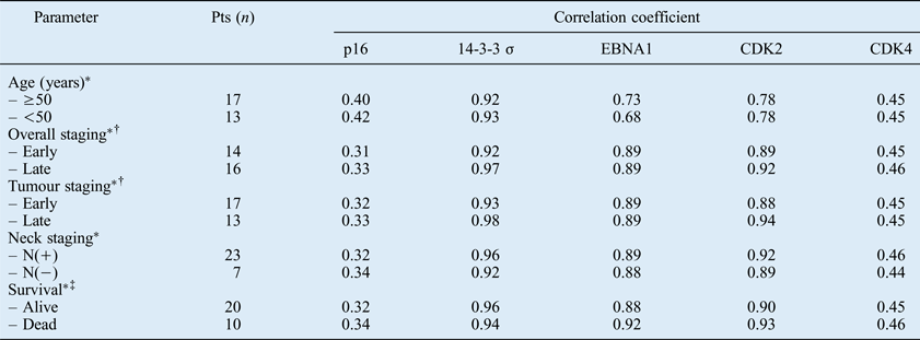
Reverse transcriptase polymerase chain reaction was used to analyse mRNA levels for 14-3-3 sigma, cyclin-dependent kinases 2 and 4, p16, and Epstein–Barr nuclear antigen 1 in NPC and normal tissue, compared with a positive control (Figure 1). There was significantly less p16 mRNA and significantly more cyclin-dependent kinase 2 and Epstein–Barr nuclear antigen 1 mRNA, comparing NPC versus normal nasopharyngeal tissue. The correlation between mRNA expression of the five tumour markers and various clinicopathological and prognostic factors was assessed; there were no statistically significant differences in these correlations, comparing patients with greater versus lesser degrees of these clinical factors (Table II).
Messenger RNA (mRNA) levels for 14-3-3 sigma, cyclin-dependent kinase 2 (CDK2), cyclin-dependent kinase 4 (CDK4), p16 and Epstein–Barr nuclear antigen 1 (EBNA1), determined by reverse transcription polymerase chain reaction assay of nasopharyngeal carcinoma (NPC) and normal tissue, and expressed as the ratio of mRNA concentrations in test sample vs glyceraldehyde 3-phosphate dehydrogenase (positive) control. Bars = means; whiskers = standard deviations
Correlation between tumour marker mRNA expression and clinicopathological parameters
*No significant difference (i.e. p ≥ 0.05), comparing correlation coefficients for both categories of parameter, for any tumour marker protein. †Early staging = stage I or II; late staging = stage III or IV. ‡At time of writing. Pts = patients; EBNA1 = Epstein–Barr nuclear antigen 1; CDK = cyclin-dependent kinase; N = node
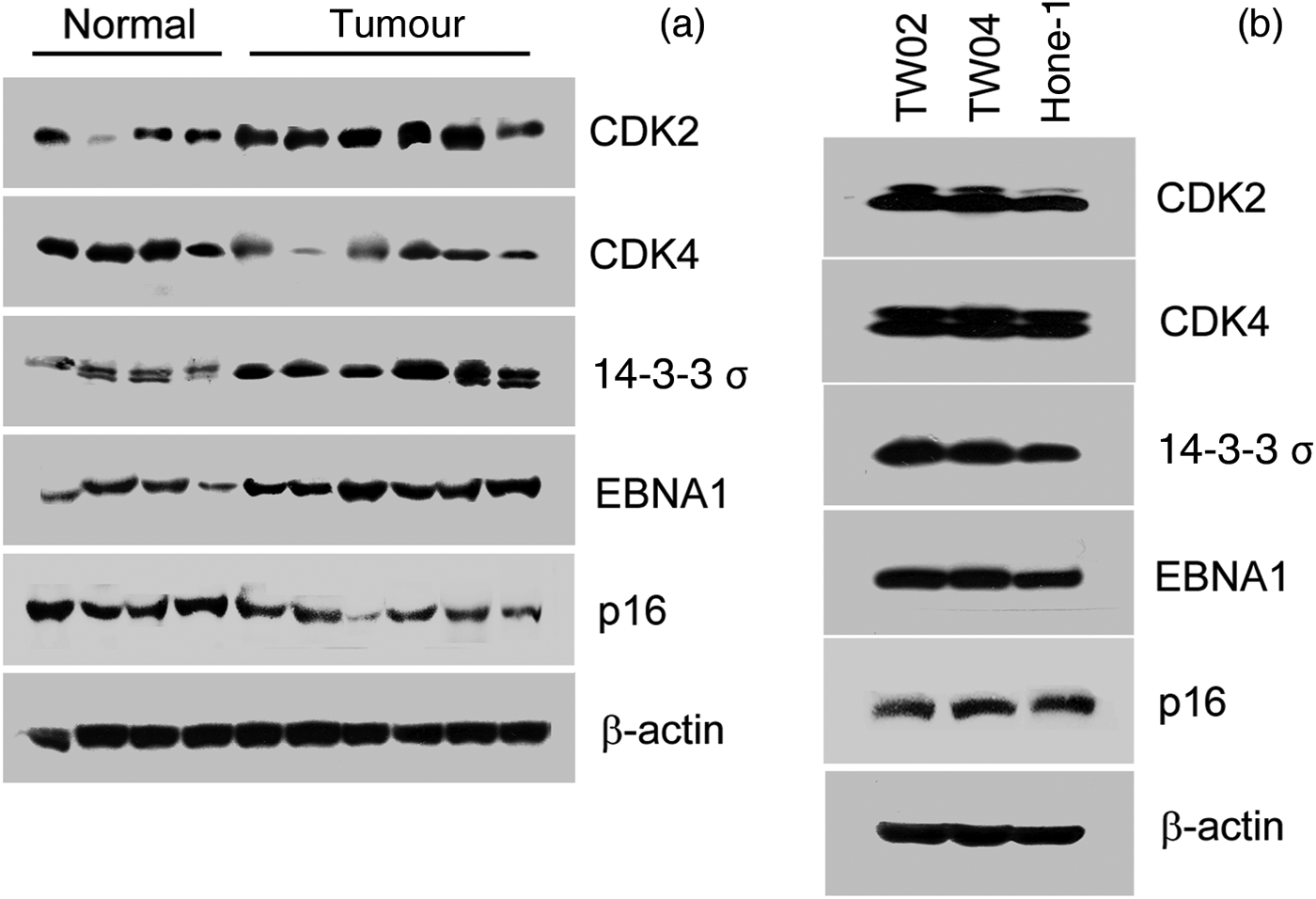
To validate the gene expression profile data for 14-3-3 sigma, immunoblot analysis was performed on six NPC tissue and four normal tissue samples. Figure 2(a) shows over-expression of 14-3-3 sigma, cyclin-dependent kinase 2 and Epstein–Barr nuclear antigen 1 in the NPC tissue samples. As shown in Figure 2(b), immunoblotting confirmed elevated expression levels of 14-3-3 sigma, cyclin-dependent kinases 2 and 4, and Epstein–Barr nuclear antigen 1 in the TW02, TW04 and Hone-1 cells.
(a) Immunoblot analysis of cyclin-dependent kinases (CDK) 2 and 4, 14-3-3 sigma, Epstein–Barr nuclear antigen 1 (EBNA1), p16 and β-actin expression, performed on 6 nasopharyngeal carcinoma (NPC) and 4 normal tissue samples; strong staining for 14-3-3 sigma, cyclin-dependent kinase 2 and Epstein–Barr nuclear antigen 1 is seen in the NPC specimens. (b) Immunoblot analysis of CDK2 and CDK4, 14-3-3 sigma, EBNA1 and p16 in TW02, TW04 and Hone-1 cells. β-actin is used as a positive control in both assays.
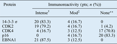
Immunohistochemical staining results are shown in Figure 3. Nasopharyngeal carcinoma samples showed intense or moderate expression of 14-3-3 sigma in 24/24 (100 per cent) samples, of cyclin-dependent kinase 2 in 23/24 (96 per cent) samples, of cyclin-dependent kinase 4 in 7/24 (29 per cent) samples, of p16 protein in 4/24 (17 per cent) samples and of Epstein–Barr nuclear antigen 1 in 24/24 (100 per cent) samples (Table III).
Photomicrographs showing immunohistochemical staining for (a) 14-3-3 sigma, (b) cyclin-dependent kinase 2, (c) cyclin-dependent kinase 4 and (d) Epstein–Barr nuclear antigen 1, in selected, positively staining biopsy specimens from patients with nasopharyngeal carcinoma. In these tumour cell specimens, there is significant, diffuse cytoplasmic and nuclear expression of 14-3-3 sigma, Epstein–Barr nuclear antigen 1, and cyclin-dependent kinases 4 and 2, while the surrounding tissue shows no expression. (×40)
Tumour marker immunohistochemistry results*
* Indicating protein expression. †More than 50% of cells; ‡10–50% of cells; ** <10% of cells. Pts = patients; Mod = moderate; CDK = cyclin-dependent kinase; EBNA1 = Epstein–Barr nuclear antigen 1
To enable further analysis of the effect of tumour stage, we compared results for patients with early stage NPC (i.e. stages I and II) versus late stage NPC (i.e. stages III and IV). No significant differences in immunohistochemical results or mRNA expression were found.
Discussion
It remains unclear how regulation of 14-3-3 sigma protein contributes to the development of cancer, and whether the loss of 14-3-3 sigma expression correlates with malignant transformation. Importantly, 14-3-3 sigma can be down-regulated or up-regulated in different types of cancer. Expression of 14-3-3 sigma may vary in different histological types of ovarian cancer.Reference Kaneuchi, Sasaki, Tanaka, Shiina, Verma and Ebina10 Other authors have reported low and high expression of 14-3-3 sigma in human cancers involving various organs.Reference Iwata, Yamamoto, Sasaki, Itoh, Suzuki and Kikuchi8–Reference Sano, Shimooka, Weixa, Segawa, Jian and Motegi13
Huang et al. suggested that 14-3-3 sigma down-regulation in nasopharyngeal carcinoma (NPC) may lead to over-expression of epidermal growth factor receptor and keratin 8.Reference Huang, Cheng, Chen, Peng, Zhang and Li24 However, we identified over-expression of 14-3-3 sigma mRNA and protein in both NPC and normal tissues. Similarly, over-expression of 14-3-3 sigma has been reported in other cancers.Reference Perathoner, Pirkebner, Brandacher, Spizzo, Stadlmann and Obrist11–Reference Sano, Shimooka, Weixa, Segawa, Jian and Motegi13, Reference Nakayama, Sano, Motegi, Oyama and Nakajima25 Perathoner et al. reported that 14-3-3 sigma over-expression was found mainly in de-differentiated colorectal carcinoma.Reference Perathoner, Pirkebner, Brandacher, Spizzo, Stadlmann and Obrist11 Kaneuchi et al. reported that some types of ovarian cancers (serous, endometrioid and mucinous), but not clear cell ovarian adenocarcinoma, showed high expression of the 14-3-3 sigma gene with minimal methylation.Reference Kaneuchi, Sasaki, Tanaka, Shiina, Verma and Ebina10 It is possible that differential expression and methylation of the 14-3-3 sigma gene characterises the various histological types of ovarian cancer.Reference Kaneuchi, Sasaki, Tanaka, Shiina, Verma and Ebina10 The various histological NPC types may therefore exhibit varying 14-3-3 sigma expression.
Similar findings have been reported in breast and oral cancers. The expression of 14-3-3 sigma has been reported to be reduced in primary breast carcinoma.Reference Umbricht, Evron, Gabrielson, Ferguson, Marks and Sukumar26, Reference Ferguson, Evron, Umbricht, Pandita, Chan and Hermeking27 However, a later study reported stronger 14-3-3 sigma immunoreactivity in myoepithelial cells than adjacent luminal epithelial cells, in both malignant and non-malignant cells.Reference Moreira, Ohlsson, Rank and Celis28 The authors concluded that the loss of 14-3-3 sigma expression was not a frequent event in breast tumourigenesis. Inactivation of 14-3-3 sigma through methylation has been reported in human oral cancer.Reference Laronga, Yang, Neal and Lee7 Using microarray technology, Villaret et al. demonstrated that 14-3-3 sigma mRNA was significantly over-expressed in primary head and neck squamous cell carcinoma (SCC) compared with normal tissue.Reference Villaret, Wang, Dillon, Xu, Sivam and Cheever29 Recent studies have reported that oral SCCs show stronger 14-3-3 sigma expression than adjacent non-malignant squamous mucosa.Reference Bhawal, Sugiyama, Nomura, Kuniyasu and Tsukinoki30 This is consistent with our findings that 14-3-3 sigma mRNA and protein are over-expressed in NPC tissue and cell lines. Taken together, our results do not support the contention that 14-3-3 sigma represents a valid marker for the early detection of NPC.
Loss of p16 expression has been reported in many different tumour types, including breast carcinoma and head and neck SCC.Reference Okami, Reed, Cairns, Koch, Westra and Wehage31, Reference Dublin, Patel, Gillett, Smith, Peters and Barnes32 Other authors have reported a high frequency of p16 inactivation in NPC.Reference Hwang, Cho, Huang, Wang, Shih and Su33 Our in vivo results indicated a loss of p16 expression in NPC patients, as assessed by both immunohistochemical analysis and mRNA assay (with a mean mRNA ratio ± standard deviation (SD) of 0.32 ± 0.05 in NPC patients and 0.67 ± 0.07 in controls; p < 0.01; see Figure 1). This suggests that inactivation of p16 expression may play an important role in NPC development. Inactivation of 14-3-3 sigma or p16 has an effect equivalent to the expression of the E6 and E7 oncoproteins of human papilloma virus, which together immortalise primary keratinocytes.Reference Sano, Shimooka, Weixa, Segawa, Jian and Motegi13 In the present study, all NPCs with p16 over-expression also showed elevated 14-3-3 sigma expression, and no cases with concomitant inactivation of both 14-3-3 sigma and p16 were observed. These findings support the hypothesis that inactivation of 14-3-3 sigma or p16 produces an effect equivalent to the expression of Epstein–Barr virus (EBV) oncoproteins.
A decrease in the expression of the p16 gene may lead to over-expression of cyclin-dependent kinase. Elevated expression of the cyclin-dependent kinase complex has been associated with carcinogenesis. Zhao et al. found that excess p16 expression arrests the cell cycle in the G0/G1 phase and suppresses cell growth. The major action of p16 protein is not associated with regulation of cyclin-dependent kinase 4 levels.Reference Zhao, Deng and Weng34 In our study, cyclin-dependent kinase 2 mRNA expression was higher in NPC than in normal control tissue. However, there was no significant difference in the expression of cyclin-dependent kinase 4 mRNA, comparing NPC patients and normal controls. Moreover, immunohistochemical analysis showed that cyclin-dependent kinase 2 was almost always positive, whereas cyclin-dependent kinase 4 was only positive in 29 per cent of NPC samples. These findings suggest that cyclin-dependent kinase 2 is more important than cyclin-dependent kinase 4 in NPC carcinogenesis.
• This study evaluated expression of 14-3-3 sigma, cyclin-dependent kinases 2 and 4, Epstein–Barr nuclear antigen 1 and p16, in nasopharyngeal carcinoma (NPC)
• Cyclin-dependent kinase 2, Epstein–Barr nuclear antigen 1 and p16 may be important in NPC carcinogenesis
• 14-3-3 sigma protein and mRNA levels were no lower in NPC than in normal tissue
Nasopharyngeal carcinoma is strongly associated with EBV, although various genetic and environmental factors have also been reported to play a role in its development. Epstein–Barr nuclear antigen 1 is usually positive in NPC with EBV, but other Epstein–Barr nuclear antigens are not. In the present study, the expression of Epstein–Barr nuclear antigen 1 mRNA was significantly greater in NPC than in normal tissue (with a mean mRNA ratio ± SD of 0.89 ± 0.08 in NPC patients and 0.18 ± 0.05 in controls; p < 0.01; see Figure 1). This is in agreement with Brooks and colleagues' report that Epstein–Barr nuclear antigen 1 mRNA was expressed in all NPC samples.Reference Brooks, Yao, Rickinson and Young35 In the present study, all cases of NPC were immuno-positive for Epstein–Barr nuclear antigen 1; this was consistent with mRNA findings.
Conclusion
In this study, protein and mRNA levels for cyclin-dependent kinase 2 and Epstein–Barr nuclear antigen 1 were significantly higher in nasopharyngeal carcinoma (NPC) compared with normal tissue, while levels of cyclin-dependent kinase 4 generally were not; results for 14-3-3 sigma varied. Nasopharyngeal carcinoma patients had diminished p16 gene expression, compared with normal tissue.
Results suggest that cyclin-dependent kinase 2 may be more important in NPC carcinogenesis than cyclin-dependent kinase 4. Further studies are necessary to clarify the role of 14-3-3 sigma and cyclin-dependent kinases 2 and 4 in NPC.
Acknowledgements
This work was supported by a grant from the Kaohsiung Chang Gung Memorial Hospital (grant number CMRPG84024). H-H Huang and C-H Chen contributed to this study equally. We would like to thank the Chang Gung Medical Foundation and the Kaohsiung Chang Gung Memorial Hospital Tissue Bank (grant number CLRPG8B0031) for technical support.