Case report
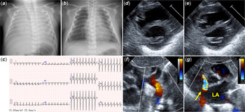
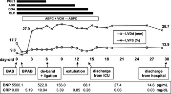
A preterm and low-birth-weight neonate (34 weeks’ gestation, birth weight of 2492 g) was born in an ambulance. Arriving at the local hospital, intratracheal intubation was performed because of severe respiratory distress. Furthermore, intratracheal bleeding was found, hence the patient was transferred to our hospital. Transthoracic echocardiography showed that left ventricular end-diastolic and end-systolic diameters were 17.7 mm (z score = 0) and 16.0 mm (z score = +3.7), respectively. Left ventricular fractional shortening was 9.6%, indicating severe left ventricular dysfunction. In addition, the direction of blood flow through the aortic arch was opposite (from distal to proximal) and the direction of blood flow through the ductus arteriosus was right to left. These findings suggested that systemic circulation was dependent on the ductus arteriosus. There was no hyperechogenicity of the endocardium or left ventricular papillary muscle, indicating myocardial ischaemia. Considering the enlarged left atrium, intratracheal bleeding was suspected to be caused by pulmonary congestion. There were no other cardiac structural abnormalities. A chest X-ray showed severe pulmonary vascular congestion, and an electrocardiogram showed no ST changes. Because of the poor general condition and prematurity, the patient was intolerant of extracorporeal membrane oxygenation placement using thoracotomy. Therefore, we developed alternative strategies. We performed balloon atrial septostomy to relieve pulmonary congestion. Before the procedure, left and right atrial pressures were 12 and 5 mmHg, respectively. After the procedure, they were 7 and 6 mmHg, respectively. The pressure gradient decreased from 7 to 1 mmHg, concomitant with an improvement in intratracheal bleeding. We used prostaglandin E1 to keep the ductus arteriosus open. At 1 day old, left heart function had not improved and blood lactate concentrations had increased. However, his condition was still poor and hypoxic therapy was considered ineffective. Therefore, we performed bilateral pulmonary artery banding to regulate pulmonary blood flow. The aforementioned strategies facilitated the supply of systemic blood flow by the right ventricle. At 2 days old, the left ventricular fractional shortening improved to 27.9%. Moreover, anterograde blood flow through the aortic arch had increased. Based on these findings, we considered that left ventricular function had recovered. At 6 days old, we performed bilateral pulmonary artery de-banding and ductus arteriosus ligation. Prior to surgery, right arm and leg SpO2 were 85–90% and 80–85%, respectively; and after surgery, both improved to over 96%. Streptococcus gallolyticus subspecies pasteurianus was detected in the blood culture obtained on admission. This confirmed the diagnosis of transient left ventricular dysfunction due to Streptococcus gallolyticus-related sepsis. The pathogen had not been detected in the vaginal culture of the mother during pregnancy. We administered ampicillin for 14 days to treat sepsis. At 30 days old, the patient was discharged without complications. Figure 1 shows the chest X-rays before and after balloon atrial septostomy (a, b), and electrocardiogram (c) and echocardiography (d, e, f, g) during admission. Figure 2 summarises the clinical course. Echocardiographic videos during admission and discharge are given in Supplementary videos 1 and 2.
(a) A chest X-ray at admission [before balloon atrial septostomy] showing severe pulmonary vascular congestion. (b ) A chest X-ray at 1 day old (after BAS) showing improvement in the pulmonary vascular congestion. (c) An electrocardiogram at admission showing no ST changes. (d)–(g) Transthoracic echocardiography during admission. (d) A short-axis view at end-diastole showing that the left ventricular end-diastolic diameter is 17.7 mm (z score = 0). (e) A short-axis view at end-systole showing that the left ventricular end-systolic diameter is 16.0 mm (z score = +3.7) and that left ventricular fractional shortening was 9.6%, which is indicative of severe left ventricular dysfunction. (f) Aortic arch view showing that the direction of blood flow through the aortic arch was opposite (from distal to proximal). (g) Left atrium view showing the enlarged left atrium and accelerated blood flow through the foramen ovale (arrow), which indicates elevated left atrial pressure. LA=left atrium.

Summary of the clinical course. LVDd=left ventricular end-diastolic diameter; ABPC=ampicillin; ADR=adrenaline; BAS=balloon atrial septostomy; BNP=brain natriuretic peptide; BPAB=bilateral pulmonary artery banding; CRP=C-reactive protein; de-band=pulmonary artery de-banding; ligation=ductus arteriosus ligation; DOA=dopamine; ICU=intensive care unit; LVFS=left ventricular fractional shortening; OLP=olprinone; PGE1= prostaglandin E1; VCM=vancomycin.

Discussion
We described the case of a preterm and low-birth-weight neonate who developed intratracheal bleeding, severe cardiac failure, and subsequent cardiogenic shock, early after birth. The neonate was successfully rescued by keeping the ductus arteriosus open and performing bilateral pulmonary artery banding instead of using extracorporeal membrane oxygenation. Intratracheal bleeding was thought to be caused by pulmonary congestion following cardiac failure. This complication had an earlier onset than expected because it did not occur until postnatal pulmonary circulation was established.
Cardiogenic shock in neonates is often associated with CHD. In contrast, neonatal shock with a normal heart structure has multiple causes, including perinatal asphyxia. Reference Bhat and Plakkal1,Reference Chan, Sanatani, Potts and Harris2 This patient was born outside the hospital. Therefore, perinatal asphyxia could be contributed to left ventricular dysfunction. On the other hand, pathogenic bacteria were detected in the blood culture in this case. Since sepsis-induced cardiomyopathy has been recognised in children, Reference Wheeler and Wong3 sepsis could directly cause left ventricular dysfunction. We considered that the cause of left ventricular dysfunction was not singular, but combinatorial. Extracorporeal membrane oxygenation facilitates the management of severe cardiac failure, even in neonates. Reference Di Nardo, MacLaren, Marano, Cecchetti, Bernaschi and Amodeo4 The use of extracorporeal membrane oxygenation is increasing in neonates with cardiac failure. Reference Roeleveld and Mendonca5 It is most commonly used during the perioperative period of CHD. However, there are several bleeding-associated complications predominantly in preterm and low-birth-weight neonates. Such complications occur in 17 and 21% of the preterm neonates at 34 and 29–33 weeks of gestation, respectively. Reference Church, Kim and Erickson6 There are guidelines on the relative contraindications to the use of extracorporeal membrane oxygenation for those aged <34 weeks of gestation or weighing <2 kg. 7
Alternative strategies to extracorporeal membrane oxygenation, in this case, included the decompression of the left atrium by balloon atrial septostomy, keeping the ductus arteriosus open, and regulation of pulmonary blood flow by bilateral pulmonary artery banding. Balloon atrial septostomy is used for decompressing the left atrium during extracorporeal membrane oxygenation in children. Reference Baruteau, Barnetche and Morin8 However, there are no reports on the strategies used in this case. The right ventricle was considered to preserve sufficient function to supply systemic circulation, and the left ventricular dysfunction was expected to improve within a few days. The above-mentioned strategy is similar to that used in place of the primary Norwood procedure for hypoplastic left heart syndrome. The implementation of this strategy on patients with hypoplastic left heart syndrome with younger gestational age and lower birth weight justifies its use in this case. Reference Karamlou, Overman and Hill9 In addition, compared to hypoplastic left heart syndrome, we expected higher SpO2 in our case because of additional oxygenated output from the left ventricle. Hence, this strategy for severe left ventricular dysfunction was considered a useful option, with the advantage of avoiding extracorporeal membrane oxygenation application on preterm and low-birth-weight neonates with high complications.
Conclusion
The combination treatment of balloon atrioseptostomy, bilateral pulmonary artery banding, and keeping the ductus arteriosus open can be an alternative to circulatory support using extracorporeal membrane oxygenation in a neonate with left ventricular dysfunction and persevered right ventricular function. Therefore, it is critical to evaluate both left and right ventricular functions. An individualised therapeutic strategy should be chosen in the management of collapsed neonates.
Supplementary material
To view supplementary material for this article, please visit https://doi.org/10.1017/S1047951120004977.
Acknowledgements
We would like to thank Editage (www.editage.com) for English language editing.
Financial support
This research received no specific grant from any funding agency, commercial, or not-for-profit sectors.
Conflicts of interest
None.
Ethical standards
We obtained written informed consent for publication from the parents of the patient.