Introduction
In recent years, the preservation and transportation of temperature-sensitive materials (TSM) have driven scientists to explore the materials with efficient thermal regulation capacity [Reference Chalco-Sandoval, Jose Fabra, Lopez-Rubio and Lagaron1]. The development of phase change materials (PCMs), which possess the advantages of high storage density in small temperature intervals, promotes maintaining TSM within the desired temperature limits [Reference Chalco-Sandoval, Jose Fabra, Lopez-Rubio and Lagaron2]. Most of the naturally existing materials that can be applied as PCMs are solid–liquid ones. During the last decade, many solid–liquid PCMs including paraffin waxes, poly(ethylene glycol)s, and fatty acids have been utilized. Paraffin waxes have been considered as one of the most promising thermoregulating materials owing to their high energy storage density and suitable phase transition temperature [Reference Sarier and Onder3]. When the ambient temperature changes, the cycle of melting and solidification in PCMs endows them with the ability of heat storage and release [Reference Kousksou, Arid, Jamil and Zeraouli4]. Therefore, PCMs can produce a stable microclimate within a narrow temperature range, which plays a great role in thermal regulation [Reference Sarier and Onder3]. However, key issues still exist in the practical applications of solid–liquid PCMs. For instance, during the phase change process, the melted PCMs can easily undergo leakage and loss. In order to maintain the shape and prevent the leakage of solid–liquid PCMs, microencapsulated phase change materials (MicroPCMs) have been developed. MicroPCMs consist of a solid–liquid PCM core surrounded by a polymer shell [Reference Chen, Xu, Shang and Zhang5]. What’s more, the core–shell structure can reduce the interaction between PCMs and the environment and increase the contact areas which are beneficial for heat transfer [Reference Zhao and Zhang6]. Various methods have been developed for the synthesis of MicroPCMs, and different types of shell materials have been used to encapsulate PCMs [Reference Palanikkumaran, Gupta, Agrawal and Jassal7]. Considering nontoxic and biodegradation request, particularly for medical and healthcare applications, natural polymers including alginate, gelatin, gum Arabic, and acacia have been employed as the shell materials in recent years [Reference Ozonur, Mazman, Paksoy and Evliya8].
Silk fibroin (SF) is a natural fibrillar polymer from silkworm larvae or Bombyx mori [Reference Baimark, Srisa-ard and Srihanam9, Reference Jin, Zhang, Hang, Shao and Hu10]. It is endowed with excellent mechanical properties, good biocompatibility, and controllable biodegradability [Reference Cao, Chen, Yao, Huang and Shao11, Reference Wei, Zhang, Shao and Hu12]. Furthermore, SF is relatively easy to be fabricated into different forms including sponges, hydrogels, films, and mats [Reference Koh, Cheng, Teng, Khin, Loh, Tee, Low, Ye, Yu, Zhang and Han13]. All these advantages make SF a potential candidate for the shell material to encapsulate PCMs. What else, SF is also capable of reducing Ag+ to Ag for the redox-active nature of the tyrosine (Tyr) residues, which is one of the eighteen amino acid residues contained within SF [Reference Xie, Lee, Wang and Ting14]. This achieves the goal of fabrication of noble metal nanomaterials by biotemplate techniques under moderate conditions.
Application of silver nanoparticles (AgNPs) in the healthcare field relies on their excellent antibacterial property toward a broad spectrum of bacterias. At the same time, AgNPs will not bring about Ag+ which has acute cytotoxicity [Reference Benn and Westerhoff15, Reference Tian, Wong, Ho, Lok, Yu, Che, Chiu and Tam16]. Generally, AgNPs can be prepared by a reduction reaction in solutions containing Ag+ [Reference Zhang, Tang and Vlahovic17]. However, AgNPs are usually in the form of colloids in the liquid system. It tends to aggregate and reduce the surface energy which may decrease the antimicrobial property [Reference Park, Neigh, Vermeulen, de la Fonteyne, Verharen, Briede, van Loveren and de Jong18]. In order to minimize such a negative impact, biotemplate techniques are employed to the fabrication of AgNPs. Xiang Fei prepared AgNPs in situ using SF as a biotemplate under light exposure [Reference Fei, Jia, Du, Yang, Zhang, Shao, Zhao and Chen19]; Semih Calamak synthesized AgNPs in situ in the electrospun fibroin nanofibers by UV reduction of silver nitrate [Reference Calamak, Aksoy, Ertas, Erdogdu, Sagiroglu and Ulubayram20].
In this study, a facile and environmental-friendly fabrication approach was introduced to prepare novel microencapsulated n-octadecane with natural SF shell incorporated with AgNPs. To our knowledge, such SF-AgNPs microcapsules obtained by self-assembly method and subsequently processed under the irradiation of an infrared lamp were rarely reported. The thermal and antibacterial properties of the resulted SF-AgNPs microcapsules were studied to explore their potential applications in thermal regulation and healthcare fields.
Results and discussion
Synthetic strategy
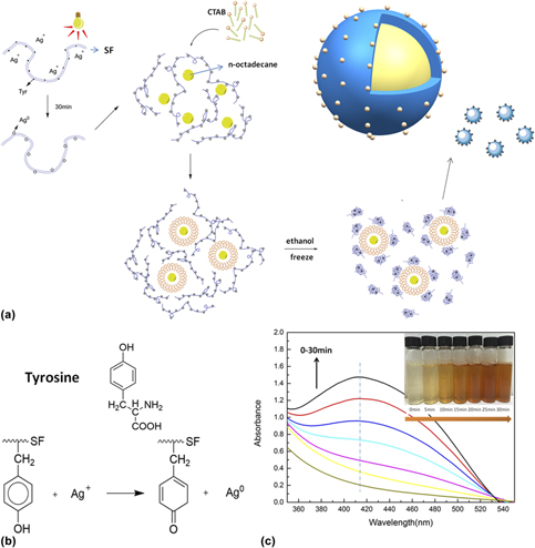
The microencapsulated n-octadecane was synthesized via a self-assembled conformation transformation on the surfaces of n-octadecane micelles in an oil-in-water emulsion system. The synthetic strategy of microcapsules is illustrated in the schematic formation mechanism shown in Fig. 1(a). The typical fabrication procedure can be described as follows: AgNPs were firstly produced from SF–AgNO3 mixture solution by exposing under the infrared lamp irradiation for 30 min at room temperature. The Tyr residues contained in SF are capable of reducing Ag+ to Ag due to their inherent redox-active nature. After mixing n-octadecane with SF-AgNPs composite, the cationic surfactant CTAB was added to help to form a stable oil-in-water emulsion since the SF-AgNPs molecules are negatively charged and the hydrophilic groups of CTAB molecules are positively charged. The SF-AgNPs molecules could be captured to the oil–water interface through the charge interaction [Reference Tian, Jiang, Chen, Shao and Yang21]. Then, certain amount of anhydrous ethanol was added dropwise into the oil-in-water emulsion. The conformation of SF-AgNPs molecules transformed from random and/or helical structure to antiparallel β-sheet, and the SF-AgNPs molecules were self-assembled at the surface of n-octadecane micelles [Reference Cao, Chen, Yao, Huang and Shao11]. In order to control the conformation transition of SF-AgNPs molecules at a proper rate, the mixtures were frozen in a refrigerator. Eventually, a thin layer of SF shell with AgNPs distributed uniformly on it was formed at the oil–water interface, and n-octadecane microcapsules with a satisfactory core–shell structure were successfully synthesized.
(a) Schematic strategy of SF microcapsule incorporated with AgNPs, (b) possible mechanism of reduction reaction of Ag+, and (c) UV-vis spectra of SF–AgNO3 reaction solution.

Based on the previous studies, either single Tyr molecule or polypeptides and proteins containing Tyr residues can be used in the synthesis of AgNPs [Reference Xie, Lee, Wang and Ting14, Reference Selvakannan, Swami, Srisathiyanarayanan, Shirude, Pasricha, Mandale and Sastry22]. SF can be also considered to reduce Ag+ to Ag by the redox-active nature of Tyr residues contained within SF. The possible mechanism of reduction reaction of Ag+ is shown in Fig. 1(b). It can be seen that the phenolic hydroxyl is reduced to carbonyl after the reduction reaction and then AgNPs is obtained. Figure 1(c) shows the time-resolved UV-vis spectra recorded from the mixture solution exposed to the infrared lamp for different time durations, and the corresponding optical observation photographs are also embedded in the figure. The main absorption peak at 414 nm increases gradually as the exposure time increases from 0 to 30 min, which means more AgNPs were produced, and this can be also speculated from the deeper color along with a longer irradiation time.
FTIR characterization
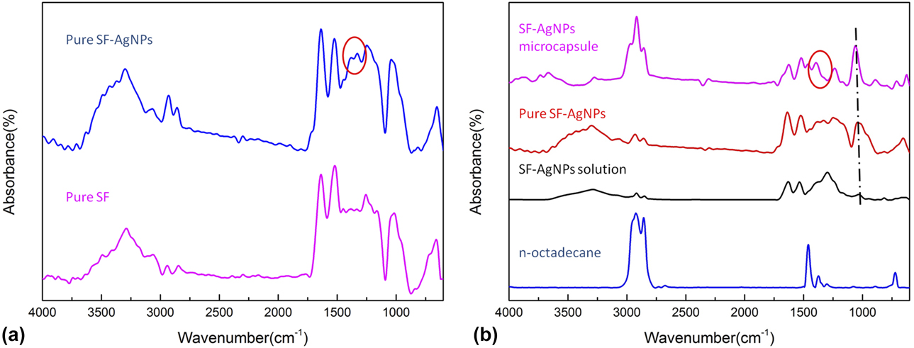
In order to further verify the production of AgNPs, the FT-IR spectra of pure SF and pure SF-AgNPs are presented in Fig. 2(a). Pure SF and pure SF-AgNPs refer to the self-assembled SF solution and SF-AgNPs solution, respectively. The absorption bands at 1296 cm−1 attribute to the carbonyl group can be obviously found in the spectrum, suggesting the successful synthesis of AgNPs by the reduction of Tyr residues.
FTIR spectra of (a) pure SF and pure SF-AgNPs, (b) n-octadecane, SF-AgNPs solution, pure SF-AgNPs and SF-AgNPs PCMs.
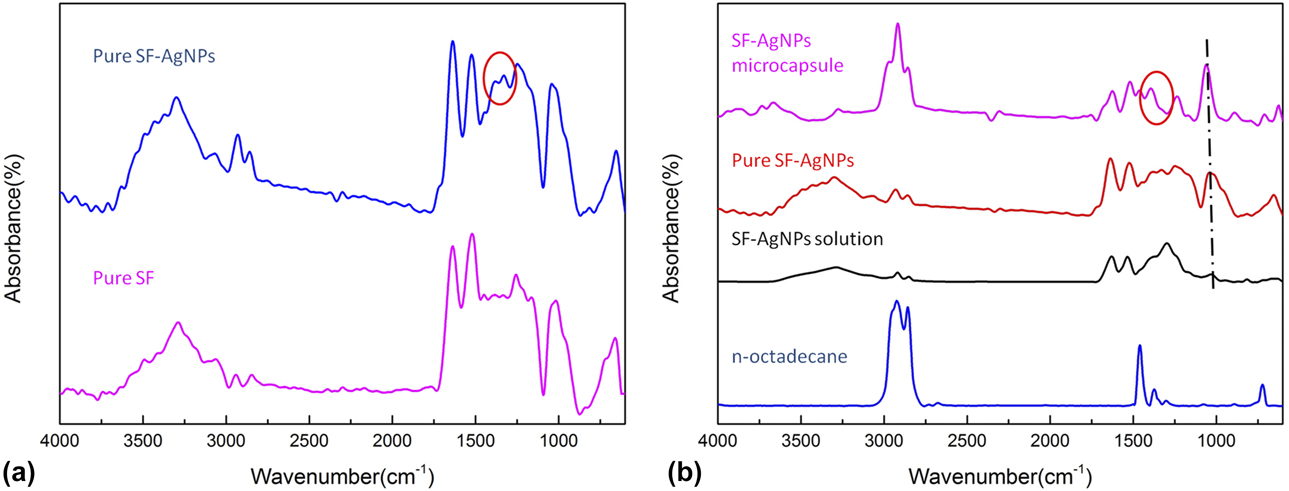
The fabrication of the resultant microcapsules was firstly confirmed with FTIR analysis. The corresponding FTIR spectra of n-octadecane, SF-AgNPs solution, pure SF-AgNPs, and SF-AgNPs microcapsule are presented as comparison groups in Fig. 2(b). In the n-octadecane spectrum, there are two strong absorption bands corresponding to the asymmetric and symmetric stretching vibrations of C–H at 2923 and 2850 cm−1. While the peaks at around 1463 and 1375 cm−1 come from the deformation vibration of –CH2 and –CH3, the peak at 721 cm−1 can be attributed to the in-plane rocking vibration of –CH2 [Reference Cao, Tang and Fang23]. As shown in Fig. 2(b), the peak at 1236 cm−1 is the typical amide absorption peak. This means the dominant conformation of pure SF-AgNPs is identified as silk II structure by comparing SF-AgNPs solution with pure SF-AgNPs. And the silk II structure is primarily composed of antiparallel β-sheets. This implies that the conformation for SF-AgNPs molecule transforms from silk I structure to silk II structure by the addition of anhydrous ethanol [Reference Taketani, Nakayama, Nagare and Senna24]. Moreover, there are no extra absorption peaks appeared in the spectrum of the SF-AgNPs PCMs compared to the absorption peaks of n-octadecane and pure SF-AgNPs. All of these results indicate the success of n-octadecane microencapsulation with SF-AgNPs shell.
Morphology of SF microcapsule and SF-AgNPs microcapsule
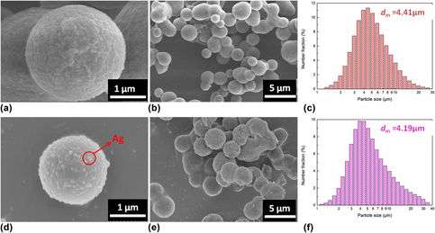
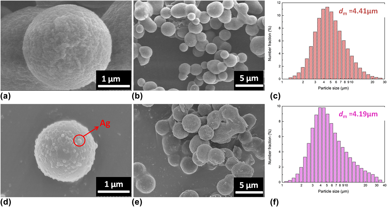
The morphologies of SF microcapsule and SF-AgNPs microcapsule are given in Fig. 3. Based on our previous study [Reference Zhao, Luo, Wang, Song and Tang25], the SF microcapsule sample was obtained according to the procedures described in section “Preparation of microcapsules” except for the production of AgNPs. The microcapsules show a regular spherical shape and uniform particle diameters [Figs. 3(b) and 3(e)]. The size distribution of SF microcapsule and SF-AgNPs microcapsule shown in Figs. 3(c) and 3(f) were recorded and collected from a laser particle size analyzer. The median diameters (d m) of both types of microcapsules are about 4 μm that are almost identical. Such a phenomenon implies that production of AgNPs has little effect on the conformation transition of SF and formation of microcapsules. The morphological details of SF microcapsule and SF-AgNPs microcapsule are shown in Figs. 3(a) and 3(d). The surface morphologies of both types of microcapsules are found to be pleated but intact. It is caused by the growth and aggregation of SF microcrystals onto the n-octadecane micelles step by step during the synthesis process. Moreover, the surface of the SF-AgNPs microcapsule is covered with AgNPs as highlighted by the red circle. This indicates that AgNPs have been successfully embedded onto the surface of SF-AgNPs microcapsule with a narrow distribution, and the particle size of AgNPs is approximately 100 nm.
The SEM micrographs and particle size distribution plots of SF microcapsule (a, b, c) and SF-AgNPs microcapsule (d, e, f).
Morphology of PCM-C and SF-AgNPs microcapsule
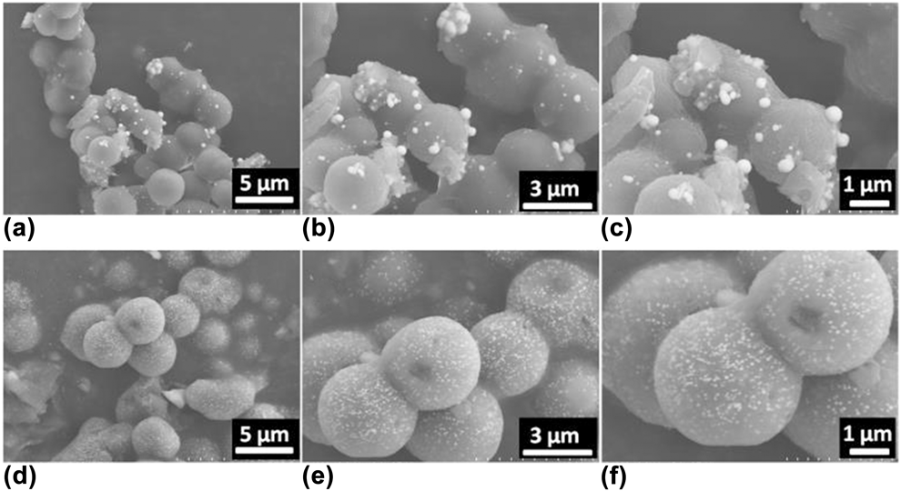
The morphologies of PCM-C and SF-AgNPs microcapsule are shown in Fig. 4. The difference between the two samples is the sequence of the reduction reaction during the synthesis process; for SF-AgNPs sample, the reduction reaction of Ag particles is prior to the formation of the microcapsules, whereas for the sample of PCM-C, the reduction of Ag+ to Ag particles has taken place directly on the surface of microcapsules. The PCM-C sample was fabricated in a typical synthesis process as below: the washed 30 g SF microcapsule was dispersed into 200 mL deionized water under ultrasonic treatment. Then, 600 mg of AgNO3 powders was added to the mixture. After being exposed under the infrared lamp for 30 min at room temperature, the resulting sample was obtained after being washed with anhydrous ethanol and water. As seen in Figs. 4(c) and 4(f), the diameters of AgNPs particle attached to the two types of microcapsules are approximately 300 nm and 100 nm, respectively. It can be found that the resulting AgNPs exhibit a narrower distribution and a smaller size by the reduction reaction before the conformational transformation of SF. Therefore, the sequence of reduction reaction in the synthesis process has great effect on the dispersion and size of AgNPs. The reason for this phenomenon can be explained as follows: with the increase in dispersion of SF microcrystals, the contact between Ag+ and Tyr residues becomes more frequent. Then, the nucleation points of AgNPs increase and spread more evenly. When the redox reaction occurs, the grain growth of AgNPs will rely on these nucleation points. Hence, the AgNPs in SF-AgNPs microcapsule exhibit a more even distribution and a relatively smaller size. Meanwhile, the SF-AgNPs microcapsule shows a regular spherical shape [Figs. 4(d) and 4(e)], and the rugged surface morphology clearly indicates that microcapsules are covered with a uniform layer of AgNPs [Fig. 4(f)].
The SEM micrographs of PCM-C (a, b, c) and SF-AgNPs microcapsule (d, e, f).

EDS spectrum
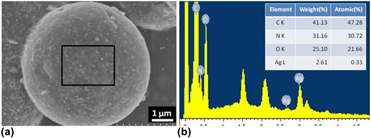
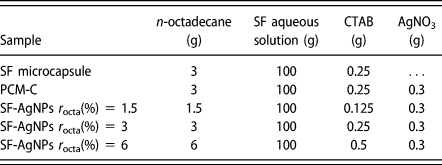
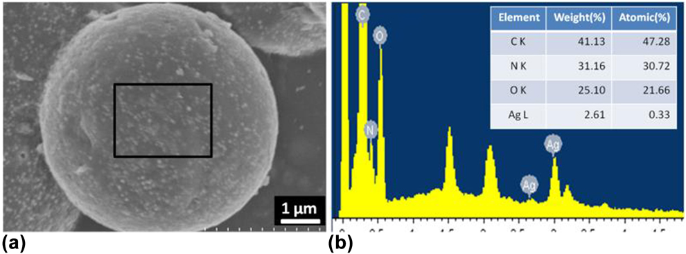
The energy dispersive spectroscope (EDS) spectrum shows the chemical composition of the SF-AgNPs microcapsule (r octa = 3%). The contents of C, N, O, and Ag atoms tested from EDS spectrum are illustrated in Fig. 5(b). The weight percentage of Ag atom is 2.61%, which is in line with the theoretical value of 2.91% calculated from Table I. This further proves that AgNPs have been incorporated onto the SF shell successfully.
(a) The SEM micrograph of SF-AgNPs microcapsule and (b) the EDS spectrum of selected area.
The formulations for the microcapsule preparation. Note: SF-AgNPs-C, fabrication of AgNPs after microencapsulation; SF-AgNPs r octa(%) = 1.5, 3, 6, fabrication of AgNPs before microencapsulation.

Thermal properties and performance of SF-AgNPs microcapsules
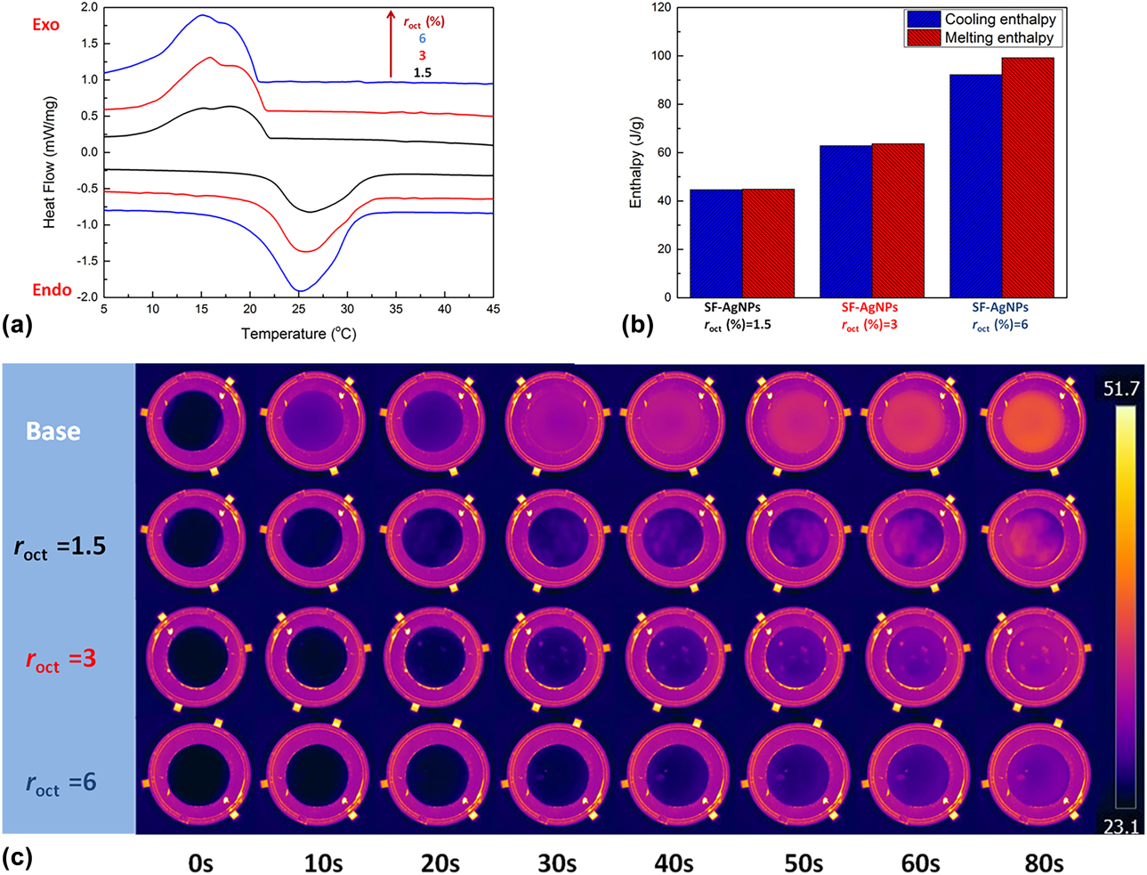
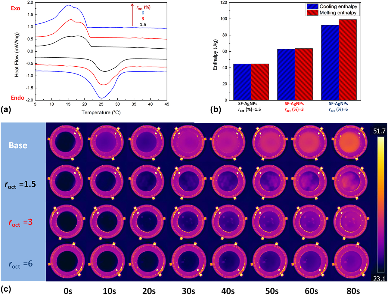
Thermal storage capacity for the overall SF-AgNPs microcapsules shown in Fig. 6(a) was determined by DSC. And the major thermal parameters of melting process and crystallization process calculated from the DSC curve are summarized in Table II. As shown in Fig. 6(a), SF-AgNPs microcapsules present two crystallization peaks at about 16 and 18 °C and a single melting peak at about 25 °C. The peak at 16 °C represents the homogeneous crystallization of n-octadecane, which is caused by its self-nucleation. The peak at 18 °C represents the heterogeneous crystallization of n-octadecane. It is caused by the nucleation on the inner side of SF-AgNPs shell. The phase change enthalpies of SF-AgNPs microcapsule are shown in Fig. 6(b). It is understandable that the latent heat of SF-AgNPs microcapsule increases with the increase in r octa. Also, it indicates that the enthalpy of SF-AgNPs microcapsule can be improved to about 95 J/g. Such phenomena imply the promotion of thermal storage capacity of SF-AgNPs microcapsule.
The thermal characteristics of SF-AgNPs microcapsule captured using DSC (a, b), and the forward-looking infrared system (c).
Thermal properties of SF-AgNPs microcapsule.

Note: T om, T oc, onset temperature on DSC curve; T pm, T pc, peak temperature on DSC curve; T em, T ec, endset temperature on DSC curve; ΔH m, ΔH c, enthalpy on DSC curve.
The base sample and SF-AgNPs microcapsules were heated from bottom at 50 °C, and an infrared camera was used to characterize the thermal regulation performance [Fig. 6(c)]. It is obvious that the temperature of SF-AgNPs microcapsules is lower than that of the base sample throughout the entire heating time range. On the one hand, it can be seen from Fig. 6(c) that the temperature of SF-AgNPs microcapsules is almost kept in the range of phase transition temperature during the heating time range. And with the increase in r octa, the temperature increases more slowly. This means the improvement in thermal regulation performance. On the other hand, the phase transition temperature is about 26 °C, which is comfortable for the human body and suitable for the potential healthcare applications.
Thermal stability of the SF-AgNPs microcapsule
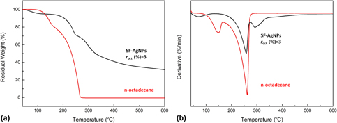
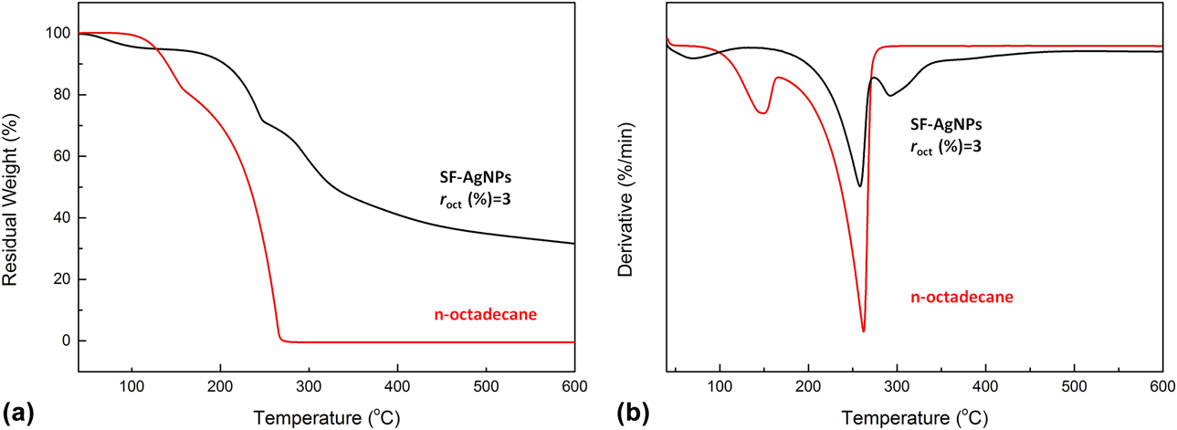
Figure 7(a) shows the TGA curve of n-octadecane and SF-AgNPs microcapsule synthesized with the r octa of 3%. And the corresponding DTG curves are shown in Fig. 7(b). It can be found from the TGA curve that there is an initial weight loss of SF-AgNPs microcapsule (r octa of 3%) below 100 °C. This is mainly caused by the evaporation of the adsorbed water within SF-AgNPs shells. However, no such phenomenon can be observed in the curve of n-octadecane. It can be noticed that the weight loss of n-octadecane starts at about 110 °C; however, the corresponding weight loss of SF-AgNPs microcapsule starts at about 200 °C. These results show that the thermal stability of n-octadecane has been greatly improved under the protection of SF-AgNPs shell.
(a) TGA curves and (b) the corresponding DTG curves of n-octadecane and SF-AgNPs microcapsule r octa(%) = 3.
Antimicrobial performance
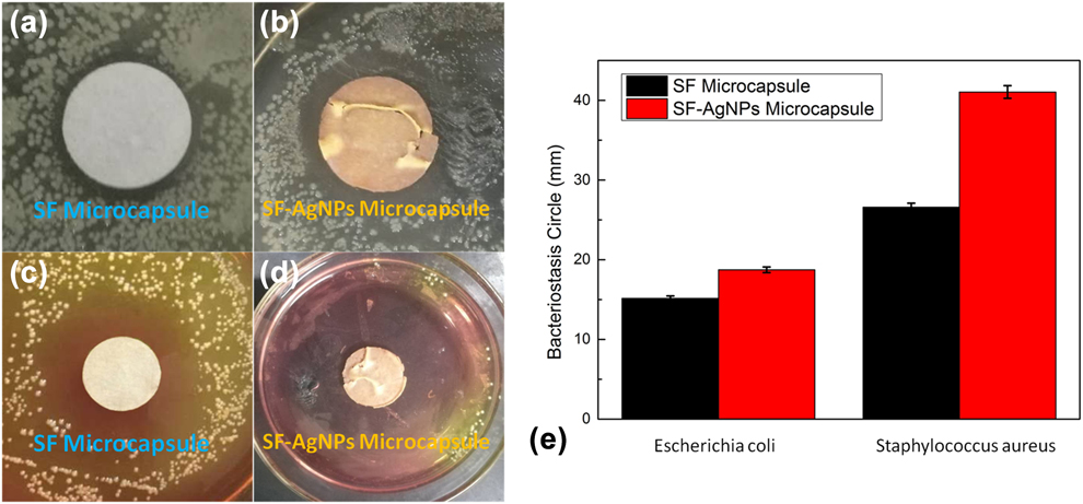
The antibacterial activity of SF microcapsule and SF-AgNPs microcapsule against Gram-positive, i.e., Staphylococcus aureus, and Gram-negative, i.e., Escherichia coli, was tested by the disc diffusion test. The relevant results are shown in Figs. 8(a)–8(d). The corresponding diameters of the inhibition zones are given in Fig. 8(e). The results show that both types of microcapsules exhibit an antibacterial activity against these two categories of bacteria. As expected, the SF-AgNPs microcapsule has much better antibacterial activity against both the bacteria in comparison with SF microcapsule. It can also be found that the inhibition zone of both types of microcapsules for Staphylococcus aureus is obviously higher compared to that for Escherichia coli.
Images of the inhibition zones for samples: (a) SF microcapsule against Escherichia coli, (b) SF-AgNPs microcapsule against Escherichia coli, (c) SF microcapsule against Staphylococcus aureus, (d) SF-AgNPs microcapsule against Staphylococcus aureus, (e) and diameters of the inhibition zones for each sample.

In other words, Staphylococcus aureus is more susceptible to AgNPs than Escherichia coli according to the above results. Xiaoyu Zhang et al. also reported the similar findings in their scientific work [Reference Zhang, Wang and Wu26]. Although the exact mechanism of action for AgNPs on Gram-negative or Gram-positive bacteria currently remains unclear, the mainstream studies propose a possible mechanism that the AgNPs might attach to the surface of the cell membrane, disturbing the permeability and respiration functions of cells [Reference Kvitek, Panacek, Soukupova, Kolar, Vecerova, Prucek, Holecova and Zboril27]. Nevertheless, the SF-AgNPs microcapsule is still found to exhibit a good antibacterial activity against these two types of bacteria.
Conclusions
In this study, novel microencapsulated n-octadecane with natural SF shell and silver nanoparticles (AgNPs) attached to the surface was successfully synthesized in oil-in-water emulsion by using a self-assembly method. The extra reductant is unnecessary over the synthesis for the reduction property of tyrosine (Tyr) residues contained in SF. The resulting microcapsules exhibit a regular spherical morphology with a median size of 4–5 μm. And the AgNPs attached to their surfaces exhibit a uniform distribution. Microencapsulated n-octadecane was used as the core material with a phase transition temperature of about 26 °C, which is a comfortable temperature for the human body. The results obtained from DSC, TGA, and forward-looking infrared system indicate that the SF-AgNPs microcapsule has energy storage capability, enhanced thermal stability, and good thermal regulation. The melting and crystallization enthalpies of the composite are −99.20 and 92.22 J/g corresponding to the heating and cooling process, and the thermal decomposition temperature has improved to about 200 °C. In addition, it is found that the SF-AgNPs microcapsule exhibits a good antibacterial performance against Gram-positive bacteria Staphylococcus aureus and Gram-negative bacteria Escherichia coli. Hence, the SF-AgNPs microcapsule could be a good potential candidate for thermal regulation and healthcare applications.
Experimental
Materials
n-Octadecane with a melting point of about 26 °C, which served as the PCM, was purchased from Tianjin Chemical Reagents Company, Tianjin, China. Silkworm (Bombyx mori) was supplied by the Sericulturist in Zhejiang province, China. Analytical-grade hexadecyl trimethyl ammonium bromide (CTAB) was purchased from Sinopharm Chemical Reagent Co., Ltd., Shanghai, China. Analytical-grade sodium carbonate (Na2CO3), lithium bromide (LiBr), silver nitrate (AgNO3), and anhydrous ethanol were supplied by Alfa Aesar, Shanghai, China. Semipermeable membrane (14 kDa cutoff) was purchased from Yuanju Co., Ltd., Shanghai, China.
Preparation of SF aqueous solution
The silk fibers were firstly degummed twice with 0.5 wt% Na2CO3 aqueous solution at 100 °C for 30 min. The degummed silk fibers were washed twice with deionized water and then dried at room temperature for 24 h [Reference Chen, Shao, Knight and Vollrath28]. After that, the silk fibers were dissolved in 9.5 mol/L LiBr aqueous solution at 80 °C for 60 min. Then, the SF solution enclosed in a semipermeable membrane was soaked in deionized water for 72 h to remove LiBr [Reference Luo, Zhang, Huang, Shao and Hu29]. The dialyzed SF solution was filtered to remove impurity, and the filtering solution was collected. The filtered SF solution was diluted with deionized water to the concentration of 3% (w/w) for further use. The concentration of SF solution was calculated through the value of index of refraction at 25 °C.
Preparation of microcapsules
SF-AgNPs microcapsule was fabricated by a self-assembled conformation transformation on the surfaces of n-octadecane micelles in an oil-in-water emulsion system. The weight ratios of n-octadecane (r octa) are 1.5%, 3%, and 6%, respectively. In a typical procedure, taking SF-AgNPs r octa(%) = 3 for example, 300 mg of AgNO3 powders was added to 100 mL of the above-obtained SF solution to form a transparent SF–AgNO3 mixture. Consequently, such SF–AgNO3 solution was exposed to infrared light (Philips infrared lamp, 375 W) for 30 min to produce SF-AgNPs composite solution. To the mixture of 100 mL SF-AgNPs solution, 3 g of n-octadecane and 0.25 g of CTAB were added, heated up to 40 °C, and then stirred at 800 rpm for 20 min to form a stable oil-in-water emulsion. To the above emulsion, 15 mL of anhydrous ethanol was added. The mixture was then frozen in a freezer at −10 ± 2 °C for 24 h. After being defrosted, the samples were washed by hot anhydrous ethanol (60 °C) and deionized water, respectively. Afterward, the samples were filtered through a filter paper to remove superfluous n-octadecane. Eventually, the samples were collected by lyophilizing.
For comparisons, the PCM-C sample was fabricated based on the SF microcapsule. In a typical synthesis process, the washed SF microcapsule was dispersed in deionized water by ultrasonic treatment. Then, AgNO3 powders were added into such suspension. After been exposed under infrared light, the PCM-C product sample was finally obtained after being washed with anhydrous ethanol and deionized water. Table I summarizes the synthesis formulations of samples.
Characterization
A spectrophotometer (S-3100, SCINCO, Seoul, Korea) was used to measure the UV-vis spectra of the as-prepared samples. The chemical structure analysis of the samples was performed by using the Fourier transform infrared spectroscopy (FT-IR) with a spectrometer (Vertex 70, Bruker, Karlsruhe, Germany) in the range 4000–600 cm−1 at a resolution of 4 cm−1. The surface morphologies of the samples were characterized by using a field emission scanning electron microscope (FESEM, S4800, Hitachi, Tokyo, Japan). The working conditions were operated at an acceleration voltage of 5–10 kV. Elemental analysis patterns were obtained via an EDS equipped with SEM. The distribution of microcapsule size was measured by a laser scattering particle size distribution analyzer (LA950V2, Horiba, Kyoto, Japan).
A differential scanning calorimeter (DSC; 823E METTLER TOLEDO, Zurich, Switzerland) was used to evaluate the thermal properties of the microcapsules under a heating rate of 5 °C/min and a constant stream of argon at a flow rate of 60 mL/min. The thermal stability was investigated from room temperature to 600 °C by using a thermal gravimetric analyzer (TGA; TGA/DSC1 METTLER TOLEDO, Zurich, Switzerland) at a linear heating rate of 10 °C/min under a constant stream of argon at a flow rate of 50 mL/min. The thermal regulation was investigated by using the forward-looking infrared system (FLIR SC6000-series, FLIR Systems Inc., Salem, Oregon) at a constant temperature of about 50 °C. The specimen discs for test were made by mixing 0.1 g of microcapsules with epoxide resins. And then the composite was cured into a 5-mm-thick and 20-mm-diameter circular plate in a Teflon mold.
Kirby–Bauer diffusion tests were performed to evaluate the effect of SF-AgNPs microcapsule (using microcapsule as control) on the proliferation of Gram-positive bacteria Staphylococcus aureus (ATCC 25293) and Gram-negative bacteria Escherichia coli (DH5alpha). On the circular plates, 2.5 mg of microcapsules was spread in a cycle area of 10 mm diameter. After being irradiated with UV for 240 min, placed over the microbial cultures, and incubated at 37 °C for 24 h, the size of the inhibition zone can be finally measured and recorded.