Introduction
Lymphangioma circumscriptum is a lymphatic malformation that is localised to an area of skin, subcutaneous tissue or sometimes muscle. This condition may be present at any age but is usually noted at birth or appears during childhood. The commonest sites are the axillary folds, shoulders, flanks, proximal parts of the limbs and the perineum.Reference Mortimer, Burns, Breathnach, Cox and Griffiths1 Very few cases of lymphangioma circumscriptum of the tongue have been reported.Reference Bansal and Sethuraman2–Reference Brandrup4 The usual treatment for such cases includes local excision or sclerotherapy.Reference Mortimer, Burns, Breathnach, Cox and Griffiths1 We herein describe a case of lymphangioma circumscriptum of the tongue which was successfully managed using intralesional injections of bleomycin alone.
Case report
A 19-year-old man presented to the out-patient department with an 11-year history of a lesion on the dorsum of the tongue. The lesion had grown in size minimally over the years; however, over the past few months, it had started bleeding spontaneously and following trivial trauma. There was no pain, respiratory difficulty or history of macroglossia.
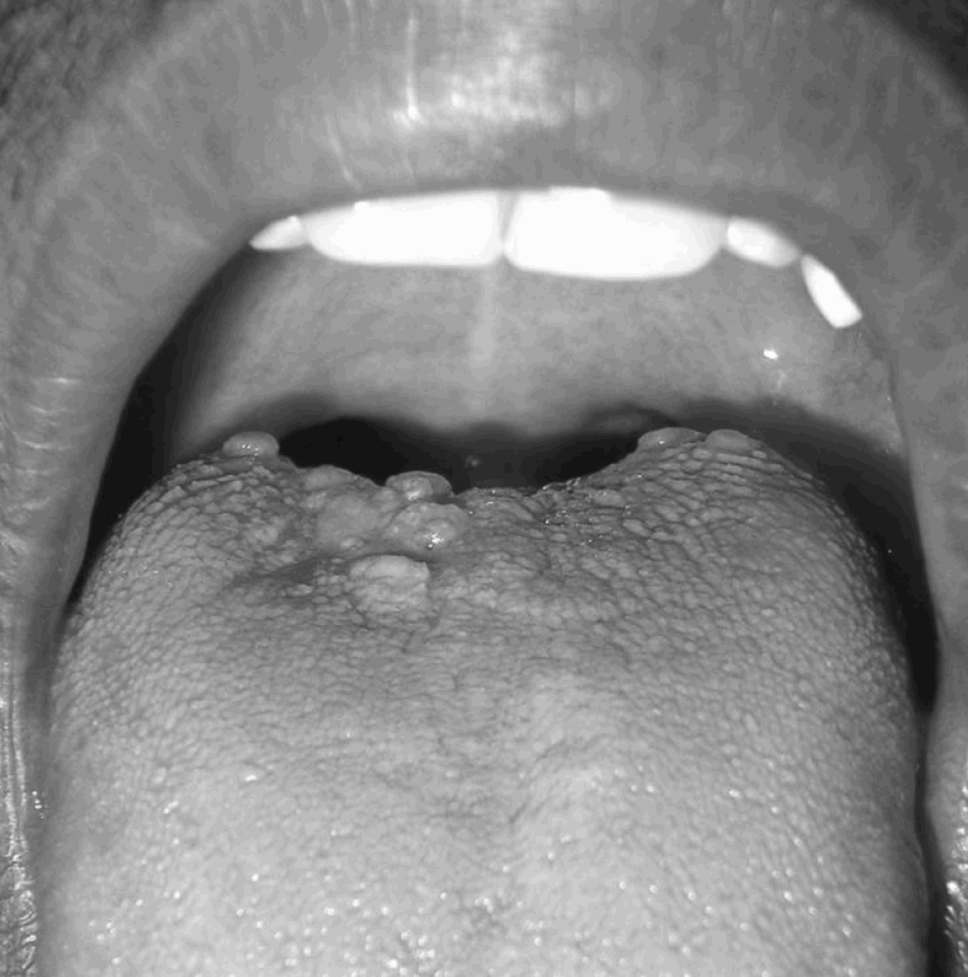
On examination, there was a cluster of small vesicles on the dorsum of the tongue, each about 1–2 mm in size, just proximal to the circumvallate papillae (Figure 1). The majority of vesicles were straw-coloured, with a few being red. No lesions were seen elsewhere in the oral cavity. There was no bleeding on palpation. The tongue was indurated in the area of the lesion.
(a) Pre-treatment photograph of the lesion in the posterior part of the oral tongue, containing multiple confluent vesicles, some of which were haemorrhagic in nature. (b) Closer view.

A biopsy was performed, which established the diagnosis of lymphangioma circumscriptum.
The patient's routine blood investigations and chest radiography were normal. Magnetic resonance imaging of the tongue was performed. This revealed the presence of an enhancing mass, approximately 3 cm in diameter, involving the dorsum of the tongue, which also had a deeper component involving the tongue musculature.
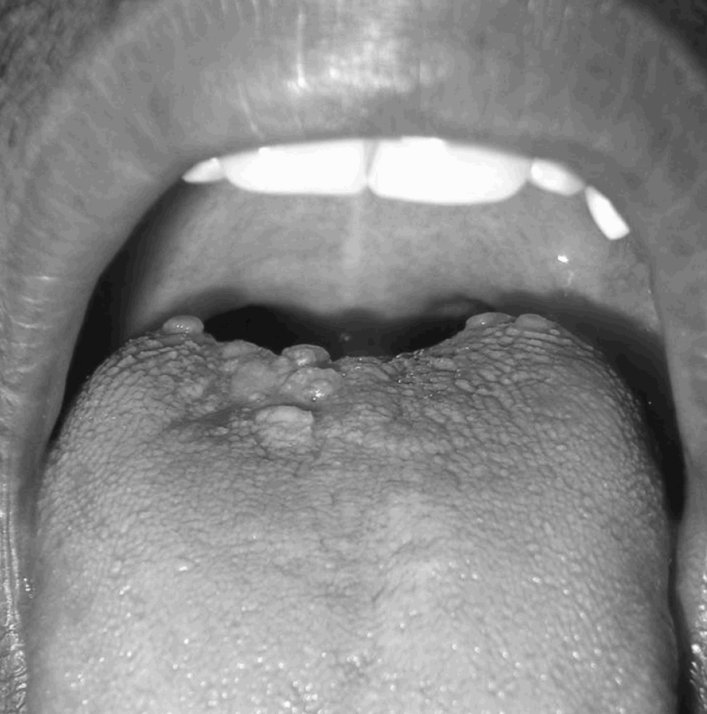
Sclerotherapy was performed with intralesional bleomycin. A total of three bleomycin injections of 0.75 U/kg each were given, two weeks apart. After each injection, the patient was observed for 24 hours for any adverse reactions such as fever or dyspnoea. No significant side effects were noted. After the first injection, there was a significant reduction in the size of the lesion. After the third injection, the patient was completely asymptomatic (Figure 2).
Photograph taken six months after completion of intralesional bleomycin injections, showing near-total resolution of the lesion, with only a few small, non-haemorrhagic, fluid-filled vesicles remaining. The patient was completely symptom-free over more than one year of follow up.
The patient was followed up for one year, without recurrence.
Discussion
Lymphangiomas are uncommon, congenital tumours of the lymphatic system which mostly appear in children under five years as lobulated masses or cysts, usually in the head and neck. Involvement of the tongue, however, is rare. Lymphangioma of the tongue was first described by Virchow in 1854.Reference Balakrishnan and Bailey5 A lymphangioma is thought to be a contiguous mass of dilated lymphatics, the cause of which is believed to be a developmental defect or primary malformation of the lymphatic channels.
Lymphangiomas can be classified histologically into four main groups, as follows. Lymphangioma simplex (capillary lymphangioma or lymphangioma circumscriptum) consists of thin-walled, vascular channels resembling capillaries, which appear as small vesicles protruding into the epidermis. Cavernous lymphangioma has larger, dilated lymphatic channels, can invade into the surrounding tissue, and is commonly seen in the tongue. Cystic lymphangioma (cystic hygroma) consists of large, multiloculated, fluid-filled chambers which represent true cysts lined by epithelium, and which may grow very large. Finally, lymphangiosarcoma (lymphoedema associated angiosarcoma) is a rare malignant tumour which develops after prolonged lymphatic obstruction and lymphoedema.
Lymphangioma circumscriptum is a term best reserved for a lymphatic malformation that is localised to an area of skin, subcutaneous tissue or occasionally muscle.Reference Mortimer, Burns, Breathnach, Cox and Griffiths1 Clinically, the condition manifests with fluid-filled vesicles; these may be well defined and discrete, or may be grouped into structures resembling frogs' spawn. The lymphangiomas may be translucent when the overlying epidermis or mucosa is very thin, or they may vary in colour from red to blue-black when they contain blood, a frequent finding. The term ‘circumscriptum’ may be misleading because, in many cases, there may be an extensive, deeper component to the malformation that is not clinically apparent. It has been postulated that the original malformation arises from deep, contractile lymphatics that are malformed and not in continuity with the normal lymph-conducting pathways.Reference Mortimer, Burns, Breathnach, Cox and Griffiths1 Tissue drainage into these abnormal lymphatics results in their gradual dilatation into lymphatic cisterns, contraction of which results in retrograde flow into the normal lymphatics. Identification of the limits of these subcutaneous cisterns prior to excision is essential for the success of surgical treatment.
Histopathologically, lymphangioma circumscriptum shows dilated lymphatics (either solitary or grouped) containing lymph or blood.Reference Calonje, Jones, Elder, Elenitsas, Johnson and Murphy6 The epidermis overlying the vesicle appears to be atrophic, with some degree of acanthosis and hyperkeratosis. The surrounding stoma shows scattered lymphocytes.
Lymphangioma circumscriptum may be present at any age but is usually noted at birth or appears during childhood. The commonest sites are the axillary folds, shoulders, flanks, proximal parts of the limbs and the perineum.Reference Mortimer, Burns, Breathnach, Cox and Griffiths1 Very few cases of lymphangioma circumscriptum involving the tongue have been reported.Reference Bansal and Sethuraman2–Reference Brandrup4
The treatment of choice is radical surgery or sclerotherapy. Simple electrocautery and vaporisation with CO2 laser are only of palliative benefit. Although some authors have reported the successful use of irradiation for treatment, this is no longer used because of its inability to completely destroy the lesion, its possible damaging effects on the growth of local structures and its potential to induce malignancy.Reference O'Cathail, Rostom and Johnson7, Reference Qin, Xin, Ren, Liu and Yao8 Surgical excision has been considered the best mode of treatment by the majority of surgeons; however patients can be affected by several complications, such as nerve injuries, prolonged lymphatic drainage, recurrence, wound infection, unacceptable scar formation, and incomplete resection due to infiltration into adjacent vital structures.Reference Feins, O'Neill, Rowe, Grosfeld, Fonklasrud and Coran9 Operative mortality is also significant, especially in the case of large lesions involving the oral cavity and those close to the airway.
Sclerotherapy is an alternative to surgery, and bleomycin and OK-432 (OK432 is a heat and penicillin-treated lyophilized powder of the Su-strain of Streptococcus pyogenes) are the two most commonly used sclerosants. Bleomycin injections have had excellent results in cases of cystic hygroma.Reference Qin, Xin, Ren, Liu and Yao8 In cases of cavernous lymphangioma and other, more complex lymphangiomatous lesions (such as lymphangioma circumscriptum and large angiosarcomas), the response to sclerotherapy is reportedly less effective.Reference Qin, Xin, Ren, Liu and Yao8 However, smaller cavernous lymphangiomas and lymphangioma circumscriptum lesions have been reported to show a good response.
The standard dosage of bleomycin as a single agent in cancer treatment is 0.25–0.5 mg/kg (0.75–1.5 U/kg), given intravenously, intramuscularly or subcutaneously, once or twice per week. For the treatment of lymphangiomas, doses of 0.25–0.6 mg/kg (0.75–1.8 U/kg) have been reported for each injection; injections are given at two-week to two-month intervals, and the cumulative amount of injected bleomycin is up to 50 mg (150 U) or 5 mg/kg (15 U/kg).Reference Sanlialp, Karnak, Tanyel, Senocak and Buyukpamukcu10, Reference Stringel and Ashcraft11 The dosage used in our patient was three injections of 0.75 U/kg each, at two-week intervals, with a total dosage of 2.25 U/kg of bleomycin.
• Lymphangioma circumscriptum, a rare variety of lymphangioma simplex, occurs even more rarely in the tongue
• Treatment is indicated only in symptomatic patients or due to cosmetic reasons
• Although complete surgical excision is the most widely used treatment, more conservative procedures such as sclerotherapy are being increasingly used for treatment of lymphangiomas
• In the presented case of lymphangioma circumscriptum of the tongue, intralesional injections of bleomycin (three injections of 0.75 U/kg body weight each, at two-week intervals) led to successful resolution
The main side effects of bleomycin are pulmonary toxicity, mucocutaneous effects (such as erythema, oedema and alopecia) and fever.Reference Dorr, von Hoff, Dorr and von Hoff12 The risk of late pulmonary toxicity, a life-threatening complication of the drug, is related to the cumulative dose. It is more common in patients over 70 years of age, those receiving prior pulmonary irradiation, those receiving supplementary oxygen, and when the total dose exceeds 400 U.Reference Dorr, von Hoff, Dorr and von Hoff12 Life-threatening pulmonary toxicity is rare at total doses below 150 mg or 450 U, occurring in 3 to 5 per cent of patients.Reference Dorr, von Hoff, Dorr and von Hoff12 Patients with pulmonary toxicity present with a persistent dry cough and exertional dyspnoea that can progress to tachypnoea, hypoxia and death. The chest X-ray typically shows reticulonodular infiltrates at the lung bases. Acute pulmonary reactions are unpredictable, and can occur at much lower doses. The total dose used in our patient was one-third of the stated danger level, and no side effects were observed for more than a year after the treatment.
Therefore, the present case demonstrates that bleomycin injection may be a simple and effective method of sclerotherapy for lymphangioma circumscriptum. However, the possible side effects and dose limitations of the drug should always be considered.