

Introduction
Microcrystal electron diffraction (MicroED) uses crystals that are a billionth of the size typically needed for x-ray crystallography.Reference Nannenga and Gonen1 For small molecules and natural products (molecules produced by natural sources such as natural drugs and metabolites), these crystals may have a powder-like appearance, which may make them seem to be amorphous to the eye.Reference Jones, Martynowycz, Hattne, Fulton, Stoltz, Rodriguez, Nelson and Gonen2,Reference Gruene, Wennmacher, Zaubitzer, Holstein, Heidler, Fecteau-Lefebvre, De Carlo, Muller, Goldie, Regeni, Li, Santiso-Quinones, Steinfeld, Handschin, van Genderen, van Bokhoven, Clever and Pantelic3
These types of samples are typically placed onto an electron microscopy grid and plunged into ethane cooled by liquid nitrogen prior to loading into the cryo-transmission electron microscope to facilitate investigation under cryogenic conditions. Although sufficient data can be collected from small molecules without cooling, the cryogenic conditions improve the obtained data by reducing the effects of radiation damage. For proteins, peptides, natural products, and samples susceptible to radiation damage, cryogenic conditions, typically liquid nitrogen or liquid ethane, are imperative for the collection of high-quality data. Once frozen, the grid is transferred under cryogenic conditions into a cryo-TEM and investigated under low-dose conditions.
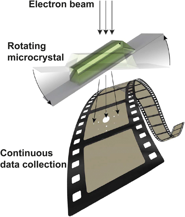
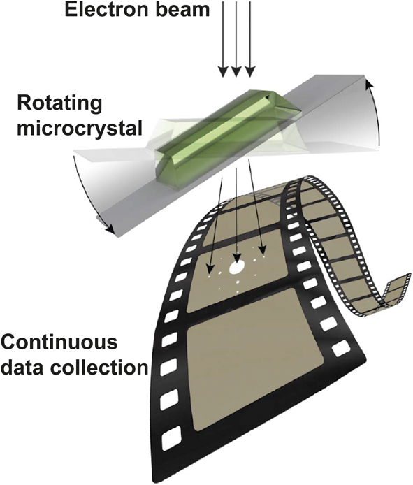
Nanocrystals in these observations appear as dark shapes, often with sharp edges.Reference Shi, Nannenga, de la Cruz, Liu, Sawtelle, Calero, Reyes, Hattne and Gonen4 When a target is identified, the electron beam is focused onto the sample, and continuous rotation MicroED data are collected by using a fast camera as the sample is continuously rotated in a single directionReference Shi, Nannenga, de la Cruz, Liu, Sawtelle, Calero, Reyes, Hattne and Gonen4,Reference Nannenga, Shi, Leslie and Gonen5 (Figure 1). It is important to note that for screening and data collection, the microscope is always operating in diffraction mode or low-magnification mode, never in higher magnification imaging modes, to limit the exposure of the sample to the damaging electron beam.Reference Hattne, Shi, Glynn, Zee, Gallagher-Jones, Martynowycz, Rodriguez and Gonen6 MicroED is analogous to the rotation method used in x-ray crystallography, and the data are processed using x-ray data processing software (e.g., MOSFLM, XDS, DIALSReference Leslie, Powell, Read and Sussman7–Reference Clabbers, Gruene, Parkhurst, Abrahams and Waterman9) after conversion using various tools.Reference Hattne, Reyes, Nannenga, Shi, de la Cruz, Leslie and Gonen10 Refinement proceeds as with any crystallography experiment and is described in greater detail next.
Microcrystal electron diffraction data collection. The electron beam is focused onto a continuously rotating sample, while data are collected.
MicroED methodology and procedures
In this section, we present an overview of the MicroED procedures—from sample preparation to structure determination. Because the descriptions are only briefly addressed, we encourage readers to refer to more detailed previously published literature.Reference Shi, Nannenga, de la Cruz, Liu, Sawtelle, Calero, Reyes, Hattne and Gonen4,Reference Hattne, Reyes, Nannenga, Shi, de la Cruz, Leslie and Gonen10–Reference de la Cruz, Martynowycz, Hattne and Gonen12
The procedure for MicroED analysis begins with the preparation of the crystalline material on a carbon-coated electron microscopy (EM) grid. For many samples, especially biological samples, it is important that the hydration of the sample be preserved; therefore, samples are applied to the carbon-coated EM grid, blotted to remove excess solution, and immediately plunged into liquid ethane to vitrify the sample. These procedures are similar to what is commonly used in single particle cryo-EM.Reference Passmore and Russo13 For samples where maintaining hydration is not important (e.g., crystals of inorganic materials or some small organic molecules), the vitrification process can be bypassed. In cases where the samples are in the form of dry powders, the sample can be directly added to the grid, shaken to remove excess material, and directly used for MicroED.Reference Jones, Martynowycz, Hattne, Fulton, Stoltz, Rodriguez, Nelson and Gonen2,Reference Gruene, Wennmacher, Zaubitzer, Holstein, Heidler, Fecteau-Lefebvre, De Carlo, Muller, Goldie, Regeni, Li, Santiso-Quinones, Steinfeld, Handschin, van Genderen, van Bokhoven, Clever and Pantelic3
Once the samples have been loaded into the cryo-TEM, they are screened at low magnification to assess the initial quality of the prepared sample. Grids that have many areas with unbroken carbon and microcrystals on the surface are considered good for further screening. The crystals are then screened using a higher magnification where the field of view consists of approximately one grid square (∼50 μm), and promising crystals (well separated from other crystals, with sharp edges) are then used to collect initial diffraction patterns by switching the microscope into diffraction mode. If a crystal shows high-resolution and high-quality diffraction, a full data set is collected from this crystal. The data are collected by continuously rotating the crystal in the electron beam as the high-speed detector records the diffraction data.Reference Nannenga, Shi, Leslie and Gonen5 It is important that the search and data collection steps be performed under a low electron dose in the TEM. Doses for data collection are typically on the order of <0.01 e–/Å2/s, and the dose during searching is even lower than that used during exposure.
Because the diffraction data collected from continuous rotation resemble the data collected by the rotation method in x-ray crystallography, the same data processing programs can be used for MicroED data.Reference Nannenga and Gonen1,Reference Nannenga, Shi, Leslie and Gonen5,Reference Clabbers, Gruene, Parkhurst, Abrahams and Waterman9,Reference Hattne, Reyes, Nannenga, Shi, de la Cruz, Leslie and Gonen10 Typically, only a single crystal or a small number of crystals are needed to generate a data set sufficient for structure determination in MicroED. For protein samples, structure solution consists of phasing the data using molecular replacement followed by refinement using electron-scattering factors in standard crystallographic refinement packages. For peptides, small molecules, or materials samples where resolution is generally much higher, phasing can be performed using direct methods followed by structure refinement. The final structures determined by MicroED are of similar quality to those determined by x-ray methods, with the important difference being the size of crystals used, which are orders of magnitude smaller for MicroED compared with what is required by x-ray crystallography.
Hardware and software for MicroED
Microscope choice and alignment are critical for a successful MicroED experiment. For proteins, we recommend the use of an electron microscope with a field-emission electron source and a cryogenic stage. For small molecules and organic and inorganic materials, a field-emission electron source is not imperative as demonstrated by recent MicroED studies.Reference Lanza, Margheritis, Mugnaioli, Cappello, Garau and Gemmi14,Reference Zhou, Luo, Luo, Li, Liu and Li15 Additionally, camera choice is important. The dynamic range and speed should be high. The physical size of the chip and pixels are also important. Hybrid pixel detectors such as TimePix and EIGER have a small sensor size and large pixels, which can be detrimental for diffraction from samples with large unit cells, however, these cameras have high speeds and high sensitivity.Reference van Genderen, Clabbers, Das, Stewart, Nederlof, Barentsen, Portillo, Pannu, Nicolopoulos, Gruene and Abrahams16,Reference Tinti, Frojdh, van Genderen, Gruene, Schmitt, de Winter, Weckhuysen and Abrahams17 The FalconIII direct electron detectorReference Hattne, Martynowycz, Penczek and Gonen18 and Direct Electron DE16 have been used successfully in MicroED experiments and appear promising. They can be shared between different cryo-EM modalities (e.g., electron tomography or single particle cryo-EM) where imaging is used.
Software for MicroED data collection
To perform MicroED experiments, the standard software that controls the TEM is used along with software that controls the camera of the TEM. The microscope software can be used to set the stage rotation rate and rotation range prior to data collection. Once this is set and the stage begins its rotation, the camera software begins to record high-speed movies and continues recording during stage rotation. Recently, SerialEM, a popular data automation suite developed for cryo-EM,Reference Mastronarde19 has been used to collect MicroED data.Reference de la Cruz, Martynowycz, Hattne and Gonen12 By using SerialEM, more than 300 MicroED data sets can be collected overnight without the need for user interaction during data collection. This will greatly improve the process of data collection and expedite structure determination using MicroED.
Software for MicroED data processing
MicroED data can be processed using standard data processing programs used for x-ray crystallography. In order to make use of these programs, parameters unique to electron diffraction experiments need to be entered (e.g., shorter wavelength and longer detector distance), and once the experimental parameters are properly described, the programs handle MicroED well. The two most popular programs used thus far for protein crystals have been XDS and MOSFLM, and XDS has been the program of choice for peptides and small molecules. Once the diffraction data have been indexed, integrated, and merged into a data set, the phases are determined using molecular replacement programs (e.g., PhaserReference McCoy, Grosse-Kunstleve, Adams, Winn, Storoni and Read20 or MOLREPReference Vagin and Teplyakov21) or using direct methods with programs such as those in the SHELXReference Sheldrick22 pipeline. For proteins and peptides, structure refinement is typically done using phenix.refineReference Afonine, Grosse-Kunstleve, Echols, Headd, Moriarty, Mustyakimov, Terwilliger, Urzhumtsev, Zwart and Adams23 or REFMAC,Reference Murshudov, Vagin and Dodson24 both of which allow for refinement against electron-scattering factors. Small molecule structure building and refinement for MicroED is performed using programs developed for small molecule x-ray crystallography such as SHELXL Reference Sheldrick25 and ShelXle.Reference Hubschle, Sheldrick and Dittrich26
Applications of MicroED for high-resolution structure determination
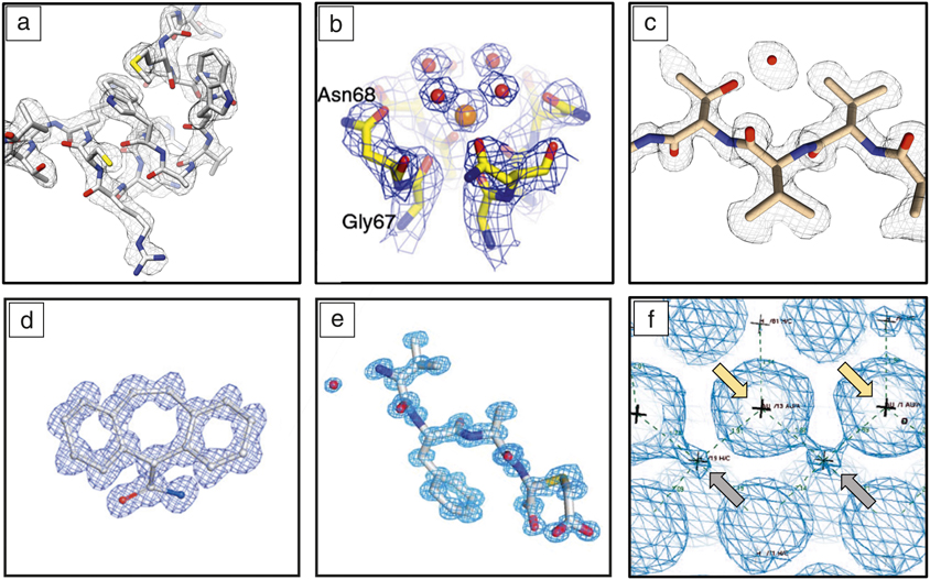
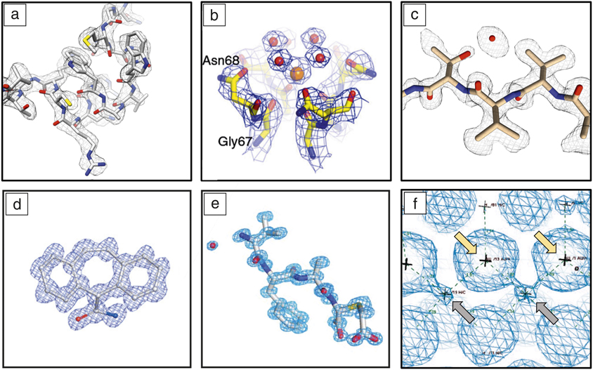
MicroED has been successfully used to study a wide variety of microcrystalline samples (Figure 2). In this section, we present and highlight some key applications of MicroED for protein, peptide, materials, and small molecule research.
Representative structures determined by microcrystal electron diffraction (MicroED). The MicroED method has been used on a variety of targets, including (a) soluble proteins such as lysozymeReference Nannenga, Shi, Leslie and Gonen5 and (b) membrane proteins such as the NaK ion channel. Asn68 and Gly67 are amino acids where Asn68 = asparagine 68 and Gly67 is glycine 67;34 (c) peptides such as the 11 residue fragment of α-synuclein;Reference Rodriguez, Ivanova, Sawaya, Cascio, Reyes, Shi, Sangwan, Guenther, Johnson, Zhang, Jiang, Arbing, Nannenga, Hattne, Whitelegge, Brewster, Messerschmidt, Boutet, Sauter, Gonen and Eisenberg53 (d) small molecules such as carbamazepine;Reference Gallagher-Jones, Glynn, Boyer, Martynowycz, Hernandez, Miao, Zee, Novikova, Goldschmidt, McFarlane, Helguera, Evans, Sawaya, Cascio, Eisenberg, Gonen and Rodriguez40 (e) natural products such as VFA-thiaGlu (valine-phenylalanine-alanine-thiaglutamate);Reference Ting, Funk, Halaby, Zhang, Gonen and van der Donk50 and (f) nanomaterials such as gold nanoparticles. Representative gold and hydrogen atoms in (f) are indicated by yellow and gray arrows, respectively. The “+” shows the location of the atoms, and the dashed lines indicate the distances between the atoms in the unit cell.Reference Vergara, Lukes, Martynowycz, Santiago, Plascencia-Villa, Weiss, de la Cruz, Black, Alvarez, Lopez-Lozano, Barnes, Lin, Weissker, Whetten, Gonen, Yacaman and Calero43
Protein crystals
MicroED was initially developed using microcrystals of soluble proteins. The first model protein used was on a hen egg white lysozyme,Reference Nannenga, Shi, Leslie and Gonen5,Reference Shi, Nannenga, Iadanza and Gonen27 which was soon followed by other model samples such as catalase,Reference Nannenga, Shi, Hattne, Reyes and Gonen28,Reference Yonekura, Kato, Ogasawara, Tomita and Toyoshima29 an antioxidant enzyme, and proteinase K, which aids in the general digestion of protein in biological samples.Reference Hattne, Shi, de la Cruz, Reyes and Gonen30 In addition to model samples used for methods development, MicroED has been used for the study of new structures, including a structure of a drug–protein complex involved in the treatment of HIV,Reference Purdy, Shi, Chrustowicz, Hattne, Gonen and Yeager31 the structure of a challenging complex transforming growth factor (TGF)-βm-TβRII, which is involved in cell growth,Reference de la Cruz, Hattne, Shi, Seidler, Rodriguez, Reyes, Sawaya, Cascio, Weiss, Kim, Hinck, Hinck, Calero, Eisenberg and Gonen32 and a novel bacterial oxidase.Reference Xu, Lebrette, Clabbers, Zhao, Griese, Zou and Högbom33 In addition to the study of soluble proteins, MicroED has been used with membrane protein microcrystals. The structure of the Ca2+-ATPase (ATP = adenosine triphosphate), a protein found in the cell plasma membrane that helps to remove calcium from the cell, was determined;Reference Yonekura, Kato, Ogasawara, Tomita and Toyoshima29 the nonselective cation channel NaK was determined to be 2.5 Å using nanocrystals.Reference Liu and Gonen34 The latter allowed the identification of two new confirmations in the channel that had not been previously identified.
Peptide structure determination
MicroED has been successful in the structural characterization of peptides, specifically those involved in neurodegenerative diseases.Reference Nannenga and Gonen1,Reference Rodriguez, Eisenberg and Gonen35,Reference Nannenga and Gonen36 The first novel structure determined by MicroED was of an 11-residue toxic fragment of the synuclein protein, which is involved in Parkinson’s disease. The structure of this peptide had resisted all other methods of structure determination; however, when MicroED was used, a 1.4 Å resolution structure was determined in a matter of days. Since this first peptide structure, several peptides have been studied using MicroED,Reference de la Cruz, Hattne, Shi, Seidler, Rodriguez, Reyes, Sawaya, Cascio, Weiss, Kim, Hinck, Hinck, Calero, Eisenberg and Gonen32,Reference Seidler, Boyer, Rodriguez, Sawaya, Cascio, Murray, Gonen and Eisenberg37–Reference Sawaya, Rodriguez, Cascio, Collazo, Shi, Reyes, Hattne, Gonen and Eisenberg42 greatly adding to our understanding of the structure of the proteins responsible for neurodegenerative disease.
MicroED with nanomaterials
In the field of materials research, researchers applied MicroED to determine the structure of a surface stabilized gold nanoparticle.Reference Vergara, Lukes, Martynowycz, Santiago, Plascencia-Villa, Weiss, de la Cruz, Black, Alvarez, Lopez-Lozano, Barnes, Lin, Weissker, Whetten, Gonen, Yacaman and Calero43 In this work, ligand capped gold nanoparticles were crystallized and then grew into large crystals. When these larger crystals were analyzed by x-ray crystallography, the resolution was limited to 1.3 Å. However, when the crystals were fragmented and analyzed in their frozen-hydrated state by MicroED, the nanocrystals produced sub-Ångstrom diffraction (0.87 Å), where a total of 146 gold atoms could be identified along with 57 ligands on the surface of the gold nanoparticle. The structure of this gold–nanoparticle cluster was able to shed light on the processes that are important for the formation of metallic nanomaterials. Electron diffraction of inorganic materials (e.g., BaSO4 and vaterite) has been performed for decades and continues to be a powerful approach.Reference Wan, Sun, Su, Hovmoller and Zou44–Reference Gemmi, Mugnaioli, Gorelik, Kolb, Palatinus, Boullay, Hovmöller and Abrahams49
Small molecules and natural products
Small organic molecules (e.g., pharmaceuticals) have been studied by MicroED extensively and are relatively straightforward due in large part to simpler sample preparation methods directly from synthesized material.Reference Jones, Martynowycz, Hattne, Fulton, Stoltz, Rodriguez, Nelson and Gonen2,Reference Gruene, Wennmacher, Zaubitzer, Holstein, Heidler, Fecteau-Lefebvre, De Carlo, Muller, Goldie, Regeni, Li, Santiso-Quinones, Steinfeld, Handschin, van Genderen, van Bokhoven, Clever and Pantelic3,Reference van Genderen, Clabbers, Das, Stewart, Nederlof, Barentsen, Portillo, Pannu, Nicolopoulos, Gruene and Abrahams16,Reference Gallagher-Jones, Glynn, Boyer, Martynowycz, Hernandez, Miao, Zee, Novikova, Goldschmidt, McFarlane, Helguera, Evans, Sawaya, Cascio, Eisenberg, Gonen and Rodriguez40 Typically, the samples are powders, seemingly amorphous; data can be collected and structures solved to atomic resolution in less than 30 min routinely. The success rate in our MicroED studies of small molecules is ∼90%.
A particularly exciting avenue of research that has already benefitted from MicroED is the field of natural product research. Typically, small amounts of natural products can be isolated from natural sources, but the amount of material can often be sufficient for investigation by MicroED. The first example of a natural product, which was amino-acid derived, was recently describedReference Ting, Funk, Halaby, Zhang, Gonen and van der Donk50 where the relative orientation of components could also be unambiguously determined experimentally by MicroED.
Summary and future view
MicroED is a relatively new method that is still in its infancy, and yet, in the short six years since the initial demonstration,Reference Shi, Nannenga, Iadanza and Gonen27 it has allowed the determination of several samples that were intractable by other methods,Reference Nannenga and Gonen1,Reference Xu, Lebrette, Clabbers, Zhao, Griese, Zou and Högbom33,Reference Krotee, Griner, Sawaya, Cascio, Rodriguez, Shi, Philipp, Murray, Saelices, Lee, Seidler, Glabe, Jiang, Gonen and Eisenberg38,Reference Krotee, Rodriguez, Sawaya, Cascio, Reyes, Shi, Hattne, Nannenga, Oskarsson, Philipp, Griner, Jiang, Glabe, Westermark, Gonen and Eisenberg41,Reference Zatsepin, Li, Colasurd and Nannenga51–Reference Guenther, Cao, Trinh, Lu, Sawaya, Cascio, Boyer, Rodriguez, Hughes and Eisenberg54 and is quickly gaining momentum in the scientific community. More than 75 structures have been deposited to the protein data bank and other depositories, and several peer-reviewed publications have resulted. Now is the time to invest in MicroED infrastructure and expertise. Service centers and user facilities similar to those at synchrotrons should be established to provide the scientific community easy, quick, and efficient access to this technology.
The first MicroED dedicated facility has opened at the University of California, Los Angeles; the MicroED Imaging Center. The center is open to both academic labs and for-profit organizations and is operated for MicroED studies similar to a synchrotron facility. Users send their samples to the center and dedicated experienced personnel prepare the cryoEM grids, collect MicroED data, and send the data back to users. Center personnel can also solve structures if needed and provide MicroED training.
As the significance of MicroED grows, we predict that several such dedicated centers will be established worldwide. A good starting point is synchrotrons and National Institute of Health funded National and regional centers that already invest in cryoEM equipment. The same equipment and cameras can be used for MicroED and other cryoEM modalities. All that remains is training, which can be achieved through workshops.
Acknowledgments
The Gonen laboratory is supported by funding from the Howard Hughes Medical Institute (HHMI). The Nannenga laboratory is supported by the National Institutes of Health (R01GM124152 and R21GM135784), and the Air Force Office of Scientific Research Grant No. FA9550-18-1-0012. MicroED was developed at the Janelia Research Campus of HHMI using HHMI funds and Janelia Visitor Program funds.

Brent Nannenga is an assistant professor of chemical engineering in the School for Engineering Matter, Transport and Energy at Arizona State University and a member of The Biodesign Institute’s Center for Applied Structural Discovery. He obtained his PhD degree from the University of Washington in chemical engineering in 2011. His research focuses on developing and using methods for high-resolution structure determination, specifically microcrystal electron diffraction, and employing structural insights gained from these methods to engineer biomolecules and biological systems. His awards include an Air Force Office of Scientific Research Young Investigator Award, and a Fulton Outstanding Assistant Professor Award. Nannenga can be reached by email at brent.nannenga@asu.edu.

Tamir Gonen is a professor of biological chemistry and physiology at the David Geffen School of Medicine of the University of California, Los Angeles, an investigator at the Howard Hughes Medical Institute, and a Member of the Royal Society of New Zealand. He obtained his PhD degree from The University of Auckland, New Zealand, in biological science, in 2002. In 2011, while leading a laboratory at the Howard Hughes Medical Institute, Janelia Research Campus, Gonen began developing microcrystal electron diffraction as a new method for structural biology. His research determined several previously unknown structures at resolutions better than 1 Å. He has authored more than 100 publications and mentored faculty from top universities around the world. Gonen can be reached by email at tgonen@ucla.edu.