Introduction
Genomic sequence along with anatomical and physiological resemblance to humans makes porcine species an excellent research model to investigate different human health-related issue (Gutierrez et al., Reference Gutierrez, Dicks, Glanzner, Agellon and Bordignon2015; Jeon et al., Reference Jeon, Kim and Lee2017; Perleberg et al., Reference Perleberg, Kind and Schnieke2018). Transgenic pigs may serve as a potential source of cells, tissues, and organs for xenotransplantation; somatic cell nuclear transfer (SCNT) is a useful technique for production of transgenic pigs with specific genetic modifications (Luo et al., Reference Luo, Lin, Bolund, Jensen and Sørensen2012; Perleberg et al., Reference Perleberg, Kind and Schnieke2018; Zhang X et al., Reference Petersen, Lucas-Hahn, Oropeza, Hornen, Lemme, Hassel, Queisser and Niemann2019). However, the production of the transgenic pigs using SCNT has a lower efficiency compared with the other livestock species (Polejaeva et al., Reference Polejaeva, Chen, Vaught, Page, Mullins, Ball, Dai, Boone, Walker, Ayares, Colman and Campbell2000). Among different factors, the age and parity (Bari et al., Reference Bari, Khalid, Haresign, Murray and Merrell2003), method of oestrus induction, and endocrine profiles (Ashworth et al., Reference Ashworth, Sales and Wilmut1989) of the surrogate mother affect the establishment of pregnancy. The level of genetic modification has also been reported to affect the production of genetically modified pigs, resulting in early death (Kim et al., Reference Kim, Jin, Lee, Taweechaipaisankul, Oh, Hwang, Ahn, Saadeldin and Lee2017; Park et al., Reference Park, Cho, Koo, Kim, Kang, Hurh, Kim, Yeom, Moon, Lee, Choi, Hong, Jang, Hwang, Yang, Lee and Ahn2014). Moreover, incomplete or improper reprogramming of donor nuclei may be responsible for the fewer numbers of healthy individuals (Campbell et al., Reference Campbell, Fisher, Chen, Choi, Kelly, Lee and Xhu2007). The dictation of reprogramming-related cues by oocytes is more natural in SCNT, with more normal DNA-methylation patterns, but in some cases DNA-methylation defects may appear in some small genomic regions (Krupalnik and Hanna, Reference Krupalnik and Hanna2014).
In this study, we generated transgenic cloned pigs with knockout of α-1,3-galactosyltransferase (GalT) and introduction of human CD55 (hCD55), human endothelial protein C receptor (hEPCR), and human transducin (hTM) genes using SCNT to supply solid organs for xenotransplantation. The GalT gene was removed to prevent hyperacute rejection by Galα1,3Galβ1,4GlcNAc-R (α-Gal) and hCD55, hEPCR and hTM genes were introduced to prevent acute humoral rejection in xenotransplantation of porcine organs to primates (Shimizu et al., Reference Shimizu, Smith, Zhao, Medof and Sykes2006; Macher and Galili, Reference Macher and Galili2008; Yazaki et al., Reference Yazaki, Iwamoto, Onishi, Miwa, Hashimoto, Oishi, Suzuki, Fuchimoto, Sembon, Furusawa, Liu, Nagasaka, Kuzuya, Ogawa, Yamamoto, Iwasaki, Haneda, Maruyama and Kobayashi2012). Donor cells for these transgenic pigs were generated by transfection of a ubiquitous F2A-bicistronic expression vector for hEPCR and hTM genes into hCD55 + GalT –/– primary fibroblasts (Perota et al., Reference Perota, Lagutina, Colleoni, Duchi, Lazzari, Cozzi, Lucchini and Galli2011). However, the modification of multiple genes in transgenic cloned pigs reduced the production efficiency compared with wild-type cell cloning (Kurome et al., Reference Kurome, Ishikawa, Tomii, Ueno, Shimada, Yazawa and Nagashima2008). To provide a stable supply of porcine solid organs for xenotransplantation studies, it is necessary to ensure a high production efficiency for the transgenic pig. To improve production efficiency, kidney cells established from the cloned fetus were used as the donor nucleus for recloning and may be beneficial for nuclear reprogramming of donor cells after SCNT because of the least differentiated donor cells in this study. Porcine kidney cells showed karyotype stability up to 71 passages and higher development rates after SCNT, described in the study by Richter et al. (Reference Richter, Kurome, Kessler, Zakhartchenko, Klymiuk, Nagashima, Wolf and Wuensch2102). Higher production efficiency of recloning in transgenic pigs has been shown in previous studies (Fujimura et al., Reference Fujimura, Murakami, Kurome, Takahagi, Shigehisa and Nagashima2008; Zhao et al., Reference Zhao, Li, Wiriyahdamrong, Yuan, Qing, Li, Xu, Guo, Jia, Zhang, Cheng, Su, Long, Wang, Zou, Kinoshita, Zhao and Wei2020) along with the same phenotypic and protein expression (Zhao et al., Reference Zhao, Li, Wiriyahdamrong, Yuan, Qing, Li, Xu, Guo, Jia, Zhang, Cheng, Su, Long, Wang, Zou, Kinoshita, Zhao and Wei2020). Recloned transgenic pigs have been reported to possess stable genetic transmission capacities and normal reproductive performances (Cao et al., Reference Cao, Li, Wen, Li, Mi, Zhang, Li and Li2014). In this study, cell lines from cloned fetal kidney derived from aborted fetuses were established and used as donor cells for recloning. Additionally, preimplantation development competence and gene expression of recloned embryos were investigated, followed by the production of recloned transgenic pigs using gilts as surrogate recipients.
Studies related to transgenic cloned pig production usually involve the use of gilts with synchronized oestrus as recipients due to their low cost and management convenience (Nagaya et al., Reference Nagaya, Watanabe, Kobayashi, Nakano, Arai, Asano, Takeishi, Umeki, Fukuda, Yashima, Takayanagi, Watanabe, Onodera, Matsunari, Umeyama and Nagashima2016; Cho B et al., Reference Perleberg, Kind and Schnieke2018). However, oestrus signs were not properly expressed at the expected time in all recipient gilts after oestrus induction and cloned embryos could not be transferred into recipient gilts. Accurate oestrus timing at the expected day in recipients can be achieved in sows by controlling the weaning time and decisions can be made at day 8 post-weaning for embryo transfer. Sows are very important for the farm and higher costs are necessary when these animals are used as recipients. However, the use of sows as recipients is advantageous for the generation of transgenic pigs in terms of their higher parturition rates and accurate oestrus timing (Clark et al., Reference Clark, Schinckel, Singleton, Einstein and Teclaw1989).
Based on the above-mentioned facts, this study was conducted to improve the conditions for generating transgenic pigs with the genotype of GalT –/– hCD55-hEPCR-hTM for xenotransplantation use. To generate this multigene-modified pigs, we compared three types of surrogate pigs (sows vs. gilts, and the gilts were either from natural or induced oestrus). Pregnancy and parturition rates were compared according to parity, oestrus induction and ovarian status, retrospectively. Additionally, recloning was also compared using two donor karyotypes (cloned fetal kidney cells vs. neonatal ear fibroblast) and the in vitro developmental competence and expression of genes related to nuclear reprogramming was assessed in the resultant recloned embryos.
Materials and Methods
All chemicals and reagents were purchased from Sigma-Aldrich Chemical Company (St. Louis, MO, USA) unless otherwise indicated. All experiments were approved and performed following the guidelines of the Institutional Animal Care and Use Committee of Chungnam National University (IACUC approval no. CNU-00624).
Experimental designs
This study was conducted to improve the conditions for generating transgenic pigs with the GalT –/– -hCD55-hEPCR-hTM genotype for xenotransplantation purposes. In the production procedure, production rates were investigated according to various conditions of recipient pigs and donor cell type. In Experiment 1, pregnancy and parturition rates were compared according to various conditions of recipient pigs including parity, estrous induction, and ovulation. In Experiment 2, in vivo developmental competence of cloned embryos after embryo transfer was compared between neonatal ear fibroblast and cloned fetal kidney cells as donor nuclei. In Experiment 3 the in vitro development rate of both the above-used donor cell groups was evaluated to investigate the reason for maintenance of pregnancy to term only in recloned groups using cloned fetal kidney cells as donor nuclei. Additionally, gene expression of reprogramming-related genes in blastocysts was also investigated between the two groups.
Donor cell preparation
Transgenic porcine donor cell lines were kindly provided by the Ceasare laboratory (Perota et al., Reference Perota, Lagutina, Colleoni, Duchi, Lazzari, Cozzi, Lucchini and Galli2011). Briefly, hTM and hEPCR genes were introduced into pig primary GalT –/– -hCD55 neonatal ear fibroblasts using a ubiquitous F2A-bicistronic expression vector. The establishment of cloned kidney cell lines for recloning was conducted as explained previously (Kim et al., Reference Kim, Roy, Fang, Hassan and Cho2019). Briefly, primary cell culture was prepared using kidneys of porcine cloned fetuses isolated at 50 days of gestation. Kidneys were cut into small pieces and centrifugation was performed several times followed by culturing for up to three or four passages. The donor cell cycle G0/G1 stage was synchronized for 48–72 h and the same passage numbers were used for each replicate (3–8 passages). Donor cells were trypsinized and suspended in HEPES-buffered Tyrode’s (TLH) medium with 0.4% (w/v) BSA.
Production of transgenic embryos
The collection of porcine oocytes and SCNT were performed as described in our previous study (Fang et al., Reference Fang, Qamar, Shin and Cho2019; Roy et al., Reference Roy, Qamar, Fang, Hassan and Cho2020). Porcine ovaries were collected from a local slaughterhouse within 4 h of slaughtering. Fluid in 3–8 mm diameter follicles was aspirated using a 10-ml syringe with an 18-gauge needle and collected into a 15-ml conical tube. After allowing the oocytes to settle for 5 min, the fluid was washed with TLH medium containing 0.05% (w/v) polyvinyl alcohol (PVA). Cumulus–oocyte complexes (COCs) with at least three layers of compact cumulus cells were selected using a stereomicroscope and were washed three times with TLH–PVA and two or three times with D-PBS modified with the addition of 0.4% BSA (mDPBS). Following washing, 50–80 COCs were selected per each well of a 4-well multidish containing 500 μl of TCM-199 for IVM including 10 IU/ml PMSG/hCG (Intervet International BV, The Netherlands). Selected COCs were cultured in a 5% CO2 in air humidified atmosphere at a temperature of 39°C. After 22 h of maturation, COCs were transferred into hormone-free IVM medium after washing with IVM medium without hormone and cultured under the same conditions for an additional 18–21 h. Matured COCs were denuded using 0.1% (w/v) hyaluronidase with gentle and repeated pipetting. Denuded oocytes were washed three times in hormone-free IVM medium, and nuclear material staining was performed by incubating matured oocytes for 15 min in 5 μg/μl Hoechst 33342 medium. In manipulation medium, enucleation of matured oocytes at the metaphase II (MII) stage was performed, removing the MII plate and first polar body (PB) with a 17-µm bevelled glass pipette (Humagen, Charlottesville, VA, USA) under an epifluorescence microscope. After enucleation, a single somatic cell derived from GalT –/– -hCD55-hEPCR-hTM neonatal ear fibroblast or cloned fetal kidney cells with the same genotype was inserted into the perivitelline space of an enucleated oocyte. Reconstructed oocytes were fused in 280 mM mannitol solution with a low Ca2+ concentration (0.001 mM) by electric cell fusion, exposing oocytes to 160 V/cm direct current (DC) of two pulses for 40 µs and 2 V/cm alternating current for 2 s using a BTX 2001 Electro-cell manipulator (BTX, San Diego, CA, USA). After 30 min of fusion, electrical activation was performed in 280 mM mannitol solution with 0.01 mM CaCl2 and 0.05 mM MgCl2 and 120 V/cm of DC of two pulses for 60 μs. After electrical activation, cloned embryos were treated for post-activation with 10 μg/ml cytochalasin B combined with 6′-dimethylaminopurine (DMAP) for 4 h. Cloned embryos were washed three times with IVC medium (PZM5; IFP0410P, IFP, Higashine, Yamagata, Japan) and cultured in 25-μl droplets. The culture medium was covered with pre-warmed mineral oil and incubated at 5% O2, 5% CO2, and 90% N2 in a humidified atmosphere at 39°C.
Preparation of recipient pigs
Recipient pigs used as surrogate mothers include sows with natural oestrus and gilts with a natural and induced oestrus. Sows from a private pig farm with three or four parities and high reproductive activity were selected as the surrogate recipients. Sows were weaned on day 0 (D0) (Thursday) and sows with oestrus expression on D6 (Tuesday) were selected as recipients. On D8 (Friday), embryos were transferred to each selected recipient. For gilts, pigs weighing 100–150 kg were selected as surrogate mothers and divided into natural oestrus group and synchronized oestrus group by feeding 20 mg of altrenogest (Regumate® Porcine; MSD Animal Health, NJ, USA) for 18 days consecutively. Embryo transfer was performed on D3 after observed oestrus in the natural oestrus group and on D5 after stopping feeding altrenogest. To compare the effect of natural and induced oestrus, only gilts were considered assuming they had similar reproductive performance and the lesser numbers of sow.
Embryo transfer and parturition
Before starting embryo transfer, anaesthesia was induced in surrogate mothers by intravenous injection of xylazine hydrochloride and ketamine, and later on anaesthesia was maintained using isoflurane. The cloned embryos cultured for 16–20 h after activation were loaded into GIFT catheters (KGIFT-110; Cook Medical Co., IN, USA) and transferred to the oviducts of the surrogate (150 embryos per recipient) after midventral laparotomy. Pregnancy diagnosis was carried out only in non-return surrogates at 25 days after embryo transfer by a transabdominal ultrasound scanner. At 114 days after embryo transfer, Caesarean section was performed for the delivery of transgenic pigs.
Genotyping
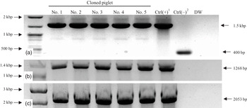
Tail biopsy in the cloned piglets was performed for genotyping to verify the gene composition of GalT –/– -hCD55-hEPCR-hTM by polymerase chain reaction (PCR) (Cho J et al., Reference Perota, Lagutina, Colleoni, Duchi, Lazzari, Cozzi, Lucchini and Galli2012). Deletion of GalT (F: TCTGACTGACCGCGTTACTCC, R: CGGGAAAACTTGTACGGC) and presence of exogenous hCD55 (F: TCTGACTGACCGCGTTACTCC, R: CGGGAAAACTTGTACGGC), hEPCR and hTM genes (F: TCTGACTGACCGCGTTACTCC, R: ACTGGCATTGAGGAAGGTCG) using each primer sets (Fig. 1).
Genotyping of five cloned piglet by genomic PCR. (a) Genotyping of GGAT1 knockout in cloned piglets. Bands of 1500 bp are shown in GGAT1-KO pigs and 400 bp are shown in wild-type pig. (b) Genotyping of hCD55 gene in cloned piglets. Bands of 1268 bp are shown in all five cloned piglets. (c) Genotyping of hEPCR-hTM gene in cloned piglets. Bands of 2053 bp are shown in all five cloned piglets. 1Positive control (Ctrl): DNA derived from donor cells. 2Negative control: DNA derived from ovary cells of wild-type pig. DW, distilled water.

Quantitative real-time PCR (qRT-PCR) analysis of gene expression profiles
The assessment of mRNA expression of genes related to nuclear reprogramming (POU5F1, SOX2 and NANOG) and apoptosis (BCL2 and BAX) was compared in the blastocysts of cloned and recloned embryos using quantitative PCR (qPCR) to investigate the different development competencies, as described in our previous study (Roy et al., Reference Roy, Qamar, Fang, Kim, Bang, De Zoysa, Shin and Cho2021). Total RNA was isolated from five porcine blastocysts on day 6 post culture using TRIzol reagent (Invitrogen USA); subsequently, cDNA was synthesized using reverse transcription 2× RT Pre-Mix (BioFACT, Daejeon, South Korea) and oligo(dT) primers (18-mers; Neoprove, Daejeon, South Korea). Real-time qPCR was performed using 1 μl of the cDNA template and a 2× real-time PCR Pre-Mix (BioFACT) containing specific primers. The reactions were performed for 39 cycles with the following cycle parameters: denaturation at 95°C for 15 min 20 s, annealing and extension at 60°C for 40 s. The primer sequences used are presented in Table 1. The expression levels of the target genes were quantified as expression relative to that of the internal control gene (β-actin). PCR specificity was checked by performing a melting curve data analysis. Relative expression quantification was based on the comparison of threshold cycles (Ct) and constant fluorescence intensity. Relative mRNA expression was quantified using the 2−(∆Ct sample − ∆Ct control) method (Livak and Schmittgen, Reference Livak and Schmittgen2001). To determine a normalized arbitrary value for each gene, every value was normalized to that of β-actin.
Sequences of specific primers used for gene expression analysis by qPCR

bp, base pairs; NCBI, National Center for Biotechnology Information; qPCR, quantitative polymerase chain reaction.
Statistical analysis
Every experiment for embryonic development was repeated at least eight times. Statistical Package for Social Sciences version 21.0 software (IBM, Armonk, NY, USA) was used to perform data analysis. Student’s t-test was used to compare the two numeric groups, while chi-squared test was performed to analyze the difference in percentages. Statistical significance was considered at P < 0.05. All experimental data percentages were presented as the mean ± standard error of the mean (SEM).
Results
Development of cloned pigs according to various conditions of recipient pigs
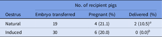
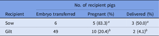
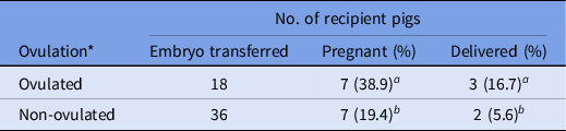
In vivo development after surgical transfer of cloned transgenic embryo was investigated according to parity, oestrus induction and ovulation. While comparing the pregnancy and parturition rates according to oestrus induction of recipients only in gilts, production of cloned piglets was observed in recipient pigs with natural oestrus (10.5%), but not in induced oestrus recipients (0.0%) (Table 2). For parity of recipient pigs, significantly higher pregnancy (83.3 vs. 20.4%) and parturition (50.0 vs. 4.1%) rates were shown when sows were used as recipients compared with that of gilts (Table 3; P < 0.05). Regarding parity, almost all the recipient sows became pregnant (83.3%) and cloned piglets were produced in three out of six sows, whereas delivery was shown for only two out of 49 recipients gilts. When pregnancy and parturition rates were compared according to ovarian status (ovulation) of the recipient at the time of embryo transfer, the recipient group with the ovulated ovary showed significantly higher pregnancy (38.9 vs. 19.4%) and delivery (16.7 vs. 5.6%) rate (Table 4; P < 0.05).
Pregnancy and parturition rates after transfer of cloned porcine embryos into recipient gilts with natural and induced oestrus

a,b Values in the same column with different superscripts differ significantly (P < 0.05).
Pregnancy and parturition rates after transfer of cloned porcine embryos into sow and gilts with natural oestrus

a,b Values in the same column with different superscripts differ significantly (P < 0.05).
Pregnancy and parturition rates after transfer of cloned porcine embryos into ovulated and non-ovulated recipients

a,b Values within the same column with different superscripts differ significantly (P < 0.05).
* Ovaries were not detected to determine ovulation status in one recipient pig.
Production of cloned pigs by donor cell type in recipient gilts
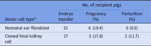
Production efficiency of transgenic cloned pigs using neonatal ear fibroblasts and cloned fetal kidney cells as donor cells was compared, as shown in Table 5, to investigate the recloning efficiency only in recipient gilts. No significant difference in pregnancy rates was observed between neonatal ear fibroblasts and fetal kidney cells: 6/31(19.3%) vs 3/17(17.6%) (P > 0.05) even though this was higher in the neonatal kidney cell donor type. In the neonatal ear fibroblasts group, all gilts (0/6) aborted before term, whereas two out of three (66.7%) in the cloned fetal kidney cells group showed normal parturition at term pregnant recipients.
Pregnancy and parturition rates after transfer of cloned porcine embryos derived from adult neonatal cells and cloned fetal cells after transfer into gilts

* One gilt has received two types of cloned embryos and removed from this comparison.
Preimplantation developmental competence of cloned embryos by donor cell type
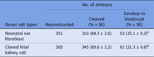
To determine the effects of cell type on in vitro development of cloned embryos, reconstructed oocytes were cultured in PZM5 medium for 6 or 7 days. The rates of cleavage and development to the blastocyst stage were calculated, and the results showed that cloned fetal kidney cells enhanced the development of blastocysts (Table 6). Cleavage rate in the cloned embryos (89.6 vs. 88.3% respectively) was not significantly different among groups, whereas the development rate to blastocyst (21.3 vs. 15.3% respectively) was found to be significantly higher in the cloned fetal kidney cell group.
Preimplantation development of porcine cloned embryos derived from transgenic neonatal ear fibroblasts and cloned fetal kidney cells

a,b Values in the same column with different superscripts differ significantly (P < 0.05).
Comparison of gene expression in cloned blastocyst by donor cell type
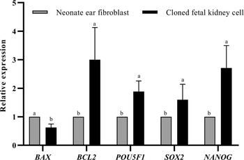
To investigate why the final term was successful only in fetal kidney cell groups, the expression of genes related to reprogramming and apoptosis was compared between the cloned blastocysts derived from neonatal ear fibroblasts and fetal kidney cells (Fig. 2). The cloned blastocysts derived from fetal kidney cells showed significantly higher expression levels of BCL2, POU5F1, SOX2, and NANOG genes compared with that of the neonatal ear fibroblasts group, whereas the BAX gene expression level was decreased significantly in neonatal ear fibroblasts.
Mean ± standard error of the mean (SEM) expression levels of BAX, BCL2, POU5F1, SOX2, and NAONG mRNA in cloned porcine blastocysts derived from both multigene-modified neonatal ear fibroblasts and its cloned fetal kidney cells. Within the same transcript, bars carrying different letters are considered statistically different at P < 0.05.

Discussion
Production of cloned pigs using nuclear transfer technology has revealed its importance especially for xenotransplantation (Polejaeva et al., Reference Polejaeva, Chen, Vaught, Page, Mullins, Ball, Dai, Boone, Walker, Ayares, Colman and Campbell2000). Various strategies have been used in the past to enhance the efficiency of cloned pig production. Briefly, these include supplementation of culture medium (Han et al., Reference Han, Baek, Lee, Lee, Yun, Lee and Lee2019), different incubation times (Betthauser et al., Reference Betthauser, Forsberg, Augenstein, Childs, Eilertsen, Enos, Forsythe, Golueke, Jurgella, Koppang, Lesmeister, Mallon, Mell, Misica, Pace, Pfister-Genskow, Strelchenko, Voelker, Watt, Thompson and Bishop2000), reprogramming through downregulation of certain genes (Zhang Z et al., Reference Polejaeva, Chen, Vaught, Page, Mullins, Ball, Dai, Boone, Walker, Ayares, Colman and Campbell2018; Weng et al., Reference Weng, Cai, Zhang, Liu, Liu and Liu2020), use of different sources, types, and stages of donor cells (Polejaeva et al., Reference Polejaeva, Chen, Vaught, Page, Mullins, Ball, Dai, Boone, Walker, Ayares, Colman and Campbell2000; Lai and Prather, Reference Lai and Prather2003; Lee et al., Reference Lee, Hyun, Kim, Kim, Lee, Lim, Lee, Kang, Lee and Hwang2003; Wei et al., Reference Wei, Qing, Pan, Zhao, Li, Cheng, Zhao, Xu, Li, Li, Ye, Wei, Li, Fu, Li, Xin and Zeng2013; Hua et al., Reference Hua, Xu, Liu, Bi, Xiao, Hua, Li, Zhang, Ren and Zheng2016). However, porcine SCNT embryos have still demonstrated poor viability with extremely low production rates compared with other species.
Three major procedures used in the production of transgenic pigs by cloning include the generation of cells of the desired gene composition, the use of these cells as the donor, and embryo transfer into surrogate mothers. In recent years, to generate cloned pigs for xenotransplantation, donor cells have required multiple genetic modifications in which many porcine endogenous genes are removed or human genes are introduced to decrease side effects (Polejaeva et al., Reference Polejaeva, Chen, Vaught, Page, Mullins, Ball, Dai, Boone, Walker, Ayares, Colman and Campbell2000; Iwase et al., Reference Iwase, Hara, Ezzelarab, Li, Zhang, Gao, Liu, Long, Wang, Cassano, Klein, Phelps, Ayares, Humar, Wijkstrom and Cooper2017; Ji et al., Reference Ji, Lee, Park, Kim, Byun, Ock, Hwang, Woo and Oh2017; Kwon et al., Reference Kwon, Kim, Hwang, Kim, Kim, Kim, Lee, Im, Lee and Hwang2017; Lee et al., Reference Lee, Park, Lee, Ji, Lee, Byun, Hwang, Kim, Ock and Oh2017). However, when multiple genes were modified in porcine cells, the viability of the cells decreased rapidly, making it difficult to obtain sufficient numbers of donor cells to be used in cloning, plus the injection process was very difficult due to many morphological changes in the donor cells. Moreover, as the number of modified genes was increased, cloning efficiency gradually decreased.
To overcome these disadvantages in the cloning of multiple gene-modified cells, we performed recloning using fetal kidney cells. Recloning, using cloned cells as donor cells, has been reported to show high production efficiency (Richter et al., Reference Richter, Kurome, Kessler, Zakhartchenko, Klymiuk, Nagashima, Wolf and Wuensch2012). In this study, we obtained enough donor cells with high proliferation ability from cloned fetal kidney cells and performed nuclear transfer more easily due to the small size and adhesion characteristics of kidney cells. Subsequently, significantly higher production and parturition rates were shown using cloned cells compared with reproductive cloning using genetically modified cells. The production efficiency of porcine recloning has been controversial among several studies. In one study, Liu and colleagues showed a lower live birth rate when cloned adult fibroblasts and cloned fetal fibroblasts were used as donor cells (Liu et al., Reference Liu, Dou, Xiang, Li, Li, Lin, Pang, Zhang, Chen, Luan, Xu, Yang, Yang, Liu, Li, Wang, Yang, Bolund, Vajta and Du2015). However, another study showed significantly higher parturition efficiency using cloned fetal fibroblasts (Fujimura et al., Reference Fujimura, Murakami, Kurome, Takahagi, Shigehisa and Nagashima2008), similar to the results of this study. In this study, pregnancy rates were not statistically significantly different between the donor cell types, but live offspring were observed only in the recloning group.
In this study, three types of recipient pigs were used: gilts with induced oestrus, gilts with natural oestrus, and sows with natural oestrus. Among them, the pregnancy rate was significantly higher in the group of sows with natural oestrus compared with gilts in natural and induced oestrus. In previous studies, the highest parturition rates have been observed in sows with a parity of two to five, and the lowest parturition rates were observed in sows with parity greater than eight and in gilts (Clark et al., Reference Clark, Schinckel, Singleton, Einstein and Teclaw1989). However, the use of sows as a surrogate recipient is associated with many difficulties. When using sows as the recipient, higher costs are necessary to support a farm breeding programme. It is difficult to fix sows on the operating table and induce anaesthesia due to their heavy bodyweight. Moreover, it difficult to incise and operate the thicker abdominal wall compared with that of gilts. However, recipient sows have several advantages that can overcome these difficulties, in addition to their higher pregnancy rates. Firstly, the date of embryo transfer can be adjusted easily and accurately by adjusting weaning time. We usually weaned the sow on Thursday (D0), and this will result in oestrus expression the next Tuesday (D6), such that Friday (D8) could be the appropriate time for embryo transfer. When using gilts with synchronized oestrus as recipients, proper oestrus expression was not observed at the expected time and in this study variable ovarian cycles were observed after abdominal incision. Moreover, using gilts with natural oestrus as the recipients negatively affected the results due to their irregular oestrous cycle. In this study, significantly higher pregnancy (83.3 vs. 20.4%) and parturition (50.0 vs. 4.1%) rates were shown in the sow group compared with that of gilts. These differences in reproductive competence between the sow and gilt make sows a more efficient recipient for the generation of transgenic cloned pigs. This may be because the sows were reproductively mature and had a previous pregnancy and parturition experienced reproductive tract compared with gilts. There is evidence that there is better development of oocytes from sows compared with gilts, and sows were better in terms of reproductive efficiency, as reported by various researchers (Onishi et al., Reference Onishi, Iwamoto, Akita, Mikawa, Takeda, Awata, Hanada and Perry2000; Polejaeva et al., Reference Polejaeva, Chen, Vaught, Page, Mullins, Ball, Dai, Boone, Walker, Ayares, Colman and Campbell2000). To target increased efficiency for production of cloned piglets, despite the disadvantages of clinical management of embryo transfer and cot, sows would be better recipients compared with gilts.
Various ovarian cycles were shown after ovulation and higher rates of pregnancy and parturition were shown in the ovulated group, except for one sow for which we had not been able to determine ovulation status. These results indicated that the ovarian cycle of the recipient is important and ovulation must occur in the recipient pig, because embryos transferred into the oviductal isthmus will experience the same environment as experienced by in vivo fertilized embryos following natural breeding. Recipients without ovulation have higher serum oestrogen concentrations that make the oviductal environment inappropriate for embryo development and movement into the uterus. This higher reproductive performance in recipient pigs with peri-ovulation and post-ovulation was similar to that found in previous studies in the generation of the transgenic cloned pigs (Petersen et al., Reference Petersen, Lucas-Hahn, Oropeza, Hornen, Lemme, Hassel, Queisser and Niemann2008; Koo et al., Reference Koo, Kang, Kwon, Park and Lee2010; Huan et al., Reference Huan, Hu, Xie, Shi, Wang, Zhou, Liu, Huang, Zhu, Liu, He, Li, Kong and Liu2015).
Production of transgenic cloned pigs was observed only in the cloned fetal cell group particularly in recipient gilts in this study. Parturition rates from gilts that were pregnant were 0/6 (0%) and 2/3 (66.7%) in adult cell and fetal cell types, respectively. This finding shows that proper nuclear reprogramming of the cloned embryo is the determining factor for the production of the cloned embryo, particularly in attaining normal parturition. We hypothesized that the less differentiated fetal cell was responsible for proper nuclear reprogramming and for pregnancy to term for fetal cells used as the donor cell type. These findings are similar to the reports in which a higher success in production of cloned pigs was achieved using fetal fibroblast cells as donor cells (Onishi et al., Reference Onishi, Iwamoto, Akita, Mikawa, Takeda, Awata, Hanada and Perry2000) and fetal fibroblast and fetal ear fibroblast (Richter et al., Reference Richter, Kurome, Kessler, Zakhartchenko, Klymiuk, Nagashima, Wolf and Wuensch2012). In our evaluation of the preimplantation development competence of cloned embryos derived from adult and fetal cells as donor cell types, the fetal cell groups showed statistically significantly higher numbers for development of embryo to blastocyst stage 82/385 (21.3%) vs 53/351 (15.1%) compared with adult cell types. This means that reprogramming of donor nuclei was more efficient through the use of least differentiated cloned fetal cells as donor cells, similar to findings of other studies (Niemann and Petersen, Reference Niemann and Petersen2016; Matoba and Zhang, Reference Matoba and Zhang2018). The findings of the current study were also similar for adult cells, as donors in SCNT had several limitations such as high rate of abortion and increased perinatal death, which was said to be caused by gaps in epigenetic reprogramming by somatic cells, where demethylation is required for fetal development (Han et al., Reference Han, Kang, Koo and Lee2003).
Moreover, as a result of comparative analysis of gene expression related to pluripotency and apoptosis, BCL2, POU5F1, and NANOG gene expression levels were significantly higher in the cloned fetal cell group. The BAX gene, which signals the onset of apoptosis, was significantly higher in the adult cell groups, whereas the BCL2 gene, which interferes with apoptosis and increases cell survival, was significantly higher in the fetal cell population. Reprogramming-related POU5F1 and NANOG genes, which are representative genes of pluripotency of cloned embryos and stem cells, were significantly more highly expressed in the cloned fetal cell groups. Although cloned fetal cells were reprogrammed cells by cloning, this was significantly advantageous in terms of nuclear reprogramming, and it can be said that it affected the efficiency of the production of cloned pigs.
Based on this finding and related studies, it is recommended, at the donor nucleus level, to use fetal cells in the early stages of pregnancy that are least differentiated or kidney cells when differentiated, rather than using adult cells that have been modified with multiple genes and have poor survival rates. For the recipient pig, it is more efficient to use sows as recipient pigs, considering the final production efficiency from the establishment of transgenic donor cells to parturition of the transgenic pig, even though costs are higher than for gilts with synchronized oestrus. In particular, it is recommended in terms of cost to supply sows that have been discharged due to poor delivery after five parities to be efficient as recipient pigs for cloned embryos.
Acknowledgements
We thank Younsang Kweon for preparation of recipient sows, Professor Byeong Cheol Kang at Seoul National University for research advisory, Jinho Cho at Chungbuk National University for the introduction of the gilt farm, and Youngju Noh, Jieun Cho and Byungsun Choi for their administrative and technical support.
Financial support
This study was supported by a grant from the Korea Health Technology R&D Project through the Korea Health Industry Development Institute (KHIDI), funded by the Ministry of Health and Welfare, Republic of Korea (grant number HI13C0954).
Declarations of interest
None.
Ethical approval
All experiments were approved and performed following the guidelines of the Institutional Animal Care and Use Committee of Chungnam National University (IACUC approval no. CNU-00624).