Introduction
Brassica oleracea is one of the three basic types of Brassica, and is an important vegetable crop. The various ecotypes and many artificial hybrids will all benefit from genetics research, both from an evolutionary angle or a breeding point of view. However, it is difficult to identify Brassica chromosomes because of their small size and also because of the greater and more uniform contraction of the chromosomes in the cell division phase than occurs in many other plants.
With the recent and rapid development of fluorescence in situ hybridization (FISH) technology, it has increasingly been used in the identification and karyotype analysis of B. oleracea chromosomes. Most are identified based on the number and location of FISH signals from the 45s (or 25s) and 5s rDNA on the chromosomes (Snowdon et al., Reference Snowdon, Kobler and Kohler1997; Hasterok et al., Reference Hasterok, Jenkins, Langdon, Jones and Maluszynska2001). Kamisugi and colleagues (Reference Kamisugi, Nakayama, O'Neil, Mathias, Trick and Fukui1998) identified five chromosomes in cabbage by using two cDNA probes and rDNA. Armstrong and colleagues (Reference Armstrong, Fransz, Marshall and Jones1998) also identified five pairs of chromosomes by using three DNA repeat sequences. Tai-Xia Wang and colleagues (Reference Wang, Wu, Huang and Wei2006) used cabbage rDNA and Cot-1 DNA to identify the nine pairs of chromosomes in Brassica. The use of FISH chromosome identification in Brassica with rDNA and other repetitive sequences as probes is limited by the distribution of rDNA repeat sequences in the chromosomes of cabbage. Karyotype analysis is difficult to conduct and the results are limited due to the few chromosome loci and lack of features in the FISH signals. GISH (genomic in situ hybridization) technology, developed from chromosome in situ suppression hybridization (CISS), comprises an important in situ hybridization tool. Molecular cytogenetics has become an important plant research tool. This technology is widely used in determining the origin and evolution of species, distant hybrid identification, detection of exogenous chromatin, establishing gene function and carrying out positioning research. Plant genomes differ in their repeated DNA sequences, the sequences repeating with high levels of specificity (Schwarzacher et al., Reference Schwarzacher, Anamthawat-Jonsson and Harrison1992). Therefore, using whole genomic DNA as the FISH probe, and an appropriate concentration of genomic DNA from a closely related species for blocking, in situ hybridization of the target chromosome, with the aim of obtaining a conserved sequence, results in high levels of specific sequences marked primarily by the hybridization probe. Because there is a high number of repeated sequences with different distributions on each chromosome in the plant genome, sufficient characteristics may be determined with GISH signals to allow identification. For each specific hybridization signal, using the signal characteristics of number, strength and location, it is relatively easy to identify the various chromosomes.
In this study, by applying GISH technology and using Brassica genomic DNA as the probe and cabbage genome as the blocking agent, it was possible to distinguish between nine pairs of chromosomes. By overcoming the limitations of using rDNA and other repetitive sequences as a probe for the FISH signals, we were able to develop a genotyping system providing a practical and accurate method of karyotype analysis for cabbage and other small and homogeneous genomes.
Materials and methods
Experimental materials
Self-incompatibile B. oleracea K-164 samples were obtained from the Agronomy and Biotechnology Department of Agriculture, Key Laboratory of Crop Quality of Southwest University (Chongqing, China). B. chinensis was provided by the Brassica Breeding and Research Landscape Horticulture of Southwest University.
Water cultivated B. oleracea and B. pekinensis seedlings, approximately 10 days old, were harvested as yellow seedlings, frozen in liquid nitrogen, and stored in a refrigerator at −80°C. Genomic DNA was extracted and prepared to synthesize the probe and blocking agents.
Chromosome preparation
Chromosomes were prepared according to the method of Li and Chen (Reference Li and Chen1985). Root tips were cultured at various temperatures and in apical darkness. When the root tip length reached 1–2 cm, it was pretreated with 8-hydroxyquinoline solution, washed with ddH2O, fixed, first low-permeability, digested with 2% cellulase and 2% pectinase at 37°C, followed by low-permeability and other steps, then stored in a refrigerator at −60°C on flame-dried slides.
Probe and blocking agent preparation
Genomic DNA from B. oleracea and B. pekinensis was extracted using the cetyltrimethyl ammonium bromide (CTAB) method (Zhu and Wang, Reference Zhu and Wang2000) and purified. Genomic DNA from B. oleracea was then used to form the random primer digoxigenin-labelled probe, with genomic DNA from B. pekinensis used as the blocking agent.
Genomic in situ hybridization (GISH)
Slide pre-treatment
The chromosomal slides were dried and incubated with 100 μg/ml RNase for 1 h at 37°C, washed in 2×sodium chloride sodium citrate (SSC) buffer, and dehydrated in 70%, 90% and 100% (v/v) ethanol.
In situ hybridization
Chromosomal slides were denatured in 70% deionized formamide at 70°C for 2 min then immediately dehydrated in pre-chilled (−20°C) 70%, 90% and 100% (v/v) ethanol and air-dried. The hybridization mixture consisted of 100% deionized formamide, 50% dextran sulphate, probe, 20×SSC, herring sperm DNA, blocking DNA and Sodium dodecyl sulphate (SDS) and was denatured at 85°C for 10 min, then immediately put on ice. The probe mixture (30 μl) was applied to every slide, covered with a cover slip, and sealed with rubber cement. The chromosomal slides were then denatured together with the hybridization mixture at 85°C for 10 min, and allowed to hybridize overnight in a humidity chamber at 37°C.
Post-hybridization washing and signal capturing
After hybridization, the chromosomal slides were washed in 2× SSC at 37°C for 10 min, 4× SSCT (SSC with Tween) for 8 min, 1× phosphate buffer saline (PBS) at 25°C for 5 min, and air-dried. The detection mixture (30 μl) was applied to every slide in dark conditions and covered with a cover slip, incubated in a humidity chamber at 37°C for 30 min, then washed and air-dried. The chromosomal slides were counterstained in Vectashield containing 2 μg/ml of DAPI 20 μl at 37°C for 30 min.
Image collecting and processing
The slides were examined with a Nikon Eclipse E600W fluorescence microscope (Nikon, Japan). The images were captured and analysed using an image analysis system, which included a Digital Sight DS-U2 charge-coupled device (CCD, Nikon) camera and NIS-Elements BR 2.20 software.
Results
Karyotype analysis of chromosomes of root tip in B. oleracea at metaphase
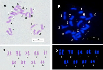
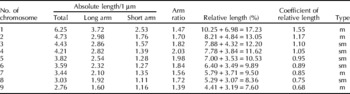
Standard karyotype analysis was applied according to the method of Li and Chen (Reference Li and Chen1985). Two kinds of staining methods were used, one with Carbol fuchsin and the other DAPI counterstaining (Fig. 1). A total of ten metaphase cells were examined to analyse the chromosomes according to the values of the assessed parameters (Table 1).
Chromosomes and karyotyping of Brassica oleracea at metaphase using (A, a) carbol fuchsin staining and (B, b) DAPI counterstaining. (See online for a colour version of this figure.)

Karyotype analysis of Brassica oleracea

m, metacentric; sm, submetacentric.
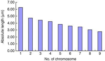
B. oleracea is homologous diploid and has nine pairs of chromosomes. From Table 1 and Fig. 2, the typical self-incompatibility in Brassica metaphase chromosomes can be seen to have the following characteristics. First, the longest and shortest chromosomes varied greatly in length, with a length ratio of 2.3:1, which is essentially consistent with the experimental results of Qi (Reference Qi2006). The first chromosome could easily be separated from the other chromosomes by virtue of its length. Second, the arm ratio of chromosomes 1, 2, 7 and 9 were below 1.70, forming the metacentric chromosomes. The arm ratios of chromosomes 3 to 6 and 8 were between 1.71 and 3.00 (chromosome 4 was above 2), and were defined as sub-metacentric chromosomes. The karyotype formula was 2n=2x=18=4m+5sm. Third, the coefficient of relative length of chromosome 1 was greater than 1.21, and it was therefore classified as a long chromosome; the coefficients for chromosomes 2 to 4 were between 1.01 and 1.21, and were therefore classified as sub-long chromosomes. Those of chromosomes 5 to 8 were between 0.71 and 0.99, indicating they were therefore sub-short chromosomes, and that of chromosome 9 was less than 0.70, which was therefore a short chromosome. Fourth, Table 1 shows that the relative lengths of all chromosomes were very similar to those in the reference of Armstrong and colleagues (Reference Armstrong, Fransz, Marshall and Jones1998), indicating the authenticity of the test results. Finally, the lengths of chromosomes 3 and 4 to 7 were similar and difficult to distinguish. Pairing between homologous chromosomes can be seen in Fig. 1a,b.
The idiogram of Brassica oleracea. (See online for a colour version of this figure.)

GISH typing of chromosomes at metaphase in B. oleracea
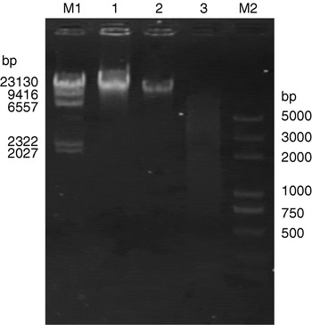
Genomic DNA of B. oleracea and B. pekinensis was extracted and purified. Genomic DNA of B. oleracea was then digested by restriction enzymes Hind III and EcoR I. Electrophoresis results showed that the majority of DNA molecules were digested into 500–10 000 bp fragments (Fig. 3). The length met the requirements of Roche's DIG kit and the DNA could be used for marking. Purified genomic DNA of B. pekinensis, following measurement of its concentration, was used as blocking agent.
Agarose gel electrophoresis of genomic DNA from B. olereacea and B. pekinensis and B. olereacea genomic DNA digested by restriction enzymes. Lanes: M1, marker λDNA/Hind III; 1, genomic DNA of B. olereacea; 2, genomic DNA of B. peknensis; 3, B. olereacea genomic DNA digested by restriction enzymes; M2, DL2000+DNA marker.

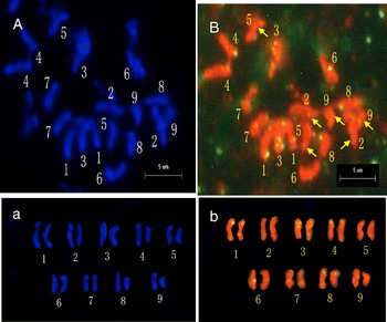
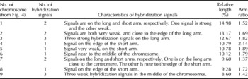
By exploring a range of conditions, the optimum conditions for GISH were finalized. Typing was conducted based on the number, location and intensity of hybridization signals from the chromosomes, enabling them to be distinguished (Fig. 4). In Fig. 4A,B, chromosomes are seen to be well scattered, fully exposed, with minimal overlapping and clear signals. Figure 4 and Table 2 clearly show the signal characteristics for each pair of homologous chromosomes. Chromosomes 1, 2 and 7 had two signals, but their location and intensity differed. The signal on chromosome 1 was strong, but on chromosome 2 it was very weak. Chromosomes 3 and 9 had three clear signals. Chromosomes 4 to 6 and 8 had one signal; the signals for chromosomes 4 and 8 were both located on the edge of the short arm and had similar intensities, but the difference in their lengths very obviously distinguished them. The features of each pair of homologous chromosomes of B. oleracea could therefore be used to distinguish between them very specifically.
Karyotype analysis of B. oleracea by GISH. (A) DAPI counterstaining (irradiation with ultraviolet light). (B) Irradiation with blue light. Arrows indicate a single chromosome. (a,b) Karyotype of B. oleracea obtained using the methods of (A) and (B) respectively. (See online for a colour version of this figure.)

Chromosomal characteristics for karyotype analysis of B. oleracea by GISH

The various parameters for the chromosomes were calculated from Fig. 4A and according to the traditional method of Li and Chen (Reference Li and Chen1985). The relative length, arm ratio and other relevant parameters in Table 2 were similar to previously obtained statistical results. The results were therefore shown to be reliable. In comparison with other methods, the GISH typing method is more reasonable and precise in terms of its results.
Discussion
GISH is suitable for use in a wide range of applications in plants. To date, researchers have used GISH to successfully identify the allotetraploid AA, BB and CC genomes of Brassica, to investigate the relationships of the AA, BB and CC genomes (Li et al., Reference Li, Li, Huang, Wu and Song2002b), to study the inter-relationships of the Brassica genome and five other genomes of related species (Li et al., Reference Li, Wu, Wang and Song2003), to analyse species that have recent relationships with Brassica and intergeneric hybrids (Li et al., Reference Li, Ceccarelli, Minelli, Contento, Liu and Cionini2002a; Zhu et al., Reference Zhu, Xu, Hua and Li2005; Cheng et al., Reference Cheng, Hua, Zhang, Chen and Wu2006; Chen et al., Reference Chen, Cheng, Liu and Wu2006b), and to establish the important role of distant hybridization in crop genetic improvement. In this study, using C genomic DNA as probe, A genomic DNA as the blocking agent, GISH technology was used on mitotic metaphase chromosomes of B. oleracea (C genome). Nine pairs of homologous chromosomes of a typical self-incompatibile B. oleracea were successfully distinguished. A further study precisely identifying the metaphase chromosomes on root tip of B. oleracea was also carried out. Although this is the first time the GISH method has been used to distinguish the chromosomes of B. oleracea, its principle is the same as previous studies, being based on the principle of complementary base pairing, choosing a specific probe and blocking agent, and analysing the signals from chromosomes to draw conclusions. This study predominantly used the number, location and intensity of hybridization signals to distinguish between chromosomes. The same signal characteristics (number, intensity and location) are found on homologous chromosomes, allowing homologous chromosomes to be accurately paired. Each pair of homologous chromosomes also has unique signal characteristics, enabling them to be distinguished from other homologous chromosomes. This method avoids measurement error, and overcomes the limitations of using rDNA as the probe, as in the FISH method. The results show that this method is reasonable and practical, and produces a good outcome, achieving precise classifications. When repeatedly applying this typing method, a slight difference was found in the locations and intensities of hybridization signals on homologous chromosomes in successive results, although most of the basic characteristics were the same. This typing method is therefore expected to play an important role in karyotype identification of small chromosomes.
In the process of species evolving from a common primitive ancestor, changes in genome size, gene number and DNA sequence must arise. Genome evolution is a long and complex process. Although the specific processes and mechanisms of evolution have not been fully elucidated to date, it is generally agreed that a wide range of point mutations and insertion or deletion mutations are the important driving force of genomic evolution (Chen et al., Reference Chen, Peng, Zhang and Tian2006a). Insertion or deletion mutation in a DNA fragment not only results in genome polymorphism, but also changes genetic structure by insertion or deletion in the exons and introns of the original gene, leading to the generation of new genes (Long et al., Reference Long, Betran, Thornton and Wang2003). In the B. rapa A genome, the interval between the two sites is 71% longer than that of the B. oleracea C genome. This indicates that B. oleracea and B. rapa, in their evolution, have undergone deletion or insertion events to generate different interval lengths, leading to differentiation. Genomic sequences contain gene and non-gene sequences, and while gene sequences undergo mutations throughout biological evolution, non-gene sequences are also mutated. Non-gene sequences mutate at a much faster rate than gene sequences, so differences between the A and C whole genome sequences were found to be greater than expected. Even so, analysis by comparative genomic mapping (Lagercrantz and Lydiate, Reference Lagercrantz and Lydiate1996) shows that the A, B and C genomes are basically the same, with a high degree of conservatism. However, in long-term evolution, the chromosomes may undergo fusion, fragmentation, segment duplication, translocation and rearrangement, resulting in them having a different chromosome structure. Previous studies (Song et al., Reference Song, Osborn and Williams1988; Warwick and Black, Reference Warwick and Black1991; Axelsson et al., Reference Axelsson, Bowman, Sharpe, Lydiate and Lagercrantz2000) have also shown that the A genome is similar to the C genome, and there is a high degree of genetic homology between the two. Studies by Li and colleagues (Reference Li, Li, Huang, Wu and Song2002b, Reference Li, Wu, Wang and Song2003) also show that the C and A genome have a high degree of homology, a close genetic relationship and low genomic differentiation. Taking as an example the S locus genes SRK and SLG, sequences of SRK and SLG of B. oleracea (CC) and B. rapa (AA), and SRK mRNA sequences of 10 were searched in GenBank using Blastn and Vector software to compare their nucleic acid sequences. From the alignment of the SRK and SLG genes it can be seen that, although individual bases mutate during long-term evolution, the sequences of the two genes are still very conserved, with a consistency rate of more than 50% and a similarity rate of 90%. Therefore, from a comparison of nucleic acids, we can conclude that the SRK and SLG genes of B. rapa can block SRK and SLG genes of B. oleracea.
It can be seen that the A and C genomes have a high degree of homology. Using B. rapa as a blocking agent will therefore preferentially block homologous parts of the chromosomes, for most combinations of homologous fragments. The differentiation between them ensures that specific sites of B. oleracea can be exposed, then hybridized by labelled probes, producing different hybridization signals. The homology and differentiation of the A and C genomes fully demonstrates the rationality and feasibility of this blocking. The size of the blocking agents has a great impact on blocking efficiency. In theory, the larger fragments display a lower blocking efficiency; when a fragment is small, the probability of combination and the blocking efficiency are both higher. Studies have shown (Li et al., Reference Li, Ceccarelli, Minelli, Contento, Liu and Cionini2002a; Chen et al., Reference Chen, Cheng, Liu and Wu2006b) that blocking agents can be used with treatment with boiling water and random cutting. Fragment sizes range between 200 and 500 bp. A very good blocking effect can be achieved. In this experiment, the genomic DNA of B. rapa was used directly as a blocking agent after extraction and purification. The experimental results show that without treatment of the DNA, B. oleracea and B. rapa are highly homologous, and as long as the ratio of blocking agents and probes is selected carefully, a very good blocking effect is achieved.
Acknowledgements
This study was financially supported by the Natural Science Foundation of Chongqing, China (9266) and the National Natural Science Foundation of China (30671429).