INTRODUCTION
Mucocutaneous leishmaniasis is an infectious disease caused by diverse flagellate kinetoplastids of the genus Leishmania, which are transmitted by Lutzomyia phlebotomines (Brasil, 2009). No vaccine is available against human leishmaniasis, and actions to combat the disease are restricted to vector control (Srivastava et al. Reference Srivastava, Shankar, Mishra and Singh2016). HIV and Leishmania co-infection occurs in 35 countries and increases the lethality of the disease due to the difficulty of clinical management and treatment (Lindoso et al. Reference Lindoso, Cunha, Queiroz and Moreira2016). More than 12 million people are infected worldwide, and 350 million people live in risk areas. In the Americas, 743 970 cases were registered between 2001 and 2013, with an annual average of 57 228 cases (OPAS/OMS, 2015). Leishmania (Viannia) braziliensis is related to this type of infection, with a geographical distribution that extends from Central America to northern Argentina (Basano and Camargo, Reference Basano and Camargo2004).
The first-line treatment for mucocutaneous leishmaniasis in Brazil is pentavalent antimonials, and the second choice drugs are amphotericin B and pentamidine. However, all of these drugs exhibit considerable toxicity. Pentavalent antimonials have many adverse reactions that often lead to discontinuation of treatment (OPS, 2013). Therefore, the search for alternative effective compounds is relevant. Natural products appear as a source for treating neglected parasitic diseases (Kayser et al. Reference Kayser, Kiderlen and Croft2003; Ndjonka et al. Reference Ndjonka, Rapado, Silber, Liebau and Wrenger2013; de Moraes, Reference de Moraes2015) and important information for the therapeutic use of plants can be gathered from popular knowledge (Anthony et al. Reference Anthony, Fyfe and Smith2005). The antiparasitic drugs artemisinin and chloroquine are examples of plant-derived products, and amphotericin B and ivermectin are important antiparasitics isolated from Streptomyces microorganisms (de Moraes, Reference de Moraes2015).
Different pepper species (Piper spp., Piperaceae) are widely used in folk medicine, and their biological activity is well documented. For instance, extracts of diverse peppers (P. claussenianum, P. nigrum, P. longum and P. aduncum) inhibit tumour growth in vitro and in vivo and even the incidence of metastasis (Sunila and Kuttan, Reference Sunila and Kuttan2004; Majdalawieh and Carr, Reference Majdalawieh and Carr2010; Marques et al. Reference Marques, Paiva, Fonseca, Capella, Guimarães and Kaplan2013; Arroyo-Acevedo et al. Reference Arroyo-Acevedo, Chávez-Asmat, Anampa-Guzmán, Donaires and Ráez-Gonzáles2015). Therefore, testing the ability of a pepper species to inhibit the growth of trypanosomatid protozoa appears to be a reasonable proposition.
Piper aduncum (matico) is a perennial bush or small tree with spear-shaped leaves that is native to southern Mexico, the Caribbean and most tropical countries in South America. Indigenous groups in the Americas, such as the Lancadones, make tea from its leaves and roots to treat a wide variety of medical conditions, such as diarrhoea, dysentery, nausea, ulcers and genitourinary tract infections (Cook, Reference Cook2016). Piper aduncum essential oil (PaEO) has the potential to be used as insect repellent (Mamood et al. Reference Mamood, Hidayatulfathi, Budin, Rohi and Zulfakar2017) and antihelminthic against the nematode Haemonchus contortus (Gaínza et al. Reference Gaínza, Fantatto, Chaves, Bizzo, Esteves and Chagas2016). Ethanolic extracts of P. aduncum leaves showed considerable antifungal and antibacterial activities in vitro (Okunade et al. Reference Okunade, Charles, Clark and Lentz1997). Additionally, these extracts were active as acaricidal agents against Tetranychus urticae (Araújo et al. Reference Araújo, Câmara, Born, Moraes and Badji2012), repellents against Aedes aegypti and Aedes albopictus (Misni et al. Reference Misni, Othman and Sulaiman2011), and reduced inflammatory interleukins in dendritic cells (Thao et al. Reference Thao, Luyen, Widowati, Fauziah, Maesaroh, Herlina, Manzoor, Ali, Koh and Kim2016), which demonstrated the wide range of biological effects of this plant species.
Essential oils (EOs) of different species of the genus Piper (P. aduncum var. ossamum, P. cernuum, P. claussenianum, P. demeraranum, P. duckei, P. heptaphylum and P. hispidum) have demonstrated activity against Leishmania amazonensis and L. infantum (Marques et al. Reference Marques, Barreto, Batista, Curvelo, Velozo, Moreira, Guimarães, Soares and Kaplan2010; Carmo et al. Reference Carmo, Amaral, Machado, Leon and Silva2012; Capello et al. Reference Capello, Martins, Farias, Figueiredo, Matsuo, Passero, Oliveira-Silva, Sartorelli and Lago2015; Houël et al. Reference Houël, Gonzalez, Bessière, Odonne, Eparvier, Deharo and Stien2015; Gutiérrez et al. Reference Gutiérrez, Montes, Scull, Sánchez, Cos, Monzote and Setzer2016). Therefore, studies on the effect of EOs against Leishmania are promising. However, few studies have investigated the interaction between EOs and L. braziliensis, which is one of the main causative agents of mucocutaneous leishmaniasis in Brazil (Jones et al. Reference Jones, Johnson, Barretto, Lago, Badaro, Cerf, Reed, Netto, Tada, Franca, Wiese, Golightly, Fikrig, Costa, Cuba and Marsden1987), primarily due to the difficulty in maintaining this parasite in vitro.
Therefore, given the variety of biological activities of P. aduncum and the need for new alternative compounds to inhibit parasite growth, we tested the effect of the EO of P. aduncum and its major components (nerolidol and linalool) against L. braziliensis. Our data showed that nerolidol had leishmanicidal activity. Ultrastructural observations and flow cytometry analysis indicated that nerolidol induced incidental cell death in the L. braziliensis promastigotes.
MATERIALS AND METHODS
Chemicals
Linalool, nerolidol, amphotericin B (250 µg mL−1), gentamicin (50 mg mL−1), PMA (phorbol-12-myristate-13-acetate), Schneider's Insect Medium, DAPI (4′,6-diamidino-2-phenylindole), MTT (3-(4,5-dimethylthiazol-2-yl)-2,5-diphenyltetrazolium bromide), CCCP (carbonyl cyanide 3-clorophenylhydrazone), Nonidet P 40 substitute (NP-40) and DMSO (dimethyl sulphoxide) were purchased from Sigma-Aldrich (St. Louis, MO, USA). RPMI 1640 medium, fetal bovine serum, propidium iodide, rhodamine 123, RNase A DNase-free and the Alexa Fluor-488 Annexin/Dead Cell Apoptosis Kit were purchased from Thermo Fisher Scientific (Carlbad, CA, USA).
Essential oil
Piper aduncum leaves were collected on March 2013 in the morning, at the Medicinal Plants Garden of the Federal University of Lavras, Minas Gerais (UFLA), Brazil. The essential oil (PaEO) was obtained at the Chemistry Department of UFLA by Clevenger distillation. The chemical characterization was performed by GC–MS, which demonstrated that nerolidol (25·22%) and linalool (13·42%) were the major components (Villamizar et al. Reference Villamizar, Cardoso, Andrade, Teixeira and Soares2017). For the assays, PaEO, linalool and nerolidol were diluted to 100 mg mL−1 in DMSO and kept at 4 °C protected from the light (Escobar et al. Reference Escobar, Milena Leal, Herrera, Martinez and Stashenko2010).
THP-1 cells
THP-1 cells (ATCC TIB-202) were kept at 37 °C in a humidified incubator with a 5% CO2 atmosphere, in RPMI 1640 medium supplemented with 10% fetal bovine serum (FBS) and 50 µg mL−1 of gentamicin. The cell cultures were maintained by weekly subculture when the growth reached 1 × 10⁶ cells mL−1.
Parasites
Leishmania braziliensis (strain MHOM/BR/75/M2903) promastigotes were maintained at 26 °C in Schneider's medium supplemented with 10% FBS and 2% male child urine with two passages per week. For the biological assays with intracellular amastigotes, differentiated THP-1 cells were infected (ratio of 10 parasites per host cell) with promastigotes from 5-day-old cultures.
Cytotoxicity assays
THP-1 cells were seeded into 96-well plates at a density of 5 × 10⁴ cells/well in RPMI 1640 medium containing 200 nm PMA. The plates were incubated for 96 h at 37 °C in a humidified incubator with a 5% CO2 atmosphere to induce differentiation into adherent macrophages (Donega et al. Reference Donega, Mello, Moraes, Jain, Tekwani and Cantrell2014). Then, the cells were incubated for 24 h with 15·625–500 µg mL−1 of the compounds, and the cell viability was evaluated by the MTT colorimetric assay. The optical density (O.D.) was read in a BioTek EL800 reader (Biotek, Winooski, VT, USA). Wells with no treatment (negative control) were used for comparison (100% cell viability). The mean cell inhibition (CI) values were obtained from three experiments performed in triplicate according to the equation CI=100−((O.D treatment/O.D. negative control)×100). Dose x response curves were used to estimate the CC50/24 h, which was the toxic concentrations for 50% of the culture after 24 h (Escobar et al. Reference Escobar, Milena Leal, Herrera, Martinez and Stashenko2010).
Alternatively, linalool, nerolidol and amphotericin B were evaluated for their haemolytic potential. For this analysis, type O+ peripheral blood was collected from a healthy donor, defibrinated for 10 min under stirring using glass beads and washed three times in glucose saline solution (0·85% NaCl and 5% glucose). The red blood cells were resuspended at 3% in the glucose saline solution and seeded into 96-well plates containing different concentrations of each compound tested. The plates were incubated for 3 h at 37 °C, and the absorbance of the supernatant was read at 550 nm in a BioTek EL800 reader. The percentage of haemolysis was correlated to the O.D. values of the positive control (red blood cell solution plus 1% Triton X-100), which was considered 100% haemolysis (Izumi et al. Reference Izumi, Ueda-Nakamura, Veiga, Pinto and Nakamura2012).
Activity against promastigotes
Four-day-old L. braziliensis promastigotes were adjusted to a density of 4·5 × 107 cells mL−1, and 100 µL well−1 of this suspension was seeded into 96-well plates. Different dilutions of PaEO, linalool and nerolidol (final concentration 1–500 µg mL−1) were added to each well (100 µL well−1). Amphotericin B was used as a positive control at concentrations ranging from 0·062 to 2 µg mL−1. Parasite viability was assessed by MTT staining after incubation with the drugs for 24 h. The mean cellular inhibition values were obtained from three experiments performed in triplicate, and dose x response curves were used to estimate the inhibitory concentration for 50% of the parasites (IC50/24 h). The statistical analysis was performed with GraphPad Prism 5 software (San Diego, CA, USA) by applying two-way ANOVA and the Bonferroni post hoc test. A P value <0·05 was considered significant.
Activity against intracellular amastigotes
Differentiated THP-1 cells were seeded into 96-well plates at a density of 5 × 10⁴ cells/well and incubated for 4 h with 5-day-old promastigotes at a proportion of 10 parasites per cell. Then, the cells were washed and incubated for another 48 h. The medium was replaced, and the monolayers were incubated with nerolidol (1·95–62·5 µg mL−1) or amphotericin B (0·062–1 µg mL−1) for 24 h (Donega et al. Reference Donega, Mello, Moraes, Jain, Tekwani and Cantrell2014). The cells were fixed with absolute methanol and stained with DAPI. The inhibitory activity was assessed by counting the number of intracellular amastigotes in 10 images randomly captured from 10 microscopic fields (20x objective). The values were expressed as the per cent inhibition (%I) calculated as: %I=100−((T×100)/C), where T is the average of all treated intracellular amastigotes and C is the average of the total intracellular amastigotes in the negative control (Guru et al. Reference Guru, Agrawal, Singha, Singhal and Gupta1989; Lakshmi et al. Reference Lakshmi, Pandey, Kapil, Singh, Samant and Dube2007).
Electron microscopy
For scanning electron microscopy (SEM), promastigotes were incubated with the IC50/24 h or 2×IC50/24 h of nerolidol or amphotericin B, fixed with 2·5% glutaraldehyde in 0·1 m sodium cacodylate buffer, washed and adhered to coverslips coated with 0·1% poly-L-lysine. The cells were post-fixed with 1% osmium tetroxide, dehydrated in an ascending acetone series, submitted to critical point drying, coated with a 20-nm-thick gold layer, and observed under a JEOL JSM 6010 PLUS-LA (Akishima, Tokyo, Japan) scanning electron microscope.
For transmission electron microscopy (TEM), after treatment with the IC50/24 h or 2 × IC50/24 h of nerolidol or amphotericin B, the promastigotes were fixed with 2·5% glutaraldehyde in 0·1 m sodium cacodylate buffer, washed and post-fixed for 1 h with 1% osmium tetroxide. Then, the samples were dehydrated in increasing concentrations of acetone and embedded in Poly/Bed 812 resin for 72 h at 60 °C. Ultrathin sections were contrasted for 30 min with uranyl acetate and for 2 min with lead citrate. The ultrathin sections were observed in a JEOL JEM-1400Plus transmission electron microscope at 80 kV.
Flow cytometry
For each type of analysis, 1 × 10⁶ L. braziliensis promastigotes were pre-treated for 24 h with nerolidol or amphotericin B (IC50 or 2 × IC50), and then labelled according to the different assays. In all cases, 10 000 events were read in an FACSCanto II flow cytometer (Becton–Dickinson, San Jose, CA, USA) and the data were analysed using the FlowJo software v10 (Treestar Software, Ashland, OR, USA). The experiments were performed in triplicate. The statistical analysis was performed in the GraphPad Prism 5 software with one-way or two-way ANOVAs and the post hoc Bonferroni test, with P < 0·05 considered significant.
To estimate the mitochondrial membrane potential (ψm), promastigotes were collected, washed with PBS and incubated for 15 min at 26 °C with 5 µg mL−1 of rhodamine 123 (Rh123) (Petit, Reference Petit1992). Changes in Rh123 fluorescence emission were quantified by the variation index VI=[(MT−MC)/MC], where MT is median fluorescence of the treated parasites and MC is median fluorescence of the control. Negative VI values indicated mitochondrial membrane depolarization (Mendes et al. Reference Mendes, Desoti, Silva, Ueda-Nakamura, Dias Filho, Yamada-Ogatta, Sarragiotto and Nakamura2016).
To estimate the cell size, promastigotes were collected, resuspended in 1 mL of PBS and immediately analysed in the cytometer to determine the size and granularity (FSC×SSC) (Jiménez-Ruiz et al. Reference Jiménez-Ruiz, Alzate, Macleod, Lüder, Fasel and Hurd2010). Histograms were generated, and the FSC-H parameter was considered representative of the cell size (Rodrigues et al. Reference Rodrigues, Ueda-Nakamura, Corrêa, Sangi and Nakamura2014).
For the cell cycle analysis, the promastigotes were resuspended in 500 µL of PBS, to which was added the same volume of permeabilizing solution and DNA staining solution (3·4 mm Tris–HCl, pH 7·4, 0·1% NP-40, 700 U L−1 of RNase A-DNase free, 10 mm NaCl and 75 µ m propidium iodide). The fluorescence reading was taken after incubation for 10 min at room temperature. The data analysis was performed from a specific gate that excluded doublets. The Dean–Jett–Fox algorithm was used to estimate the percentage of cells in the G0/G1, S and G2/M phases (Kessler et al. Reference Kessler, Gradia, Rampazzo, Lourenço, Fidêncio, Manhaes, Probst, Ávila and Fragoso2013). In apoptotic cells, DNA fragmentation was perceived as a sub-peak formation in cell fluorescence at the G0/G1 stage (Khademvatan et al. Reference Khademvatan, Gharavi and Saki2011).
To measure cell membrane integrity and phosphatidylserine (PS) exposure, promastigotes were resuspended in annexin-binding buffer (140 mm NaCl, 5 mm CaCl2 and 10 mm HEPES-Na, pH 7·4) and labelled with Alexa Fluor 488-conjugated Annexin-V and 100 µg mL−1 of propidium iodide (PI). A 0·25% concentration of saponin was used as the positive control (Lazarin-Bidóia et al. Reference Lazarin-Bidóia, Desoti, Martins, Ribeiro, Ud Din, Rodrigues-Filho, Ueda-Nakamura, Nakamura and Silva2016). In apoptotic cells, PS is exposed and can be detected by its reactivity with Annexin-V. Annexin-V (PS exposure) labelling alone is interpreted as evidence of apoptosis, whereas PI labelling is perceived as necrosis (i.e., loss of membrane permeability/damage to the plasma membrane). Double-labelled cells (Annexin-V and PI) are viewed as cells with a lack of distinction between late apoptosis and necrosis (Islamuddin et al. Reference Islamuddin, Sahal and Afrin2014; Chouhan et al. Reference Chouhan, Islamuddin, Want, Ozbak, Hemeg, Sahal and Afrin2015; Masic et al. Reference Masic, Hernandez, Hazra, Glaser, Holzgrabe, Hazra and Schurigt2015; Awasthi et al. Reference Awasthi, Kathuria, Pant, Kumari and Mitra2016; Corpas-López et al. Reference Corpas-López, Merino-Espinosa, Díaz-Sáez, Morillas-Márquez, Navarro-Moll and Martín-Sánchez2016). Apoptotic cells are also recognized by positive labelling with Annexin-V regardless of the positive or negative PI status (Jimenez et al. Reference Jimenez, Paredes, Sosa and Galanti2008).
RESULTS
EOs are mixtures of saturated and unsaturated hydrocarbons, alcohols, aldehydes, esters, ethers, ketones, terpenes and phenolic oxides that can produce characteristic odours (Ali et al. Reference Ali, Al-Wabel, Shams, Ahamad, Khan and Anwar2015). Linalool and nerolidol are sesquiterpenes that are present as major components in the EO of the P. aduncum pepper analysed here. First, we assessed the inhibitory effect of PaEO, nerolidol and linalool on the growth of L. braziliensis promastigotes after 24 h of treatment. The IC50/24 h value was determined by the colorimetric MTT method (Supplementary Fig. 1), which is an easy, inexpensive method that is suitable for drug screening (Fumarola et al. Reference Fumarola, Spinelli and Brandonisio2004). The activity profiles of the PaEO and nerolidol on the L. braziliensis promastigotes were similar, with comparable IC50/24 h values (77·9 and 74·3 µg mL−1, respectively). In contrast, linalool demonstrated little activity (IC50/24 h = 430 µg mL−1). Determination of the per cent inhibition of intracellular amastigote growth after incubation with nerolidol or amphotericin B (Supplementary Fig. 2) showed that the IC50/24 h value for nerolidol against amastigotes was 47·5 µg mL−1 and for amphotericin B was 500 ng mL−1 (Table 1).
Cytotoxicity (CC50/24 h) and leishmanicidal (IC50/24 h) activity of the Piper aduncum essential oil (PaEO), linalool and nerolidol on the Leishmania braziliensis promastigote (pro) and amastigote (ama) forms compared to the reference drug amphotericin B (Amph-B). All data are in μg mL−1.

CC50, cytotoxic concentration for 50% of macrophage cultures after 24 h; HC50, hemolysing concentration for 50% of red blood cells after 24 h; IC50, inhibitory concentration for 50% of parasites after 24 h; SIMф, selectivity index for macrophages; SIRBC, selectivity index for red blood cells; ND, not done.
After the anti-leishmanial potential of PaEO and nerolidol was confirmed in vitro, next we assessed the toxic concentration in a mammalian cell line (macrophage differentiated THP-1 cells). Linalool had lower cytotoxicity (CC50/24 h > 500 µg mL−1) than PaEO or nerolidol (87·14 and 104·55 µg mL−1, respectively) (Supplementary Fig. 3). We estimated the Selectivity Index, which expresses the drug-specific selectivity of parasites rather than mammalian cells. Leishmania braziliensis intracellular amastigotes were more susceptible (SIMф = 2·20) than promastigotes (SIMф = 1·41) when incubated with nerolidol. Additionally, the toxicity of nerolidol was investigated on human red blood cells (RBCs). The selectivity index (SI) showed that nerolidol was more selective for RBCs (SI = 20·19) than for macrophages or promastigotes (Table 1).
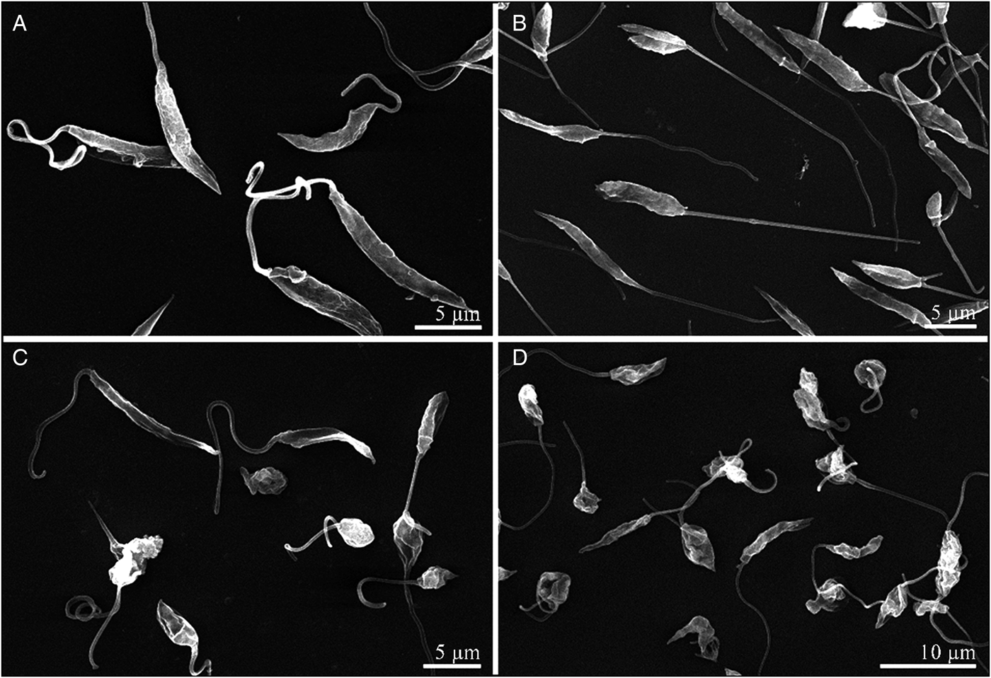
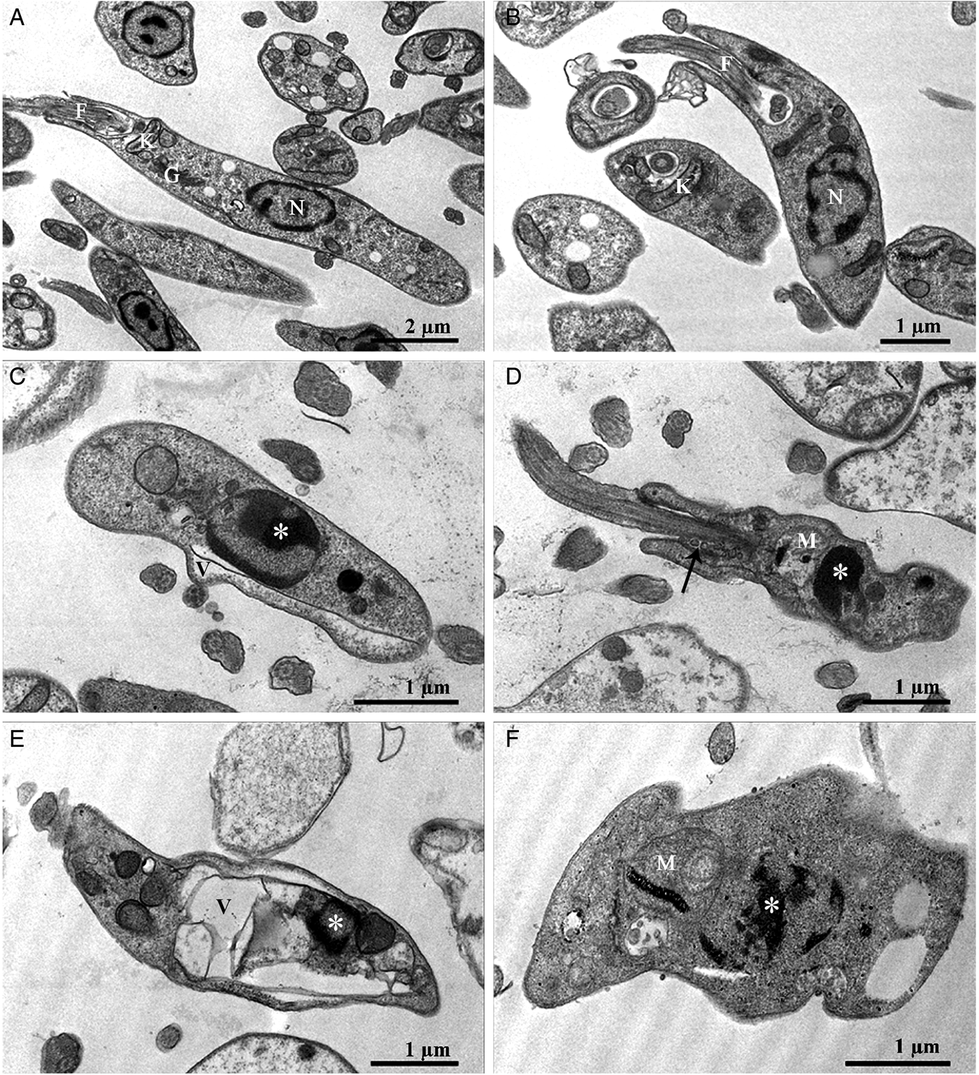
Promastigotes were treated with nerolidol (IC50/24 h or 2 × IC50/24 h) and then processed for scanning (SEM, Fig. 1) and transmission (TEM, Fig. 2) electron microscopy. Severe morphological changes were observed by SEM, including reduction, shrinkage and roundness in the parasite cell body (Fig. 1). With respect to the ultrastructure (TEM), mitochondrial damage was noted, with mitochondrial swelling and disorganization, in addition to the presence of internal vesicles and concentric membranes in the organelle (Fig. 2D, F). Other alterations were frequent, such as the occurrence of cytoplasmic vacuolization (Fig. 2C, E), vesicles at the flagellar pocket (Fig. 2D), and chromatin condensation (Fig. 2C–F). Little or no difference was noted by SEM and TEM after treatment with the IC50/24 h of amphotericin B (Figs 1B and 2B) compared with the untreated control (Figs 1A and 2A). Because nerolidol was responsible for ultrastructural damage, its activity was further investigated by flow cytometry. The presence of multiple mitochondrial morphological alterations led us to hypothesize that the mitochondrial membrane potential was altered, thereby contributing to cell death.
SEM of L. braziliensis promastigotes. (A) Untreated control; (B) Treatment with IC50/24 h of amphotericin B; (C) Treatment with IC50/24 h of nerolidol; (D) Treatment with 2 × IC50/24 h of nerolidol. Although no morphological alteration was noted after incubation with amphotericin B, nerolidol treatment caused shrinkage and roundness of the parasite cell body.

Transmission electron microscopy of L. braziliensis promastigotes. (A) Untreated control; (B) IC50/24 h of amphotericin B, showing no morphological alterations; (C–F) Incubation with IC50/24 h (C, D) or 2 × IC50/24 h (E, F) of nerolidol. Note cytoplasmic vacuolization (V), chromatin condensation (*), vesicles at the flagellar pocket (arrow) and mitochondrial swelling and disorganization (M). F: flagellum; G: Golgi complex; N: nucleus.

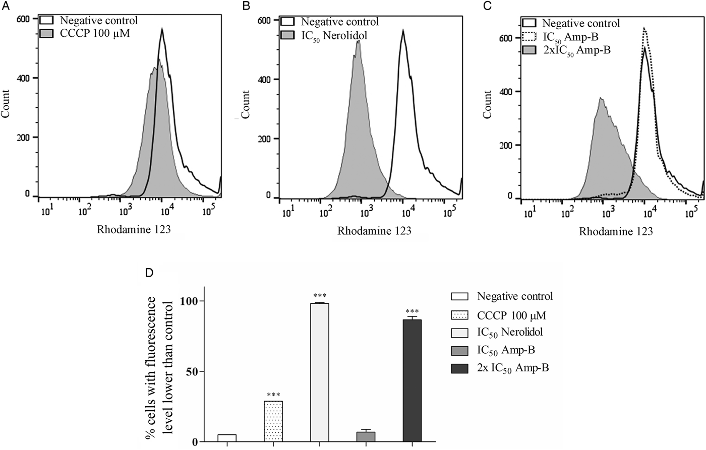
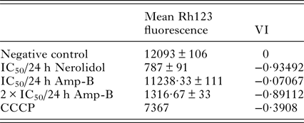
Incubation with Rh123 after nerolidol treatment (IC50/24 h and 2 × IC50/24 h) led to a reduction in fluorescence of nearly 100% in the promastigotes (Fig. 3B). Both treatments gave similar results and confirmed the mitochondrial damage observed by TEM. Parasites incubated with amphotericin B at the IC50/24 h value presented variation in the mitochondrial membrane potential in only 7% of the cells, which was not significantly different from the negative control. In contrast, higher amphotericin B concentrations (2 × IC50/24 h) induced a reduction in fluorescence in 80% of the cells (Fig. 3C). Treatment with 100 µ m CCCP (positive control, Fig. 3A) reduced the Rh123 signal in 30% of the cells. The VI calculations showed that nerolidol treatment led to mitochondrial membrane depolarization (Table 2).
Mitochondrial membrane potential (ψm) of L. braziliensis promastigotes incubated for 24 h with the IC50 of nerolidol and the IC50 or 2 × IC50 of amphotericin B. Graphs in white correspond to the untreated (negative) control. (A) Positive control – 100 µ m CCCP; (B) IC50 of nerolidol; (C) IC50 of amphotericin B and 2 × IC50 of amphotericin B; (D) Percentage of parasites with Rh123 fluorescence level lower than the negative control. *P < 0·05; **P < 0·01; ***P < 0·001.

Mean and standard deviation of the variation index (VI) to assess the mitochondrial membrane potential of promastigotes labelled with rhodamine 123 (Rh123).

Amph-B, amphotericin B; CCCP, positive control.
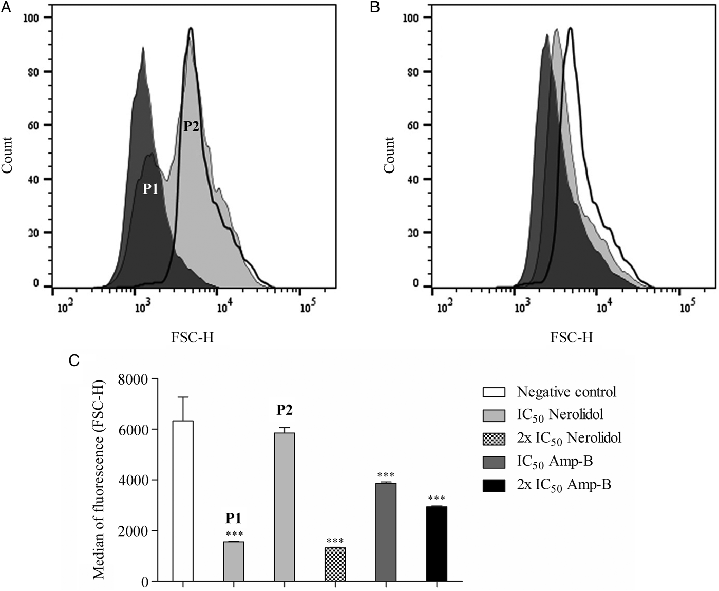
The cell size of promastigotes incubated with the IC50/24 h value of nerolidol was evaluated by flow cytometry by taking into account the FSC-H parameter (Fig. 4). This experiment confirmed the SEM results, with observations of roundness and diminished parasite cell bodies. Two subpopulations were formed after this treatment. The first subpopulation (P1) comprised 47·78% of the cells and displayed smaller sizes compared with the control parasites. Another subpopulation (P2) included 49·85% of the promastigotes and featured a median cell size that did not significantly differ from the negative control (Fig. 4C), indicating that no changes in cell size occurred in this subpopulation. Treatment with the 2 × IC50 of nerolidol resulted in a unique population with almost 100% of the cells smaller in size than the control cells. Parasites treated with amphotericin B exhibited a reduction in volume in a dose-dependent manner that significantly differed from the negative control.
Cell size of L. braziliensis promastigotes treated with nerolidol (A) or amphotericin B (B). P1 and P2 represent two distinct populations formed after treatment with the IC50 of nerolidol. In both histograms, the IC50 is represented by the light grey filled curve and the 2 × IC50 with the dark grey filled curve. The thick and unfilled line curve represents the negative control. (C) Median autofluorescence related to the cell size (FSC-H). *P < 0·05; **P < 0·01; ***P < 0·001.

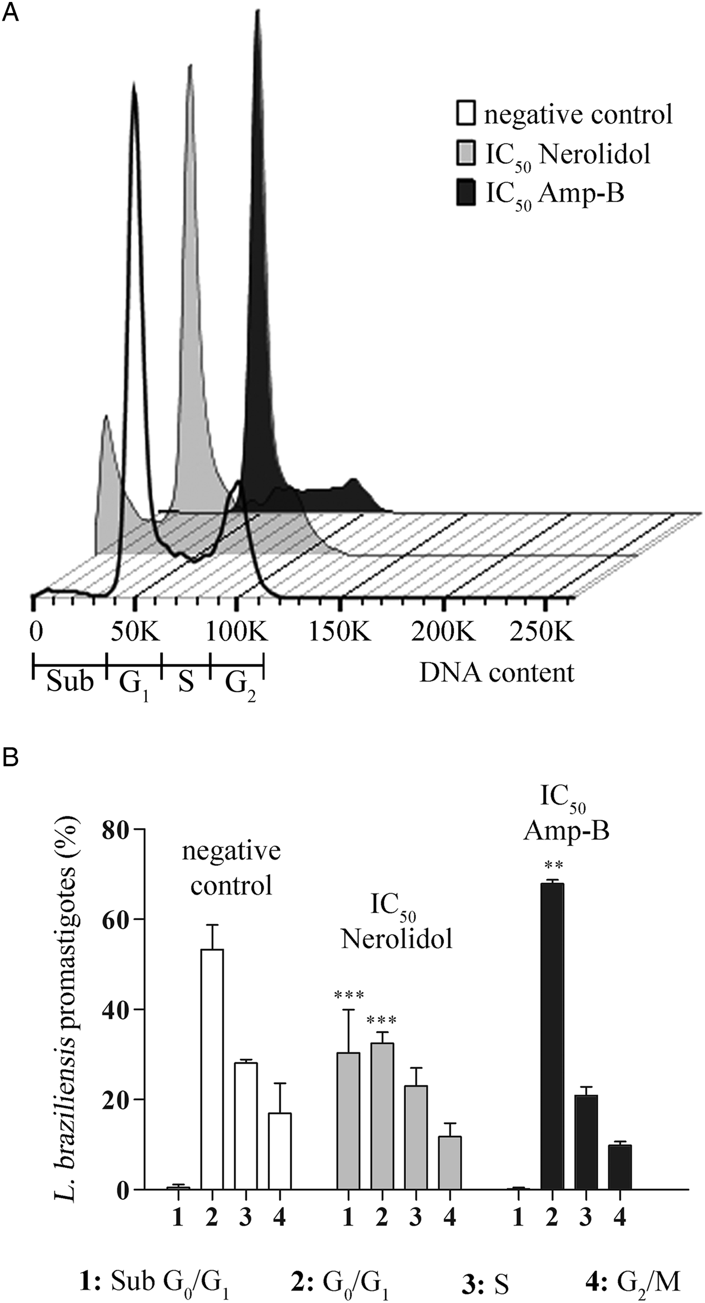
Cell cycle analysis after treatment with the IC50/24 h of nerolidol and PI labelling showed the formation of a G0/G1 sub-peak by 30·36% of the cells, which was in contrast to the 0·53% of the population that formed this peak in the negative control cells (Fig. 5). Consequently, fewer cells were in the remaining steps of the cell cycle. No peaks were observed when the 2 × IC50 of nerolidol was used, suggesting intense DNA degradation (data not shown). Treatment with amphotericin B did not result in G0/G1 sub-peak development. However, a significant increase was observed in the number of cells at the G0/G1 phase, which consequently reduced the cells in the S and G2/M phases (Fig. 5).
(A) Histogram of DNA content in the different cell cycle stages (sub G0/G1, G0/G1, S and G2/M) in L. braziliensis promastigotes treated with IC50 of nerolidol or amphotericin B (Amp-B). Sub: sub G0/G1; G1: G0/G1; G2: G2/M. (B) Percentage of cells at the different cell cycle stages. *P < 0·05; **P < 0·01; ***P < 0·001.

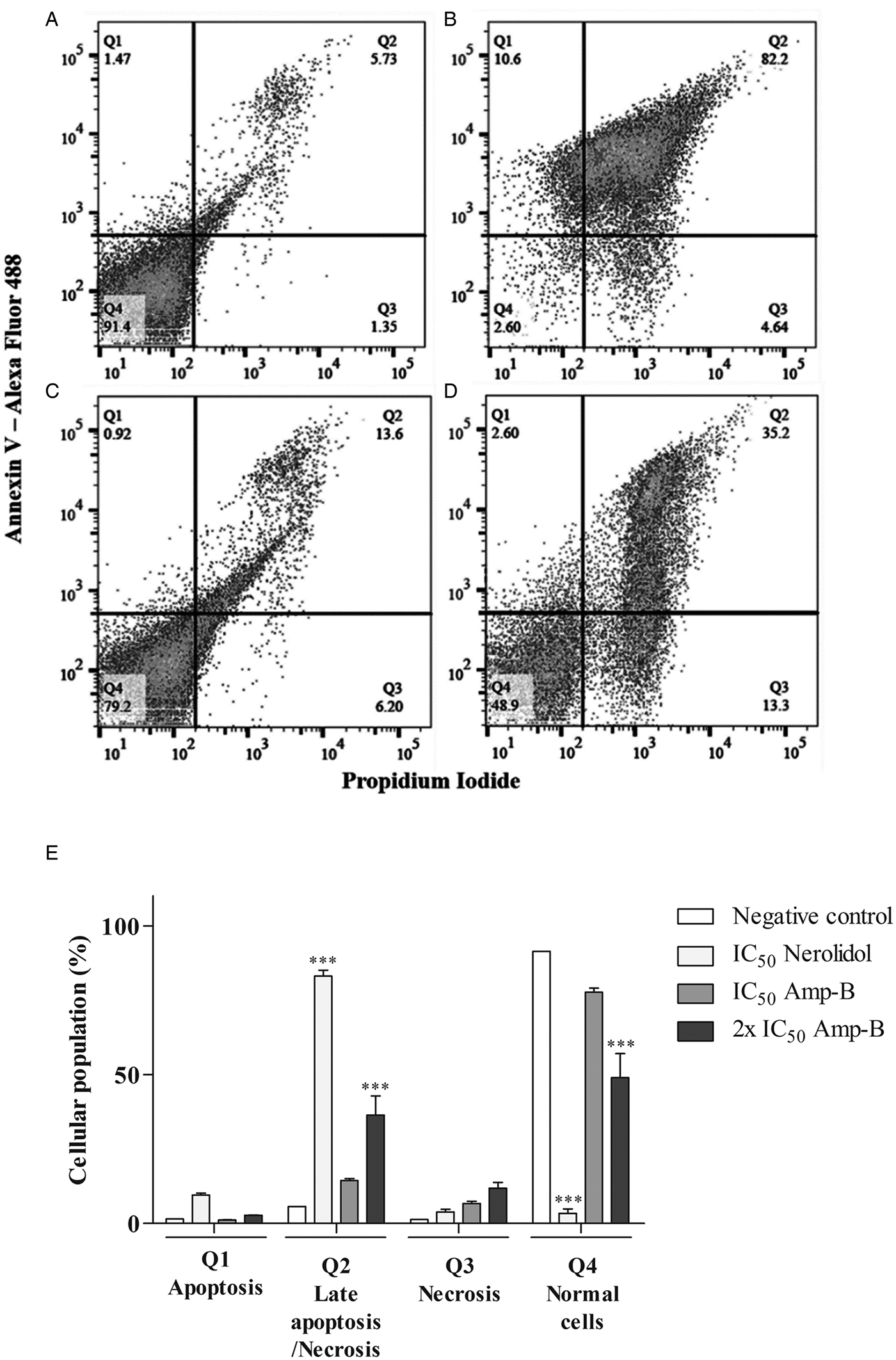
To confirm cell death induction by nerolidol in L. braziliensis, the cells were double-labelled with Annexin-V and PI. Our results showed that 82·2% of the parasites treated with the IC50/24 h of nerolidol displayed positive labelling for both Annexin-V and PI (Fig. 6B). Nevertheless, 4·64% of the cells were positive exclusively for PI, which did not differ significantly from the negative control (1·35% of cells positive for PI). Amphotericin B treatment at the IC50/24 h concentration (Fig. 6C) did not result in significant differences in Annexin-V/PI staining compared to the negative control. However, 35% of the cells were double-labelled only after 2 × IC50 treatment (Fig. 6D).
Assessment of the membrane permeability and phosphatidylserine exposure of L. braziliensis promastigotes. (A) negative control; (B) IC50/24 h of nerolidol; (C) IC50/24 h of amphotericin B; (D) 2 × IC50/24 h of amphotericin B; (E) cell population present in each quadrant (Q1: apoptosis; Q2: late apoptosis/necrosis; Q3: necrosis and Q4: live cells). *P < 0·05; **P < 0·01; ***P < 0·001.

DISCUSSION
Our data show that PaEO was effective against L. braziliensis promastigotes. The result (IC50/24 h = 77·9 µg mL−1) was similar to that obtained for the P. demeraranum and P. duckei EOs on L. amazonensis promastigotes, with IC50/24 h values of 86 and 46 µg mL−1, respectively (Carmo et al. Reference Carmo, Amaral, Machado, Leon and Silva2012). Several EOs have already been tested against different Leishmania spp. (Supplementary Table S1), most assays being performed for 72 h, which leads to lower IC50 values (Misra et al. Reference Misra, Kumar, Khare, Gupta, Kumar and Dube2009; Monzote et al. Reference Monzote, García, Montalvo, Scull and Miranda2010; Esperandim et al. Reference Esperandim, Ferreira, Rezende, Magalhães, Souza, Pauletti, Januário, Laurentz, Bastos, Símaro, Cunha and Andrade e Silva2013; Bernuci et al. Reference Bernuci, Iwanaga, Fernandez-Andrade, Lorenzetti, Torres-Santos, Faiões, Gonçalves, Amaral, Deschamps, Scodro, Cardoso, Baldin and Cortez2016). We carried out tests for 24 h to minimize the effects of nutrient starvation and toxicity due to occasional parasite lysis. Our data confirmed the leishmanicidal activity of Piper spp. EOs.
Nerolidol, the main constituent of PaEO (Villamizar et al. Reference Villamizar, Cardoso, Andrade, Teixeira and Soares2017), showed anti L. braziliensis activity (IC50/24 h = 74·3 µg mL−1). This finding validates the antimicrobial potential of compounds obtained from a natural source (Anthony et al. Reference Anthony, Fyfe and Smith2005). More research is needed to clarify the mechanism of action of the active compounds derived from plants, which may to lead the development of potent anti-leishmanial drugs in the near future (Ullah et al. Reference Ullah, Nadhman, Siddiq, Mehwish, Islam, Jafri and Hamayun2016).
The nerolidol activity was previously tested against L. amazonensis, L. braziliensis and L. chagasi promastigotes, resulting in IC50/24 h values of 85, 74 and 75 µ m, respectively (Arruda et al. Reference Arruda, D'Alexandri, Katzin and Uliana2005). For L. braziliensis, the SI for mammalian cells was 1·69, which was similar to the result obtained in our research (SI = 1·41). Nerolidol showed decreased toxicity against erythrocytes (SI = 20·19). One possible explanation is the absence of mitochondria in red blood cells, which suggests that nerolidol acts on intracellular targets rather than via inducing cell membrane disruption.
Low density compounds, such as EOs or small hydrophobic molecules (e.g., nerolidol, which is a sesquiterpene alcohol) can easily cross the plasma membrane to reach intracellular targets (Anthony et al. Reference Anthony, Fyfe and Smith2005). Terpenes do not affect the membrane protein dynamics; instead, their activity is mostly restricted to the lipidic membrane components, thus altering the membrane fluidity (Camargos et al. Reference Camargos, Moreira, Mendanha, Fernandes, Dorta and Alonso2014). Together, these data suggest that nerolidol may damage the parasite membrane, thereby increasing its permeability while inducing cell death mechanisms consistent with an apoptosis-like process in Leishmania.
An apoptosis-like process has been suggested as a cell death mechanism in Leishmania following drug treatment. For instance, eupomatenoid-5, which is derived from Piper regnellii, leads to mitochondrial membrane depolarization, PS exposure and cell size reduction (Garcia et al. Reference Garcia, Lazarin-Bidóia, Ueda-Nakamura, Silva and Nakamura2013). The leishmanicidal activity of racemoside-A was evidenced by morphological alterations, such as cell shrinkage, ovoid cells, absence of flagella and chromatin condensation. These effects were attributed to cell death induction detected via Annexin-V, PI and the loss of mitochondrial membrane potential, which culminated in DNA fragmentation and permanency of the cells in the sub-G0/G1 phase (Dutta et al. Reference Dutta, Ghoshal, Mandal, Mondal, Banerjee, Sahu and Mandal2007). DNA fragmentation into multiple 200 bp pieces was observed in L. amazonensis promastigotes treated with 40 µ m miltefosine for 24 h. Together with PS exposure and the permanence of the cells in the sub-G0/G1 phase, this result was interpreted as programmed cell death (Marinho et al. Reference Marinho, Gonçalves, Oliveira, Oliveira, Bellio, d'Avila-Levy, Santos and Branquinha2011).
We found several morphological evidences of an apoptosis-like process in nerolidol-treated L. braziliensis, such as mitochondrial membrane depolarization, PS exposure and cell size reduction (cell shrinkage). These evidences indicate that treatment with nerolidol would lead to L. braziliensis cell death through an apoptosis-like process, a nomenclature used in several studies on Leishmania cell death (e.g.: Jiménez-Ruiz et al. Reference Jiménez-Ruiz, Alzate, Macleod, Lüder, Fasel and Hurd2010; Gannavaram and Debrabant, Reference Gannavaram and Debrabant2012; Islamuddin et al. Reference Islamuddin, Sahal and Afrin2014; Awasthi et al. Reference Awasthi, Kathuria, Pant, Kumari and Mitra2016; Shadab et al. Reference Shadab, Jha, Asad, Deepthi, Kamran and Ali2017). Our morphological data do not indicate necrosis, since oncosis (cell swelling) and plasma membrane rupture were not observed.
Proto et al. (Reference Proto, Coombs and Mottram2013) reviewed the evidences for unregulated/regulated cell death pathways in parasitic protozoa and proposed that unregulated cell death in these organisms can be classified into just two primary types: necrosis or incidental cell death. According to these authors, regulated necrosis and (regulated) extrinsic apoptosis are precluded in parasitic protozoa owing to the absence of established death receptors and caspases. Trypanosomatids have metacaspases, which have significant structural similarities with caspases, but are not caspases and cannot be considered as mediators of a similar regulated cell death (Proto et al. Reference Proto, Coombs and Mottram2013). Therefore, according to Proto et al. (Reference Proto, Coombs and Mottram2013), in absence of robust biochemical evidence on mechanism, parasitic protozoan caspase independent/metacaspase dependent death should be classified not as apoptosis, but as incidental cell death. In the absence of biochemical evidences, we then conclude that nerolidol-induced incidental cell death in Leishmania braziliensis.
The biochemical events leading to cell death may provide key novel therapeutic targets that could be exploited in the design of specific chemotherapeutic agents for pathogenic trypanosomatids (Welburn et al. Reference Welburn, Macleod, Figarella and Duzensko2006). Leishmania cells contain a single mitochondrion, similar to other kinetoplastids. Therefore, this organelle appears to be an interesting chemotherapeutic target, with characteristics that are distinguishable from those of mammalian cells (Inacio et al. Reference Inacio, Canto-Cavalheiro, Menna-Barreto and Almeida-Amaral2012).
In a L. donovani study, parasites treated with pentostam or amphotericin B presented evidence of cell death, with discontinuation in one DNA strand, DNA fragmentation, diminished mitochondrial membrane potential and increased cell permeability (Lee et al. Reference Lee, Bertholet, Debrabant, Muller, Duncan and Nakhasi2002). The mechanism of action of amphotericin against Leishmania spp. can be explained by its affinity for the sterols present at the parasite membrane. Transmembrane channels are formed following interactions with the membrane, leading to cation loss and cell death (Brajtburg and Bolard, Reference Brajtburg and Bolard1996). Thus, nerolidol and amphotericin B appear to play roles in both parasite membrane permeability and cell death.
Currently, only two methods are available to treat and control leishmaniasis: rational use of drugs and vector insect control. A limited number of drugs are available, and these drugs are expensive, have side-effects and are inefficient (Srivastava et al. Reference Srivastava, Shankar, Mishra and Singh2016). A study in Latin America to evaluate differences in responses to pentavalent antimonial treatment showed that the cure afforded by the use of this drug differs between distinct Leishmania species (Arevalo et al. Reference Arevalo, Ramirez, Adaui, Zimic, Tulliano, Miranda-Verástegui, Lazo, Loayza-Muro, De Doncker, Maurer, Chappuis, Dujardin, Llanos-Cuentas and Llanos-Cuentas2007). Therefore, the search for alternative effective compounds from natural sources is relevant. Terpenes are powerful skin permeation promoters (El-Kattan et al. Reference El-Kattan, Asbill, Kim and Michniak2001; Camargos et al. Reference Camargos, Moreira, Mendanha, Fernandes, Dorta and Alonso2014). The sesquiterpene nerolidol showed an anti-L. braziliensis effect as described here. Nerolidol has low toxic potential in vivo (Lapczynski et al. Reference Lapczynski, Bhatia, Letizia and Api2008) and presents anti-tumorigenic activity in female rats (Arroyo-Acevedo et al. Reference Arroyo-Acevedo, Chávez-Asmat, Anampa-Guzmán, Donaires and Ráez-Gonzáles2015). Obtaining nerolidol derivatives with better SI can be the source for promising candidates in the search for new compounds to kill Leishmania parasites.
SUPPLEMENTARY MATERIAL
The supplementary material for this article can be found at https://doi.org/10.1017/S0031182017000452.
ACKNOWLEDGEMENTS
The authors thank the Program for Technological Development in Tools for Health-PDTIS FIOCRUZ for the use of its facilities (Platforms RPT08L – Flow Cytometry-PR and RPT07C – Confocal and Electron Microscopy-PR).
FINANCIAL SUPPORT
This work was supported by Conselho Nacional de Desenvolvimento Científico e Tecnológico (CNPq), Fundação de Amparo à Pesquisa do Estado de Minas Gerais (FAPEMIG) and Fundação Oswaldo Cruz (Fiocruz).