I. INTRODUCTION
More than often, studies on natural materials have provided insights into design and development of new classes of materials. Reference Mayer1,Reference Mayer2 Examples include studies performed on biological shells and marine crustaceans. Reference Chen, Peng, Wang and Wu3–Reference Melnick, Chen and Mecholsky15 In line with this, the discovery of the deep sea shrimp species such as hydrothermal vent shrimp (Rimicaris exoculata) that exists near high-temperature deep sea vents Reference Fricke, Giere, Stetter, Alfredsson, Kristjansson, Stoffers and Svavarsson16–Reference Connelly, Copley, Murton, Stansfield, Tyler, German, Van Dover, Amon, Furlong, Grindlay, Hayman, Huhnerbach, Judge, Le Bas, McPhail, Meier, Nakamura, Nye, Pebody, Pedersen, Plouviez, Sands, Searle, Stevenson, Taws and Wilcox20 (∼500 °C) has spurred new research activity. The key studies so far have focused on understanding the changes in the physiological and biological activities of such shrimps at high temperatures and at high-temperature gradients. Reference Clarke and Fraser21–Reference Oliphant, Thatje, Brown, Morini, Ravaux and Shillito26 Researchers have published extensive data on the metabolism and oxygen consumption rate of hydrothermal vent shrimps. Studies suggest that both metabolism rate and, therefore, the oxygen consumption rate increase with the increase in the surrounding temperatures. Reference Smith, Brown, Mestre, Reed and Thatje22–Reference Allan, Froneman and Hodgson24 In the current study, the focus is on understanding how the exoskeleton of Rimicaris exoculata deforms as a function of temperature. The measurement results are compared with the properties of the exoskeletons of a shallow water shrimp species (Pandalus platyceros). Rimicaris exoculata lives near high-temperature deep sea vents with temperatures of volcanic sites up to 500 °C and the temperature of nearby region up to 90 °C at a depth of 2300 m. Reference Fricke, Giere, Stetter, Alfredsson, Kristjansson, Stoffers and Svavarsson16–Reference Connelly, Copley, Murton, Stansfield, Tyler, German, Van Dover, Amon, Furlong, Grindlay, Hayman, Huhnerbach, Judge, Le Bas, McPhail, Meier, Nakamura, Nye, Pebody, Pedersen, Plouviez, Sands, Searle, Stevenson, Taws and Wilcox20 Properties of focus, therefore, are nanoindentation hardness and creep strain rate measured at temperatures ranging from 25 to 80 °C.
Shrimp exoskeleton belongs to a subclass of marine exoskeletons. It consists of chitin-based fibrils coated in proteins at nanometer level which further bind together to form fibers. These fibers are then woven together to form chitin–protein layers which are stacked in a twisted plywood structure known as the Bouligand pattern. Reference Hepburn, Joffe, Green and Nelson4–Reference Chen, Lin, McKittrick and Meyers11 The spacing between such woven structures is filled with proteins and biominerals. An extensive examination of the literature regarding marine crustacean exoskeletons mechanical property measurements will reveal that there has been no study focusing on nanomechanical properties of coastal and deep sea shrimp exoskeleton as a function of temperature. Since Rimicaris exoculata survives at unusual ocean depths and at high temperatures, a comparison of its exoskeleton properties with those of shallow water shrimp (Pandalus platyceros) can reveal important insights regarding design principles of biological materials that survive at high temperatures. There have been several studies to find chemical composition of shrimp exoskeleton. Reference Ravichandran, Rameshkumar and Prince27–Reference Ibrahim, Salama and El-Banna33 Shahidi and Synowiecki Reference Shahidi and Synowiecki30 compared chitin, protein, and carotenoid contents of shrimps and crabs. Protein content in shrimp shell was found to be almost double as compared to that in crab shell. This study aims to understand how the high-temperature deformation correlates to exoskeleton structure in the examined shrimp species. Since such shrimps survive at varying ocean depths under constant pressure, studies focus on creep deformation to provide an assessment of the two types of exoskeleton for the role of the proteins and minerals to cause differences in behavior and properties between the two shrimp species. Hardness measurements are performed to correlate noncreep plastic deformation strength measurements (hardness) with the creep strength measurements.
II. METHODS
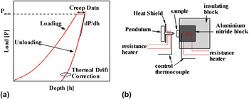
The shrimp exoskeleton has micrometer scale thickness as well as significant heterogeneity. In such materials, traditionally performed uniaxial tests will only provide overall mechanical strength information. Nanoindentation measurements have the capability to provide site specific data with minimal sample preparation. This makes nanoindentation a standard experimental technique to find the modulus and hardness of heterogeneous materials at nano and micro scales. Figure 1(a) shows a standard indentation curve and parts of the curve used for material property calculations.
(a) Indentation curve showing parts of curve used for stiffness, creep, and thermal drift calculations with load being denoted as P and depth being denoted as h (the figure does not have any units as P and h can have different units at different scales) and (b) schematic of the instrument setup for high-temperature indentation tests.

The experimental procedure involves indenting the surface of the material being tested by increasing the force in small steps until the predetermined peak load (P max) or peak depth (h max) is achieved. A Berkovich indenter is used in the present work. The unloading part is used for predicting the material properties using a framework based on contact mechanics. Reference Oliver and Pharr34,Reference Pharr35 Unlike normal indentation tests without dwell, the indentation creep tests also involve viscoelastic deformation during unloading. The nanoindentation hardness involves total deformation including viscous deformation. Therefore, hardness alone is not a true indicator of material behavior. Therefore, in this work, indentation creep behavior is also analyzed by developing an understanding of time-dependent deformation curves based on earlier work reported by authors. Reference Gan and Tomar36–Reference Verma and Tomar40
A. Overall experimental procedure
Experiments were performed on dry samples as a function of temperature in a multi module mechanical tester (NanoTest, Micro Materials Ltd.). Reference Gan and Tomar36–Reference Verma and Tomar40 Figure 1(b) shows schematic of hot stage. The instrument consists of a vertical pendulum pivoted on a frictionless spring. It incorporates a feedback mechanism to accurately stabilize the temperature during experiments. An indenter is attached to the pendulum that indents sample during experiment horizontally. The force on the pendulum is applied through magnetic coils which allows for a very high sensitivity during experiment. The depth of indentation during experiments is measured by capacitor plates located behind the indenter. Before experiments, calibrations are performed to get accurate measurements of load and depth. Depth calibration, load calibration, and frame compliance experiments are performed before conducting actual experiments. A Berkovich indenter was used to perform all the experiments. The tip radius of the indenter is approximately 20 nm. During indents, samples were mounted on stage firmly to avoid any movement during experiments.
Experiments were performed on 10 different exoskeleton samples for each examined species to capture statistical property differences possibly due to biological changes in samples. Experiments were performed in the temperature range from 25 to 80 °C. This range was chosen based on two criteria: First, temperature range should include the room temperature (25 °C) conditions at which shallow water shrimps generally live and second, it should be high enough to give reasonable temperature activated energy for failure of both species. It is always a challenge to simulate exactly the same experimental condition while performing experiments for long time intervals. To avoid discrepancies in the collected data, the best approach used by most researchers is to perform all experiments using an identical experimental setup. In the present study, all experiments were conducted using the same indenter, the same hot stage, the same mounting technique at all temperatures. All high-temperature indents were performed at the peak load of 2 mN and at the loading rate of 0.05 mN/s. These values were chosen based on indentation size effect analyses. Around 50 indentations were performed for each data point. Indentation depth for samples was around 300 nm.
B. Thermal drift measurements
The indentation instrument was kept in a chamber and its temperature during experiments was maintained at a constant value by a feedback mechanism. The correct measurements are dependent on thermal stabilization of indentation instrument during experiments. Reference Feng and Ngan41 The errors generated during indentation due to thermal stabilization are termed as thermal drift. To correct this error, thermal drift data were collected at 10% of the peak load of indentation. Thermal drift rate was calculated from collected data and was used to adjust indentation data. A time period of 200 s was allowed to collect thermal drift data during experiments performed. This time was based on the total time of each indentation experiment to account for the fluctuations that might have occurred during the whole indentation process. This step ensures that hardness calculated from indentation curves is free from thermal drift errors. This setup of thermal drift measurements has been verified earlier in Refs. Reference Gan and Tomar36–Reference Verma and Tomar40.
C. Substrate effect
During the measurements of hardness values of thin exoskeleton samples, substrates could introduce major errors. A general rule is that indentation depth should be less than 10% of sample thickness. Reference Saha and Nix42–Reference Kramer, Volinsky, Moody and Gerberich44 Thickness of analyzed samples was around 10 µm and indentation depth at the load of 2 mN was 300 nm which satisfies the 10% rule. This analysis verifies that substrate effect has been taken into account and does not interfere with the measured values.
D. Sample preparation
The whole shrimp samples of Rimicaris exoculata were provided by Dr. Juliette Ravaux from the mission Momardream in 2007 from the depth of 2300 m. The Rimicaris exoculata shrimps were maintained for 10 h at 10 °C with a 1 h heat shock at 30 °C in pressurized aquaria on board. Shallow water shrimp (Pandalus platyceros) samples were procured fresh and kept frozen after supply. Both specimens were stored in the same way after procurement. They were stored in liquid nitrogen to help decrease any degradation that could happen in the normal environment.
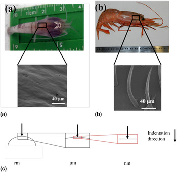
Figure 2 shows the picture of the whole shrimps and the samples prepared for nanoindentation and energy dispersive x-ray (EDX) tests. The sample preparation steps were as follows: (i) shrimps were taken out from the liquid nitrogen storage container, (ii) the shrimps were allowed to settle down to room temperature and then the carapace was removed from them. Carapace is the head part of the shrimp as shown in Figs. 2 and 3, the smaller samples for nanoindentation and EDX tests were cut from the carapace with a sharp blade for a clean cut without mechanically straining the samples. The indents were placed on the surface of the samples. The surface is shown in the scanning electron microscopy (SEM) images in Fig. 2. The tip of the indenter used is 20 nm. This is small enough to ensure that the surface that indenter sees is flat as shown in Fig. 2(c). The surface being indented in current experiments was visually checked with inbuilt microscope in the indentation instrument to make sure that it was free from any cracks, voids, and other effects that could potentially affect indentation procedure. Carapace is the exoskeleton of shrimp that protects cephalothoracic region of shrimp. Cephalothoracic is the front part of shrimp that contains vital parts of shrimp including mouth, eyes, antennas, etc. Carapace is referred to as shrimp exoskeleton further in this study. The exoskeleton was then dried in open for 3 days to prepare samples for high-temperature tests.
Picture of whole shrimps and the SEM images of the samples for experiments: (a) Rimicaris exoculata and (b) Pandalus platyceros, (c) surface curvature with respect to testing length scale.

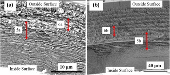
SEM images of full cross section of shrimp exoskeletons: (a) Rimicaris exoculata and (b) Pandalus platyceros.

III. RESULTS AND DISCUSSION
A. Structure of exoskeleton
The structural design of the examined shrimp exoskeletons was studied by obtaining SEM images of samples. EDX analysis data were also obtained to get a detailed understanding of composition. Figures 3(a) and 3(b) show the SEM images of full cross section of Rimicaris exoculata and Pandalus platyceros, respectively. Figures 3(a) and 3(b) show the inner side of the exoskeleton at the bottom and the outer side of the exoskeleton on the top. There is a clear difference in stacking density and thickness of layers across the cross section.
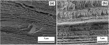
In Figs. 3(a) and 3(b), the layers at the top of the exoskeleton are much thicker as compared to the layers at the bottom. Figure 3 also shows that the bottom region has low porosity with dense stacking. The maximum thickness of the layer at the top of the exoskeleton Rimicaris exoculata is around 1 μm and minimum thickness at the bottom most region is of the order of 20–30 nm. While in Pandalus platyceros, the maximum thickness of the layers is up to 2 μm and minimum thickness at the top layer drops down to 500 nm. Each visible plane is a layer of woven α-chitin–protein fiber network, the characteristic feature of exoskeleton of crustaceans. Reference Boßelmann, Romano, Fabritius, Raabe and Epple6–Reference Raabe, Sachs and Romano8 Each layer consists of different mixes of proteins and minerals. A closer look at Figs. 3(a) and 3(b) shows that the thickness of the layers changes as a function of depth. Starting from the bottom of the exoskeleton, these layers increase in thickness gradually until there is a stacking of layers of similar thickness at the point corresponding to half of the total thickness. At this point, there is an interface where the thickness of the layers increases. Such distribution of the layer is of particular interest and suggests the possible contribution of stacking sequence in the overall properties of the shrimp exoskeleton. To investigate this issue further, Fig. 4(a) shows the middle interface region in the case of Rimicaris exoculata and Fig. 4(b) shows similar middle interface region in the case of Pandalus platyceros. The interface region of shallow water shrimp (Pandalus platyceros) shows a sudden change in the thickness of Bouligand Reference Bouligand9 layers whereas that of Rimicaris exoculata shows a more gradual change.
SEM images of interface of shrimp exoskeletons: (a) Rimicaris exoculata and (b) Pandalus platyceros.

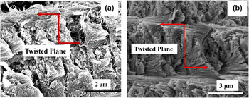
Figure 5(a) shows the top twisted layers of the Rimicaris exoculata exoskeleton structure revealing a Bouligand pattern. These layers are formed by helicoidal stacks of the α-chitin–protein planes. The thickness of each layer has 180° rotated stacked layers of α-chitin–protein planes. For comparison, Fig. 5(b) shows the Bouligand structure of the Pandalus platyceros exoskeleton. The similarity in Bouligand pattern is evident. However, the thickness of protein layers in Pandalus platyceros is found to be approximately double of that in Rimicaris as noted from the red vertical line of the twisted planes in Figs. 5(a) and 5(b).
Bouligand structure of shrimp exoskeletons: (a) Rimicaris exoculata and (b) Pandalus platyceros.

Rimicaris exoculata lives at high temperature and pressure at depths of 2300 m. The environment at those depths is much different as compared to the shallow water. Several studies have been performed to understand the effect of these environmental changes on these shrimps that include measuring the difference in metabolic activities. From the structural point of view, to sustain those high pressures and temperatures there must be structural difference in these shrimps. The SEM images in Figs. 3–5 show similar pattern in the structural design with difference being in the position of change in the thickness of the layers. To further investigate these changes in structure, EDX spectrums for both shrimps are compared to reveal information about differences at compositional level.
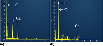
Figure 6 shows the EDX spectrums for Rimicaris exoculata and Pandalus platyceros. Both the spectrums were collected using the same QUANTA 3D SEM (FEI Company, Hillsboro, OR). Figure 6(a) shows the collected spectrum from Rimicaris exoculata and Fig. 6(b) shows spectrum from Pandalus platyceros. Figure 6 shows that exoskeleton is high in oxygen and carbon which are main constituents of proteins. Reference Seki, Schneider and Meyers13 Figure 6(a) shows a peak corresponding to calcium and a peak corresponding to phosphorus. These are two key differences that we observe in both spectrums. There is one extra peak showing noticeable amount of phosphorus and also peak for Ca is higher as compared to Pandalus platyceros spectrum.
EDX analysis of cross section of shrimp showing elemental spectrum in the case of (a) Rimicaris exoculata and (b) Pandalus platyceros.

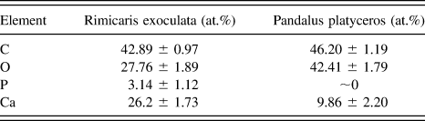
These changes in composition can be further verified from Table I giving quantitative elemental composition. The composition given in Table I is comparable to the chemical composition obtained by several researchers on coastal shrimps. Reference Ravichandran, Rameshkumar and Prince27–Reference Ibrahim, Salama and El-Banna33 Deep sea shrimp Rimicaris exoculata shows some variations in composition with an extra phosphorus peak. Table I gives relative quantitative analysis of individual elements present in the exoskeleton of both the shrimps. The presence of Ca supports the fact that Rimicaris exoculata survives at very high pressures and needs to have more structural strength to survive. Phosphorus is one of the materials from volcanic vents in nearby habitat of Rimicaris exoculata. Phosphorus also became part of the shrimp exoskeleton with evolution of shrimps near deep sea vents possibly via food sources while its presence is not significant in Pandalus platyceros as shown in Fig. 6(b). A combination of observations from Figs. 3–6 reveals that the Rimicaris exoskeleton has higher content of minerals and reduced protein layer thicknesses in comparison to the Pandalus platyceros exoskeleton. Higher mineral content allows for better structural strength of the Rimicaris exoculata exoskeleton at such extreme environments.
Comparison of quantitative data by EDX analysis on cross section of shrimp exoskeleton for Rimicaris exoculata and Pandalus platyceros.

B. Intra/inter layer crack propagation in the exoskeleton
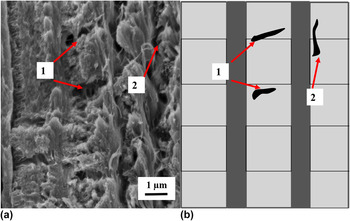
Long-term failure of polymeric matrix composites is normally a result of the accumulation of microscale damage at matrix/fiber level. Reference Koyanagi, Yoneyama, Nemoto and Melo45 Time-dependent strength of the matrix/fiber interface is expected to play an important role in time-dependent strength of fiber-reinforced composite materials. As a basic model, shrimp exoskeleton is a stacked layered structure consisting of woven layer made up of protein fibrils, minerals stacked together in a Bouligand pattern. Thickness of layers is highest at the outer side of the exoskeleton, Fig. 3. Layer thickness keeps decreasing as we move toward the inner side with thinnest layers at the bottom region. One reason for this characteristic design can be understood from crack propagation behavior observed in shrimp exoskeleton. Figure 7 shows a cross section image of a Rimicaris exoculata exoskeleton with visible cracks formed after hardness measurements at room temperature.
(a) A SEM image showing the cracks in a Rimicaris exoculata exoskeleton observed after hardness measurement at room temperature and (b) a schematic showing crack in regions corresponding to the SEM image with (1) crack propagation inside planar layer and (2) crack at the interface of two layers.

As shown in Fig. 7(a), most of the cracks are confined in the multilayered design indicated by number one. There are some cracks along the interface region but it is hard to see any cracks crossing the interface between layers. The cracks are confined in the Bouligand layers. The high number of layers in Bouligand pattern gives layers extra strength against failure through crack-induced defects. Thicker layers allow propagation of cracks better than thin layers. Outer layers are thicker facilitating movement of crack to the outer side as compared to the inner side. Vital organs of shrimp are located just below exoskeleton. The high number of very thin layers restricts movement of crack in a top-down direction of shrimp exoskeleton that could be fatal to shrimp. The job of thin layers at the bottom appears to deflect the cracks and allow them to be confined in a plane as much as possible. Combined with a large number of such planes, the probability of crack crossing the shrimp shell in a top-down direction is considerably lower than that of the same crack growing in a lateral fashion.
C. Hardness of the exoskeletons at high temperatures
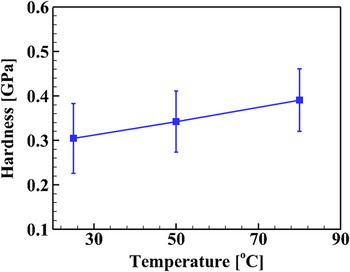
Figure 8 shows the change in hardness of the exoskeleton of Rimicaris exoculata with temperature. The hardness values increase with increase in temperature. Unlike elastic modulus which depends on properties of constituent materials, hardness depends on a variety of different factors such as grain size and packing density in the crystalline structures, packing density of fibers, and constraint offered by filler material to movement of fiber chains in polymers. A look at chemical composition of shrimp exoskeleton from previous researchers and also EDX analysis shown in the current study reveals that the major constituents of the examined exoskeletons are protein, moisture, and calcium. All such constituents show change in their properties as a function of temperature. Temperature increase of proteins has been shown to increase their volume. Reference Tilton, Dewan and Petsko46,Reference Frauenfelder, Petsko and Tsernoglou47 It has also been shown that with increased temperature proteins tend to lose water molecules. Boßelmann et al. Reference Boßelmann, Romano, Fabritius, Raabe and Epple6 have shown that organic matter loses its mass with increase in temperature.
Hardness variation in the temperature range of 25 to 80 °C in the case of Rimicaris exoculata.

It has already been established that the structure of the exoskeleton is highly hierarchical. Hardness depends on structural packing and interaction of individual components with each other. The overall properties of the exoskeleton will also depend on interface arrangement. In the current study, an increase in hardness observed with temperature can be related to the change in the structural packing changes with temperature increment. This increment could be possible because as temperature increases, fibers lose strength giving space for mineral phase to become more compact and subsequently making it harder.
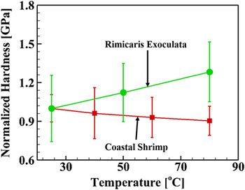
The hardness variation with temperature for both Rimicaris exoculata and Pandalus platyceros is plotted in Fig. 9. Hardness values are normalized by room temperature hardness values to better compare trends as a function of temperature. Hardness decreases with temperature in the case of Pandalus platyceros whereas it increases in the case of Rimicaris exoculata as a function of temperature increase.
Variation in normalized hardness of Rimicaris exoculata and Pandalus platyceros in the temperature range of 25 to 80 °C.

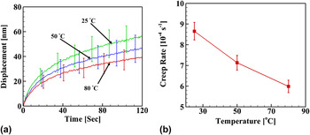
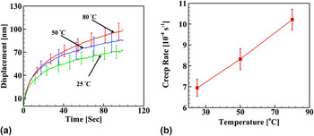
Increase in hardness for Rimicaris exoculata can be further verified from creep tests as shown in Fig. 10(a). Creep rate is decreasing with temperature increase as plotted in Fig. 10(b). It shows that the exoskeleton of Rimicaris exoculata is better adapted to changes in temperature to survive at deep sea hydrothermal vents. Figures 11(a) and 11(b) show the dwell plots and creep rate for Pandalus platyceros, respectively. Pandalus platyceros shows an increase in creep rate with increase in temperature. The reason for this behavior lies in differences in the composition and structure of the exoskeleton. More discussion related to this behavior in hardness is provided in the next section.
(a) Plot of dwell data during creep tests and (b) plot of creep rate in the temperature range of 25 to 80 °C for Rimicaris exoculata.

(a) Plot of dwell data during creep tests and (b) plot of creep rate in the temperature range of 25 to 80 °C for Pandalus platyceros.

D. Trends regarding temperature dependence of hardness between Pandalus platyceros and Rimicaris exoculata
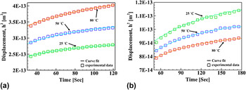
Basic building blocks of the Rimicaris exoculata and the Pandalus platyceros exoskeleton are polymer chains made up of chitin proteins and calcium-based biominerals. Matrix material is made up of chitin, protein fibers woven together in a planar structure. Spacing between woven fibers is filled with biominerals, mostly of CaCO3. These layers are then arranged to form twisted plywood structure known as Bouligand pattern. Reference Raabe, Romano, Sachs, Fabritius, Al-Sawalmih, Yi, Servos and Hartwig7,Reference Raabe, Sachs and Romano8 Such hierarchy in this structure makes these materials similar to polymer composites. Hardness of a material is its ability to resist deformation under applied stress. In the present material, hardness exhibited with temperature follows the trend shown in Figs. 8 and 9. Nanoindentation data have been used by several researchers in the past for improved predictions of the material properties. For instance, the study of the effect of conical or spherical indenter to improve the modulus calculations by instrumented indentation measurements in linear viscoelastic solids (Cheng et al.), Reference Cheng, Ni and Cheng48 creep corrections in the contact mechanics formulations for modulus calculations (Tang and Ngan) Reference Tang and Ngan49 and viscoelastic corrections during unloading part of nanoindentation curve to correct modulus measurements to eliminate creep effects (Feng and Ngan and Ngan et al.). Reference Feng and Ngan41,Reference Ngan, Wang, Tang and Sze50 There are several other formulations to fit the nanoindentation creep given by Oyen et al., Reference Oyen51–Reference Cook and Oyen53 Lu et al. Reference Lu, Wang, Ma, Huang and Viswanathan54 The model chosen in the current study is the most fit for the nanoindentation data of the creep during viscoelastic deformation. This model uses the dwell data over time for the holding period to calculate creep compliance parameters. To model the load–deformation behavior exhibited by examined shrimp exoskeletons, the creep data are fitted with the creep function given for viscoelastic materials by Oyen Reference Oyen51 as:
 $${h^2} = {{{\gamma ^2}} \over {\pi \tan \psi }}\left\{ {{C_0}k{t_{\rm{R}}} - \sum {{C_i}k{\tau _i}\exp \left( {{{ - t} \over {{\tau _i}}}} \right)} \left[ {\exp \left( {{{{t_{\rm{R}}}} \over {{\tau _i}}}} \right) - 1} \right]} \right\}\quad .$$
Here, h is the indentation depth during dwell period, ψ is the indenter included angle, γ is a constant relating contact displacement to total displacement, k is the loading rate, t
R is the start time of dwell period, C
i
and τ
i
are creep compliance coefficients, and t is the time period of loading. One term approximation of Eq. (1) is fitted to the experimental creep data reported in Figs. 10 and 11 to find the coefficients C
i
and τ
i
. The respective coefficients for both Rimicaris exoculata and Pandalus platyceros are tabulated in Table II. Fitted curves to the experimental data are also shown in Fig. 12. Maximum indentation depths,
 ${\bar h_{{\rm{max}}}}$
, for a corresponding maximum load, P
max, as calculated from Eq. (2) are given in Table II along with the creep coefficients as:
 $$\mathop {{{\bar h}_{{\rm{max}}}}}\nolimits^2 = {{{\gamma ^2}{P_{{\rm{max}}}}} \over {\pi \tan \psi }}\left\{ {{C_0} - \sum {{C_i}\left( {{{{\tau _i}} \over {{t_{\rm{R}}}}}} \right)} \left[ {1 - \exp \left( {{{ - {t_{\rm{R}}}} \over {{\tau _i}}}} \right)} \right]} \right\}\quad .$$

Plot of dwell data during creep tests and corresponding fit using Eq. (1) for (a) Pandalus platyceros and (b) Rimicaris exoculata.

Maximum indent depth decreases with increase in temperature as given in Table II for the case of Rimicaris exoculata. This directly corresponds to hardness values measured in indentation experiments. Hardness of material depends on area under indent at maximum load. With decrease in maximum depth, area under indenter decreases, which results in hardness increase of the material.
Elastic modulus depends on the overall material composition including matrix and fiber material. However, in the case of hardness, it is the resistant provided by material to applied force. In the present case, hardness increases with temperature in the case of the Rimicaris exoculata exoskeleton. This behavior can be explained by looking at composition of shrimp exoskeleton as given by EDX analysis in Fig. 6. The presence of additional CaCO3 in the exoskeleton of Rimicaris exoculata gives it extra hardness when the temperature is increased. In general, polymers with added constraints due to additional filler material show decrease in creep behavior. Reference Shokuhfar, Zare-Shahabadi, Atai, Ebrahimi-Nejad and Termeh55 This behavior is further related to the thermal stability of a given material. Polymers with filler material that is thermally stable show better resistance to thermal degradation and better creep at higher temperatures. Reference Nikolaidis, Achilias and Karayannidis56,Reference Leszczyńska, Njuguna, Pielichowski and Banerjee57 These differences in biomineral content explain better thermal resistance to change in properties for Rimicaris exoculata. This also explains selectivity of nature while evolving exoskeleton of similar species in different environments. At sea level, Pandalus platyceros does not need to sustain high pressures whereas the deep sea habitat of Rimicaris exoculata is at very high pressures. From the experimental observations, we conclude that the higher amount of biominerals in Rimicaris exoculata is a way to adapt itself to sustain high pressure–temperature environment as compared to Pandalus platyceros but this correlation does not imply causation. It is the result for this particular case, more controlled experiments need to be conducted on different species to evaluate the role of minerals, pressure, and other environmental factors to make a general conclusion for all species.
IV. CONCLUSIONS
A series of experiments were performed at 25, 50, and 80 °C on the exoskeleton of Rimicaris exoculata to understand its temperature-dependent deformation behavior. The exoskeleton structure was also studied using SEM. Hardness dependence as a function of temperature is discussed. A comparison is drawn between the exoskeletons of Rimicaris exoculata and Pandalus platyceros. The following are key results:
-
(1) Comparison between exoskeleton of Rimicaris exoculata and Pandalus platyceros shows Rimicaris to be better adapted to temperature changes, especially higher temperatures.
-
(2) The better thermal stability shown in terms of nanoindentation hardness of the deep sea shrimps is explained via the presence of higher mineral content (with EDX analysis) that helps it survive at high temperature–pressure at deep sea.
-
(3) Hardness variations as a function of temperature are further analyzed by fitting a viscoelastic creep function to dwell data of experiments.
-
(4) SEM images reveal structural hierarchy of exoskeleton. The exoskeleton structure consists of layers with thicknesses from around 20 nm to 1 μm. Stacking density changes along cross section as variation of layer thicknesses. Bouligand structure is observed in layers filled with biominerals. Rimicaris exoskeleton has higher packing density than that in the case of Pandalus platyceros.
ACKNOWLEDGMENTS
The authors express their sincere thanks to Dr. Juliette Ravaux, Université Pierre et Marie Curie for providing the samples of Rimicaris exoculata. Also, the authors would like to acknowledge the excellent technical assistance of Dr. Christopher J. Gilpin, Chia-Ping Huang, and Laurie Mueller with the scanning electron microscopy at Purdue University. Lastly, Devendra Verma would like to thank his colleagues Dr. Devendra Dubey, Dr. Ming Gan, Dr. You Sung Han, Dr. Hongsuk Lee, Tao Qu, and Yang Zhang for helpful discussions. This research was supported by US-NSF grant CMMI-1131112.