Attention deficit disorder with or without hyperactivity is a common psychiatric disorder, affecting about 5% of children and adolescents and 3% of adults.Reference Polanczyk and Rohde 1 , Reference Schlander, Trott and Schwarz 2 Frequently, attention deficit disorder implicates a challenge for social and academic development of affected children and leads to increased rates of life events in adulthood and developmental comorbidities. The most prominently and successfully used substances in the pharmacotherapy of attention deficit disorder are the psychostimulants methylphenidate and amphetamine, with increasing numbers of prescriptions.Reference Faraone, Spencer and Aleardi 3 , Reference Schubert, Koster and Lehmkuhl 4
Potential side effects of stimulants are in the focus of scientific discussions, intensified by the higher number of prescriptions. An extensive number of studies support efficacy and tolerability of stimulants. However, in 2006, the United States Food and Drug Administration added a black box warning that stimulants may cause serious cardiovascular effects like high blood pressure up to sudden death. A general contraindication for children with known serious heart rhythm abnormalities, or other severe cardiac problems, which may increase their vulnerability to the sympathomimetic effects of stimulants, was stated (http://www.fda.gov/ohrms/dockets/ac/oc06.htm). However, a recently published large retrospective cohort study showed no evidence that current use of an attention deficit disorder drug was associated with an increased risk of serious cardiovascular events in children,Reference Cooper, Habel and Sox 5 and young and middle-aged adults.Reference Habel, Cooper and Sox 6
To minimise eventual risks, most clinical guidelines currently recommend pre-treatment screening including thorough history and physical examination. Cardiologic referral and electrocardiogram are advised only in cases of abnormal findings in cardiac examination, personal history of cardiac symptoms, or family history of sudden cardiac death.Reference Leslie, Alexander and Trikalinos 7
Basal regulation of heart rate by the autonomous nervous system is one potential factor modulating cardiovascular side effects of stimulants, and altered vagal and sympathetic activation levels were reported for children affected by attention deficit disorder. Recently, we could demonstrate the lack of physiological maturation of autonomic function with low parasympathetic tone in children with attention deficit disorder at the average age of 10 years.Reference Buchhorn, Conzelmann and Willaschek 8 From a pathophysiological point of view, patients with low parasympathetic activity have an increased risk for sudden cardiac death, independent of other risk factors.Reference La Rovere, Bigger and Marcus 9 , Reference Vaseghi and Shivkumar 10
We now publish our data about ventricular arrhythmias found in Holter electrocardiograms from 100 children with attention deficit disorder since 2006.
Patients and methods
All children in the age group of 6–16 years referred from January, 2006 to April, 2012 for Holter electrocardiogram examination to the outpatient clinic for paediatric cardiology of Caritas hospital Bad Mergentheim by paediatricians or child and adolescent psychiatrists were consecutively included in the analysis. A total of 100 children fulfilled the inclusion criteria for the diagnosis of attention deficit disorder with or without hyperactivity. These diagnoses were confirmed before referral by child and adolescent psychiatrists. The motivation for cardiac examination were anamnestic signs for potential cardiac disease, conspicuous standard electrocardiograms, or concerns of the parents about cardiac safety of medical treatment. We obtained 24-hour electrocardiogram recordings for data analysis from 72 attention deficit disorder children treated with methylphenidate and 28 children without medication – some children had more than one Holter electrocardiogram examination. More than 120 complete Holter electrocardiogram files were screened for ventricular arrhythmias.
We assumed a ventricular arrhythmia if a child had more than 20 premature ventricular beats per hour or more than one consecutive premature ventricular beat. The diagnosis of ventricular parasystole was reached on the basis of classical electrocardiogram criteria: (1) a varying coupling interval of the ectopic ventricular beats to normal QRS complexes; (2) interectopic intervals identical, or multiples of a common denominator; and (3) ventricular fusion beats. Accelerated idioventricular rhythm was diagnosed by the appearance of regular monomorphic ventricular rhythm at rates up to 10% faster than the prevailing sinus rate on electrocardiogram.
All children received a cardiac ultrasound from a paediatric cardiologist and a standard 12-channel electrocardiogram. All children with ventricular arrhythmias had exercise testing on a treadmill ergometer.
Processing and analysis of 24-hour Holter recordings
Autonomic control of cardiovagal function was tested by time domain analysis of 24-hour ambulatory digital recordings of the electrocardiogram. A two-channel Holter monitor (Pathfinder™, Spacelabs Healthcare, Washington, USA) was used for recording while the children followed their normal daily routines. All Holter recordings were reviewed by an experienced cardiologist (R.B.) and were edited to validate the system's QRS labelling in order to exclude artefacts. Measures of heart rate variability were calculated using only normal to normal intervals. The Holter electrocardiograms were analysed as average values from the entire 24 hours of analysable data and additionally for circadian rhythm analysis as mean hourly values with regard to the time of day.
Measurement and physiological interpretation of heart rate variability parameters was performed according to the standards of the Task Force of the European Society of Cardiology and the North American Society of Pacing and Electrophysiology. The parameter RMSSD (the square root of the mean of the sum of the squares of differences between adjacent NN intervals) and heart rate reflect predominantly a response to changes in the vagal tone.
Chronobiological analysis of hourly values of premature ventricular beats, the vagal parameter RMSSD, and heart rate were performed via the single Cosinor method. In this model, a cosine function is fitted to the 24-hour profile of heart rate variability parameters, with the circadian rhythm defined by three main parameters of the curve: the average value (mesor), the amplitude, and the time until the curve reaches the highest value (acrophase).
The study protocol was approved by the ethics committee of the Ärztekammer Baden-Württemberg (F-2012-056).
Results
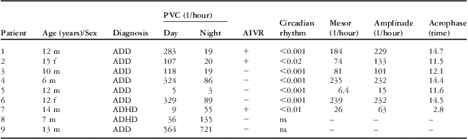
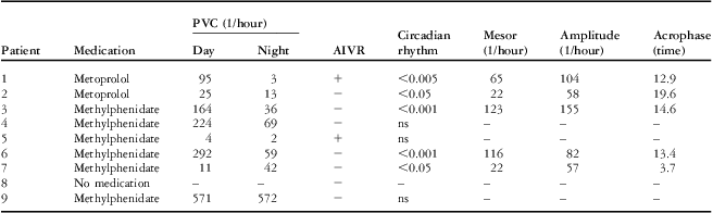
Out of 100 children with attention deficit disorder, nine children had a significant ventricular arrhythmia (mean age 11.4 ± 3.1 years, 77% male, 77% receive methylphenidate, Table 1). All these children had ventricular parasystole – four of them together with an accelerated idioventricular rhythm. The parasystolic beats had a left bundle branch block pattern in seven patients and are unpredictable in two patients without premature ventricular contractions in the standard electrocardiogram. Only one girl (Patient 2) suffers from palpitations that improve during metoprolol therapy. None of the children suffer from syncope. As shown in Table 2, the number of premature ventricular contractions seems to be unaffected in six children who received methylphenidate before diagnosis. However, one boy (Patient 5) had only accelerated idioventricular rhythm during the beginning of methylphenidate therapy. Premature ventricular contractions were abolished in all children during exercise testing on a treadmill ergometer.
Patient characteristics and results of cosinor analysis of PVC circadian rhythm.

ADD = attention deficit disorder; ADHD: attention deficit hyperactivity disorder; AIVR = accelerated idioventricular rhythm; f = girl; m = boy; PVC = premature ventricular contractions
Medical treatment and cosinor analysis of PVC circadian rhythm.

AIVR = accelerated idioventricular rhythm; PVC = premature ventricular contractions
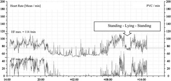
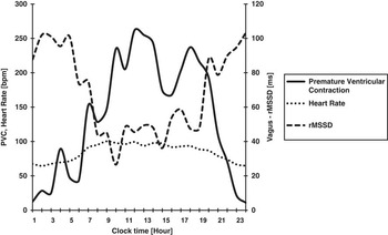
We observed a significant reduction of premature ventricular contractions during metoprolol succinate therapy (1 mg/kg/day) in two children by more than 50% (Patients 1 and 2, Table 2). These two children with the highest activity of the ectopic ventricular focus showed a reproducible orthostatic effect. As shown in Figure 1, the premature ventricular contractions were nearly abolished in a lying position compared with many premature ventricular contractions while standing. We further observed a highly significant circadian rhythm of premature ventricular contractions in seven children and a maximum ectopic ventricular activity between 11:00 pm and 3:00 am in patients 1–7. Figure 2 shows an inverse correlation between the number of hourly premature ventricular contractions within a 24-hour period and vagal cardiac activity, measured by the heart rate variability parameter RMSSD (r = −0.83; p < 0.001). However, compared with our recently published data,Reference Buchhorn, Muller and Willaschek 11 the mean RMSSD value of children with arrhythmias and attention deficit disorder (42 ± 20 ms) was comparable to that of healthy children (44 ± 10 ms) but higher than the RMSSD value of children with attention deficit disorder without arrhythmias and methylphenidate (26 ± 4 ms).
Original reprint from a 24-hour Holter electrocardiogram in a 12-year-old boy (Patient 1). The chart shows the heart rate and the number of premature ventricular contractions. The boy was told to lie down in an awake state between 12:00 pm and 1:00 pm. PVC = premature ventricular contractions.

Mean hourly values of premature ventricular contractions, heart rate, and the heart rate variability parameter RMSSD that represents the vagal tone in seven children with a significant circadian rhythm. Premature ventricular contractions significantly correlate with heart rate (r = 0.77; p < 0.0001) and RMSSD (r = −0.83; p < 0.0001). PVC = premature ventricular contractions; RMSSD = root mean square of the successive differences.

Discussion
In a survey of spontaneously occurring paediatric ventricular tachycardias with a reported incidence of 1.1 episodes/100,000 childhood years in a stable population, Roggen et alReference Roggen, Pavlovic and Pfammatter 12 found that essentially half (13/27) of the patients had no associated or functional heart disease. Wang et alReference Wang, Zhu and Hamilton 13 reported about 19 children with accelerated idioventricular rhythm in a single-centre retrospective review of patients younger than 18 years, which represents – as the authors stated – the largest single paediatric series reported to date. This 26-year retrospective review was undertaken at the Hospital for Sick Children in Toronto, a tertiary paediatric care institution serving a regional catchment population of ∼5 million, as well as patients referred from elsewhere in Canada. Compared to these prevalence data, 4 cases with accelerated idioventricular rhythm within 6 years in a regional paediatric care institution serving a population of ∼250,000 people or 21,000 children with an age between 0–16 years, is very high. All these four cases were found in Holter electrocardiograms from 100 children with attention deficit disorder. Owing to the fact that our patients are highly selected, with respect to the reported incidence of circa 0.5 episodes/100,000 childhood years in children with structural normal hearts, we should find a maximum of one child with ventricular tachycardia within 6 years in our population.
However, accelerated idioventricular rhythm occurs infrequently in patients without demonstrable heart disease and, in this case, it has a good prognosis.Reference MacLellan-Tobert and Porter 14 , Reference Riera, Barros and de Sousa 15 These cases and further five children of our population had a frequent number of ventricular parasystole.
Autonomic imbalance has been identified as a trigger of accelerated idioventricular rhythm and ventricular parasystole.Reference Kinoshita, Ogawa and Mitsuoka 16 – Reference Lanza, Lucente and Rebuzzi 18 The effect of standing and exercise on ventricular parasystole was well described by Kinoshita et alReference Kinoshita and Mitsuoka 17 , Reference Lanza, Lucente and Rebuzzi 18 who show that parasystolic cycle length was prolonged after exercise, in contrast to a shortening of the sinus cycle length. Parasystolic cycle length and sinus cycle length both shortened on standing. These studies explain our observation in six children that ventricular parasystole were predominantly recognisable while standing and were suppressed by lying and exercise. This may be a reason that these arrhythmias were not detected by others despite thousands of electrocardiograms in children with attention deficit disorder in clinical studiesReference Prasad, Furr and Zhang 19 or routine pre-treatment examinations like recommended from several national societiesReference Leslie, Alexander and Trikalinos 7 routinely measured in a lying position. To the best of our knowledge, no other group published 24-hour Holter electrocardiogram examinations in children with attention deficit disorder or methylphenidate treatment.
The parasystolic foci discharge rate may undergo the effects of circadian variations of the autonomic nervous system. Undoubtedly, these ectopic ventricular foci are sensitive to variations of the sympathetic tone,Reference Fazio, Novo and Sutera 20 whereas their responsiveness to vagal stimuli was in question. The traditional view was that the vagus did not send a significant number of fibres to the ventricular myocardium and that it exerted poor influence on the electrical properties of the ventricles.Reference Rosen and Hoffman 21 However, some studies have sufficient evidence that cholinergic innervation in the ventricular myocardium is considerably above the cardiac specific conduction system. Furthermore, a vagal effect upon ventricular arrhythmias has been demonstrated by clinical studies.Reference La Rovere, Bigger and Marcus 9 Our data clearly indicate that vagal activation in the lying position indicated by lower heart rates and higher RMSSD values (Figs 1 and 2) suppress ventricular parasystole and an accelerated idioventricular rhythm by a lower parasystolic discharge rate compared with sinus rhythm. Figure 2 shows that circadian variation of premature ventricular contractions in our children with attention deficit disorder depends on the vagal tone indicated by RMSSD. Recently, we describe an autonomic imbalance in children with attention deficit disorder measured by an analysis of heart rate variability in 24-hour Holter electrocardiograms that showed lower RMSSD values compared with healthy controls.Reference Buchhorn, Muller and Willaschek 11 The surprisingly high rate of ventricular arrhythmia with signs of autonomic influence supports the idea of autonomic imbalance as an underlying condition in attention deficit disorder. However, probably for methodological reasons – due to arrhythmias, <90% data of these children are analysable – the mean RMSSD of children with arrhythmias and attention deficit disorder is not significantly reduced compared with healthy children and higher than in children with attention deficit disorder without arrhythmias. We speculate that only a specific subgroup of children with attention deficit disorder develops ventricular arrhythmias. These children are not characterised by significantly lower average RMSSD values per day, but by a significant correlation of hourly premature ventricular contractions and RMSSD values within the individual patient.
A lower number of premature ventricular contractions during metoprolol succinate therapy in two patients indicates that the parasystolic foci also better respond to β1 receptor blockade than the sinus node. However, ventricular parasystole and accelerated idioventricular rhythm seem to be an arrhythmia with a good prognosis and need no therapy in asysmptomatic patients.
Methylphenidate and other psychostimulants should not be given to children with known serious heart rhythm abnormalities or other severe cardiac problems that may increase their vulnerability to the sympathomimetic effects of stimulants.Reference Wilens, Prince and Spencer 22 However, we see no significant increase of ventricular arrhythmias in six children who receive methylphenidate before diagnosis. Despite the current recommendation, six families decided to continue with methylphenidate medication because of escalating psychological problems after stopping this therapy.
We urgently need more data about children with ventricular arrhythmias or congenital heart diseaseReference Batra, Alexander and Silka 23 who, after profound information, decide on receiving psychostimulants despite the current recommendations.