BACKGROUND
An 87-year-old female with WHO performance status 0, presented to accident and emergency with a 10-day episode of vaginal bleeding. Examination revealed a 10 mm palpable mass in the upper anterior vagina. Colposcopy obtained a tissue biopsy and the lesion was cauterized. Histology confirmed an endometrial recurrence.
Eight years previously, the patient had a total abdominal hysterectomy and bilateral salpingo oophorectomy for stage 2b, grade 1 endometrial carcinoma. Adjuvant external beam radiotherapy (EBRT) was not prescribed. The patient had shown no evidence of recurrence during active surveillance and was discharged from surgical follow-up at year 5.
A staging computed tomography (CT) scan failed to clearly demarcate the vaginal recurrence, although indicated that there was no evidence of distant metastases. A pelvic magnetic resonance (MRI) scan described a 17 × 9 × 19 mm right sided soft tissue mass involving the upper third of the vagina, with indiscrete definition inferiorly and stranding into the paravaginal soft tissues.
Radical EBRT to a limited pelvic volume was considered, although a history of diverticular disease raised the concern of the iatrogenic effects on the bowel. Due to her asymptomatic status following cauterization, the patient wanted to avoid significant acute toxicity as she hoped to remain self-caring. Intracavity brachytherapy was suggested as an alternative, with the potential for good local control, minimal hospital attendances and reduced toxicity.
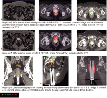
Due to the tumour extent being difficult to quantify with CT, an alternative imaging modality was required. We have experience registering CT radiotherapy planning (RTP) scans with RTP MRI for our cervical cancer image-guided brachytherapy. However, for this case, MRI-guidance had disadvantages. First, MRI is not geometrically accurate, which has greater significance for small target volumes(Figure 1). Second, the image quality and resolution is compromised, reducing the target delineation accuracy, where the target volume abuts the brachytherapy applicator. Finally, the applicator required was not MR compatible.
Imaging and volume delineation.

How could this tumour be accurately delineated to ensure brachytherapy target volume coverage whilst keeping the normal tissue dose to a minimum?
PROPOSED SOLUTION
UCLH RT department has 40% use of a Positron emission tomography (PET)-CT scanner, used as the sole planning modality for a wide range of EBRT, which can greatly improve the accuracy of tumour delineation.Reference Chiti, Kirienko and Grégoire1 [18F] Fluoro-2-deoxyglucose (FDG) is administered and the PET-CT is acquired in the treatment position. The CT is used for dosimetric planning, removing the need for an additional RT-CT and the associated image registration. This enables smaller planning target volumes (PTV) to be considered and a less complex patient pathway with fewer hospital visits for multiple scans. The biological target volume (BTV) is delineated by an experienced nuclear medicine physician (NMP) using visual thresholding, from which the subsequent target volumes are delineated by the clinical oncologist (CO).
UCLH has PET-CT planned more than 170 patients, with efficient pathways and successful interdepartmental working. It was therefore considered appropriate to consider this case for PET-CT image-guided brachytherapy.
PET-CT brachytherapy planning of gynaecological cancers has been described as beneficial for the guidance and evaluation of dosimetry due to the visualisation of the treatment applicators in direct relation to the BTV.Reference Belhocine and Grigsby2 Chiti (2010)Reference Chiti, Kirienko and Grégoire1 reports that 89–97% of endometrial tumours are FDG-PET sensitive, with a specificity of 50%, meaning that the majority will enhance, although tumour size may be over-estimated. Belhocine & Grigsby (2005)Reference Belhocine and Grigsby2 describe the over-expression of glucose transporters and elevated activity of glycolytic pathway enzymes as responsible for this high sensitivity. Thorwarth (2010)Reference Thorwarth, Geets and Paiusco3 also states that PET-CT can result in over-estimation of small tumour volumes due to a 5–7 mm spatial resolution and partial volume effects. Our team concluded that any additional information provided by PET-CT that would reduce the risk of tumour miss, justifies a small over-estimation of target volumes.
Lee (2010)Reference Lee4 supports PET-CT as a planning modality, identifying the importance of an experienced physician when using visual thresholding. We considered that our extensive experience working with the consultant NMP for over 170 patients satisfies this requirement, reducing the risk of inaccurate image interpretation. This is a significant concern when treating a small target volume with a high dose gradient modality such as brachytherapy.
Despite the evidence being supportive of this proposed solution, some concerns remained. First, inserting the applicator post-FDG injection could result in a high staff dose. Second, the local trauma from moving the patient from the uptake bay to the scanner with the applicator in situ may increase the signal at the applicator surface. Finally, hyperintense urine in the bladder could obscure the PET-avid disease.
To overcome these issues, the patient would be catheterised and have the applicator inserted on the PET-CT couch prior to FDG administration, remaining there during the 60 minute uptake period prior to the scan acquisition.
EQUIPMENT
PET-CT
• GE Discovery DST
Brachytherapy unit & applicator
• Nucletron Microselectron Vs 3, 30 channel Iridium-192 afterloader, source reference activity 370 GBq (10 Ci).
• Nucletron Miami vaginal applicator with six circumferential channels
Treatment planning systems
• GE Advantage Windows (ADW) with functional PET-CT mode for BTV visual thresholding.
• Oncentra Masterplan V4.2 (OMP) utilising the standard brachytherapy AAPM TG-43 calculation algorithm.
RESULTS
RT-PET-CT acquisition
The patient attended as an outpatient. The 60 minute uptake period was uneventful. The scan range was L5 to 5 cm below the vagina; 2.5 mm slice thickness, 120kV, modulated mA, and 4 minutes per bed position for the 2D-PET acquisition. Data was exported via DICOM (Digital Imaging and Communications in Medicine) to ADW. The catheter and applicator were removed and the patient returned home.
Volume delineation
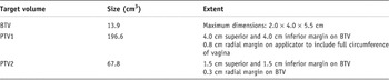
The NMP delineated the BTV using visual thresholding. There was some PET avidity that was symmetric and uniform at the applicator surface, indicating false positive uptake related to localised trauma from the applicator insertion and was therefore excluded from the BTV. The CO delineated a phase 1 and 2 PTV on the RTP-PET-CT (see Table 1) using the BTV and diagnostic MRI. The rectum, urethra and bladder were delineated as organs at risk (OAR).
Target Volumes

Treatment prescription
Phase 1 – 4 × 5.75 Gy fractions to PTV1 over 2 weeks.
Phase 2 – 2 × 5.75 Gy fractions to PTV2 over 1 week.
The prescription was calculated to give a total biologically equivalent dose (BED) of 101 Gy3 and 54 Gy10 using the calculation:
![[{{\rm BED}} = \it Nd[ {1 + {d \over {( {\alpha /\beta } )}}} ]\matrix{ {N =\rm 6(4{\rm{Ph1, 2 ph2)}}} {d = \rm 5.75{\rm{ Gy }}}}](https://static.cambridge.org/binary/version/id/urn:cambridge.org:id:binary:20151128095645299-0576:S1460396911000264_eqn1.gif?pub-status=live)
The dose is comparable to a typical prescription dose of 60 Gy given at a continuous low dose rate of 0.5 R (Gy/hr). BED = 103 Gy3 and 73 Gy10 using the calculation:

Equations as given by Dale and Jones (1998)Reference Dale and Jones5 where: D = total dose; T = irradiation time; R = dose rate; Isodose is relative to prescription dose (not basal dose rate); N = number of HDR fractions; d = dose per HDR fraction; μ = repair constant; T 1/2 = corresponding repair half-time = 1.5 hr; α/β = alpha-beta ratio. HDR assumes complete repair between doses.
Treatment planning
A circumferential channel applicator was selected over a standard vaginal applicator as the target volume was not uniformly localised around the vagina.
Phase 1
The aim was to cover PTV1 with ≥90% isodose. All of the applicator dwell positions within PTV1 were activated. The initial plan was optimised based on dose points placed evenly over the surface of PTV1, and then adjusted for improved PTV1 coverage within the TPS. Prior to approval, the individual dwell times were checked to ensure a reasonable dwell time gradient between active positions.
Phase 2
The phase 1 plan was adapted by deactivating dwell positions not located within PTV2. The initial plan was optimised based on dose points placed evenly over the surface of PTV2, and then adjusted for improved PTV2 coverage.
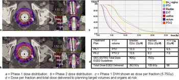
A composite of the phase 1 and 2 plans was evaluated with respect to GEC-ESTRO guidelines of OAR tolerance doses for gynaecological brachytherapy (Figure 2).Reference Pötter, Haie-Meder, van Limbergen, Barillot, De Brabandere, Dimopoulos, Dumas, Erickson, Lang, Nulens, Petrow, Rownd and Kirisits6 The 2 cc total dose in equivalent 2Gy fractions (EQD2) for bladder and rectum significantly exceeded the GEC-ESTRO guidelines. The clinical justification for this was based on the acknowledgement that brachytherapy was to be the sole treatment modality, and the patient had had no previous radiotherapy exposure. The relationship between the patient’s specific pelvic anatomy and the location of the tumour recurrence superior and anterior within the vagina, resulted in the close proximity of the target volume to the bladder. This made further avoidance of the bladder unfeasible, without significantly under-dosing the target and compromising tumour control probability.
Phases 1 and 2 dose distributions and DVH.

Treatment delivery
The first fraction was delivered 9 working days after the PET-CT acquisition to allow all planning stages to be completed.
For each fraction, the brachytherapy applicator was inserted by the specialist radiographer. The orientation of the treatment catheters was verified by land-marking one of the catheter locations to the ‘12 O’clock’ position. The depth of insertion was maintained by ensuring the patient was in the same position for each fraction, that the applicator was parallel to the treatment couch and the protruding length of the applicator measured and clamped for each fraction. The patient voided her bladder prior to each fraction to replicate the empty bladder status as per the PET-CT scan.
Treatment was delivered without anaesthesia or sedation on an out-patient basis.
There was some bleeding (from the tumour), which had completely resolved by the final insertion, although the patient did report increasing discomfort. End of treatment side-effects were pruritus and moderate diarrhoea, which was controlled effectively with oral anti-motility medication.
Follow-up
One week post radiotherapy, the patient reported vaginal soreness and mild dysuria, although was able to self care. At 2 weeks, the vaginal soreness was improving and the dysuria had resolved and at 6 weeks post-RT, only a small amount of vaginal erythema remained. At 7 months, there was no evidence of late toxicity, with minimal dysuria. Speculum examination was difficult due to some vaginal stenosis, although no vaginal mass could be palpated or visualised. A repeat MRI pelvis was performed at 9 months and demonstrated no residual or recurrent vaginal disease, a normal rectum and urethra and an oedematous bladder.
LESSONS LEARNT
• Effective intra- and interdepartmental communication facilitated an efficient patient pathway and treatment delivery. Now that a process has been successfully established for this site, the time between PET-CT and first fraction could be reduced to 5 working days. Fraction 1 delivery is unlikely to be feasible for the same day as PET-CT acquisition, due to the multiple-stage planning process, requiring contributions from the wide multi-disciplinary team.
• The accessory equipment (clamp) became contaminated with radioactive urine, requiring decontamination. This could have been avoided by using a protective cover.
• A greater range of applicator sizes would be ideal as a smaller applicator may have been better tolerated by the patient.
• An increased bladder volume would have reduced the volume of bladder irradiated, although would not have impacted the 2 cc dose. This may have been possible if the impact on the PET-avid disease was explored further.
• Verification of applicator position could be achieved with greater accuracy by CT scanning the applicator in situ prior to delivery of each fraction. However, transporting a patient between CT verification and the brachytherapy suite may be associated with some applicator movement.
• Follow up—due to there not being a ‘baseline’ PET-CT acquired i.e. without the applicator in situ, follow-up PET-CT’s were not performed. Instead, follow-up MRI was compared to the baseline pre-treatment MRI. This could be an area for patient pathway development.
• This technique would be considered for similar referrals where additional PET data could contribute to target definition.