The persistently patent arterial duct is a common congenital malformation. Excluding premature newborns, it accounts for up to one-tenth of all the congenital malformations associated with the heart. Since its first description by Gross in 1939, surgical closure of the patent arterial duct remained for many decades the only available treatment. While the outcome of surgical ligation is excellent, it is an inherently invasive surgical procedure, requires general anaesthesia, and therefore involves the risks and complications of both. Recently developed transcatheter devices have enriched the options for closure of persistently patent arterial ducts. One of the foundations for successful development of these devices lies in our better understanding of the anatomy and the geometry of the patent arterial channel. Transcatheter closure saves patients from tracheal intubation, surgical incision and pain, bleeding, relatively longer hospital stay, and prolonged convalescence. Today, we have access to a variety of devices, such as non-detachable and detachable coils, and the Amplatzer device, allowing us to close almost any type or size of the duct. For some devices, long-term results are now available and compare favourably to surgical ligation.
All these new modalities for treatment have led to changes in strategies of management. The purpose of this review is to evaluate the contemporary role of cardiac surgery in the presence of the new therapeutic initiatives. Furthermore, we try to show that, in the current era, transcatheter closure is the therapy of choice. We suggest that the remaining indications for surgical intervention are in premature babies, for closure of huge ducts not suitable to accommodate devices, and after complications arising during a trial of intervention.
Historical review
Surgical closure
It was not Leonardo Botallo, who lived from 1530 through 1600, but probably Galen who, even before the birth of Christ, first described the persistently patent arterial duct. It was not until 1939, however, that Gross and Hubbard1 first described successful surgical ligation of a duct in a boy aged seven years. Over the subsequent years, the efficacy and safety of the surgical procedure improved, and excellent results were achieved even in infants.2 Later, ligation and division were showed to be the optimal surgical management. A significant step forward in surgical treatment came in 1993, when Laborde et al.3introduced video-assisted thoracoscopic ligation. This new technique appears to be effective and safe, even as an outpatient procedure.4
Transcatheter closure
Since the first clinical attempt to close a patent arterial duct using a transluminal approach, made by Porstmann et al.5 in 1967, various investigators have refined the technique and developed better devices. By the end of the 1970s, many devices had undergone studies in animals, but only a few were accepted clinically. Among these was the double umbrella device reported by Rashkind and Cuaso.6 Initial success with the Rashkind device suggested that it might well be the management of choice. The double umbrella, however, was unwieldy, and required a large bore sheath for its introduction. When used to close large ducts in small children, there was also a risk of causing left pulmonary arterial stenosis or aortic narrowing. Moreover, the device was suitable only if the narrowest diameter of the duct was less than 9 millimetres. About half of patients with persistently patent ducts, therefore, would have still needed surgical ligation.7
By the early 1990s, Cambier et al.8 had led the way in using Gianturco coils to close small ducts. Such coils were much less cumbersome than the umbrellas, and hence found rapid worldwide acceptance. But such coils were still of use only in smaller ducts measuring less than three millimetres at their narrowest diameter. The closure of larger ducts using multiple coils was appealing, and often proved to be successful, but was associated with an increased risk of left pulmonary arterial stenosis,9 and a relatively high rate of inadvertent embolization.10
The introduction of the Cook detachable coil provided an even safer option,11 and this device almost entirely replaced the Gianturco coil for closing small duct. Then, in 1993, the PFM coil was introduced for clinical use. It has since undergone several modifications, but clinical experience is now encouraging when it is used for closing the small and moderate patent arterial duct.12
Despite these developments, the large ducts still remained in the domain of the surgeon. Rao et al.,13 as well as later Grifka et al.,14 designed devices to close such larger ducts, but neither device emerged as a significantly marketable option. It was the emergence of the Amplatzer duct occluder that expanded the options of the interventional cardiologist. This device is currently used safely and effectively for closure of ducts measuring up to 16 millimetres at their narrowest diameter.
Present state and clinical indications
Establishing the diagnosis
Prior to the advent of echocardiography, the diagnosis and outcome of ligation was evaluated based exclusively on physical examination. Echocardiography, however, revolutionized the diagnosis and management of the persistently patent arterial duct. Motion-mode echocardiography provided a sophisticated method for diagnosing ducts by evaluating the signs of left-sided volume overload. With the advent of cross-sectional echocardiography, the duct became visible. With Doppler echocardiography, the continuous murmur was audible. With the help of colour Doppler, the shunt across the duct could be visualized, and the narrowest diameter measured with considerable accuracy.
General indications for closure
Besides congestive heart failure, indications include prophylaxis against endarteritis. The question remains as to whether asymptomatic patients with small to moderate ducts, or those with relatively larger ducts but with satisfactory control of congestive heart failure, should be submitted to closure? The answer, in our opinion, is positive, but this needs some elaboration. For those with relatively large ducts, the argument is simple, since it is accepted universally that closure eliminates the need for life-long medications, the risk of endocarditis, and the development of the Eisenmenger syndrome. In asymptomatic patients with a small duct, in contrast, the only argument in favour of closure is avoiding the risk of developing endarteritis and endocarditis. This risk has been said to be 0.45% per patient year, but a recent report from Sweden challenges this estimate,15 finding no instances of endarteritis in this setting.
Indications in infants and children
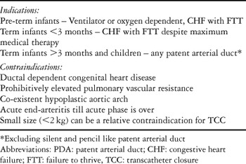
In preterm newborns, closure of the duct using indomethacin, or more recently Ibuprofen, or surgical ligation should medical therapy fail, is warranted in the presence of congestive heart failure, or when it is not possible to wean the patient from the ventilator or oxygen. In newborns at term, an arterial duct is regarded as persistently patent if it remains present beyond 3 months of age. In this situation, it is deserving of closure as soon as the diagnosis is established. Infants younger than 3 months, in whom congestive heart failure is not responsive to medical treatment, also qualify for closure.
Indications in adults
The indications are similar to those discussed above for the younger patients. The risk of surgical closure, nonetheless, can be increased in adults because of calcification and increased friability of the ductal ampulla. There are no randomized studies to underpin firm recommendations, but the American Heart Association recommends that, apart from those with a silent duct, all other patients without an irreversible elevation in pulmonary resistance should be subjected to closure.16
General contraindications for closure
These include co-existing duct-dependent congenital cardiac disease, a prohibitive elevation in pulmonary vascular resistance, and a co-existing hypoplastic aortic arch. Closure should also be deferred in those with endartritis or endocarditis, at least until the acute phase is over.
Indications and contraindications for surgery
Surgery is still indicated for premature babies, and for very small infants unresponsive to medical therapy, those with very large ducts not suitable for closure with a device, those with very short ducts of the so-called aorto-pulmonary window variant, and in cases when an attempt at transcatheter closure has failed. Contraindications are no different than the general ones. In cases with aneurysmal dilation of the duct, a calcified ductal ampulla, or when the patient is at high risk for general anaesthesia, surgery can be risky, and a transcatheter approach is to be preferred.
Indications and contraindications for transcatheter closure
Presently, at least four different devices are in clinical use. Occasionally, the use of these devices is not mutually exclusive (Table 1). Contraindications to catheter-based closure devolve on the size of the patient. Very small premature infants with relatively large ducts still require surgical closure, primarily because of the feared risk of thrombosis and permanent injury to the femoral artery and vein. Those with the aorto-pulmonary window variant, and those with large ducts but smallish aortic arches, may also not be suitable for closure with a device because of the possibility of developing obstruction to flow in the aorta (Table 1).
Table 1. Indications and contraindications for closure of the patent arterial duct.

Technique and equipment
General issues pertaining to transcatheter closure
Transcatheter closure should ideally be performed under conscious sedation. General anaesthesia is sometimes used in haemodynamically unstable patients, or when it is felt to be safer for the patient. Vascular access is obtained through the femoral artery and vein. The duct can be accessed in anterograde fashion through the pulmonary trunk, or retrograde via the aorta. Intravenous heparin, in a dose of 100 international units per kilogram of body weight, is often used for thromboprophylaxis at the site of vascular access. Second generation cephalosporine, in a dose of 30 milligrams per kilogram of body weight, is given intravenously during the procedure, and twice within the next 24 hours thereafter.
Devices, equipment, and technique
Gianturco coil.
The Gianturco coil, manufactured by William Cook, from Bjaeverskov, Denmark, is made out of stainless steel with attached Gortex filaments. It is available in 10 sizes, from 1 to 10 centimetres length of the stretched coil by 1 to 10 millimetre loop diameter (Fig. 1). A sheath of 4 French dimension is recommended for access to the femoral artery, and one of either 4 or 5 French dimension for accessing the femoral vein. A pigtail or NIH catheter of 4 French dimension is used to record aortic pressure, and to perform aortography so as to evaluate the ductal anatomy and to measure its size. For the venous side, a 4 or 5 French catheter is used for diagnostic evaluation. After the decision is made to close the duct, an end-hole catheter is advanced in anterograde fashion across the duct into the descending thoracic aorta. Using the soft end of a 0.038 inch Teflon wire, the chosen coil is advanced into the guiding catheter positioned in the ductal ampulla. The loops of the coil are then deployed in the ampulla until less than one loop is left for the pulmonary artery, where the last part of the coil is deployed by gently pushing the remaining loop. When approached in retrograde fashion, a 4 French end-hole catheter is advanced into the duct. The wire pushes out one loop of the coil into the pulmonary artery. The remaining coil is released by pulling back the catheter and extruding the coil(Fig. 2).

Figure 1. Some of the various sizes of Gianturco coils.

Figure 2. Angiography in lateral view of a long, but not very large, conical duct, shows before and after deployment of a Gianturco coil. There is no residual shunt.
Cook detachable coil.
The detachable coil made by William Cook is stainless steel, but coated with dacron filaments to increase its thrombogenicity. It is available in 8 different sizes. They differ in the number of loops, either 3, 4 or 5, and the diameter of the loop, which can be 3, 5, 6.5 or 8 millimetres (Fig. 3).The equipment and the technique used are similar to those described for the Gianturco coil, albeit that the Cook coil is loaded by screwing it on the delivery system. A mandril present inside the system is then advanced inside the coil to keep it straight. When approaching anterogradely, which we prefer, the tip of the catheter is positioned a few centimetres below the ductal ampulla. The coil is pushed out of the guiding catheter, and the mandril is pulled back simultaneously to deploy one loop after the other. An aortogram is performed to ensure the coil is positioned satisfactorily, and the delivery wire is then turned counterclockwise, pulling out the mandril completely to deploy the coil (Fig. 4).

Figure 3. The Cook detachable coil attached to its delivery system.

Figure 4. Angiography in the lateral view, showing a conical, and superiorly oriented duct, before and after release of a detachable coil. There is no residual shunt. The coil was deployed anterogradely. Its loops are compact, and occupy most of the ampulla.
PFM coil.
This coil is made of woven stainless steel wire, without any filaments attached to it. It comes in 3 different types and 9 different sizes (Fig. 5). The delivery system is bulkier than for the other coils and this coil is designed only for an antegrade approach. The delivery system consists of the delivery wire, which has two black marks at its middle part. Pushing the delivery system, along with the coil, inside the long sheath brings the marks closer to the sheath. A few centimetres after the marks, there is a bulky black device attached to the delivery wire. This device is for deployment and release. The long sheath, which is positioned in the duct prior to advancing the delivery system, also comes with the package. The first part of the coil is deployed in the descending aorta until the first black mark reaches close to the delivery sheath. The whole system is then pulled back inside the ductal ampulla. Once the tip of the catheter is pulled across the narrowest diameter of the duct, holding the delivery system in place and pulling back the sheath deploys the loop meant to be left in the pulmonary artery. An aortogram is performed to confirm the position. The coil is released by rotating the round device in a clockwise fashion until it cannot be pushed any further. Another device is then pushed to release the coil (Fig. 6).

Figure 5. When deployed, the PFM coil attains a conical shape.

Figure 6. Angiography in lateral view of a conical duct with medium length, before and after deployment of a PFM coil. There is no residual shunt. The typical appearance of this coil is well seen, and it adapts itself to the shape of the duct.
Amplatzer duct occluder.
The device made by the AGA Medical Corporation, of Golden Valley, Minnesota, United States of America, is made from a 0.004 inch mesh of Nitinol wires, producing a cage-like structure which is filled with polyester fibres. It is available in sizes of 5/4, 6/4, 8/6, 10/8, 12/10, 14/12, and 16/14 millimetres (Fig. 7). The device has an additional retention skirt, which is 4 millimetres larger than the largest diameter in the first three sizes, and 6 millimetres larger in the rest of the series. The length of the device also varies, with the device of 5/4 millimetres measuring 5 millimetres, the 6/4 and 8/6 devices measuring 7 millimetres, and the others measuring 8 millimetres in length. These dimensions should be remembered when selecting the size of the device.

Figure 7. The Amplatzer occluder attached to its delivery system.
For occlusion, after having advanced an end-hole catheter anterogradely through the duct, a 0.035 inch stiff exchange wire is advanced through this catheter deep into the descending aorta. This catheter is then exchanged with a long sheath, which comes with the delivery system, making sure that the sheath is one size larger than that recommended by the manufacturer. A back bleed device is attached to the sheath to prevent loss of blood. The device is loaded by screwing it onto the delivery system, and then pulling it inside a loader that comes with the sheath. The device is advanced inside the sheath, and the first half is pushed into the thoracic aorta. The whole system is pulled back until the tip of the sheath, and hence the middle part of the device, reaches the narrowest part of the duct. Then the proximal part of the device is deployed. An aortogram is performed to assure good position of the device, which is then released by rotating the delivery system counter-clockwise (Fig. 8).

Figure 8. Angiography in the lateral view of a long conical shaped duct, before and after deployment of a Amplatzer device. There is no residual shunt. It is obvious that the selected device is overstretching the ampulla of the duct.
Special issues pertaining to procedure
Issues pertaining to decision-making, and choice of the appropriate device.
The anatomic type, size, and the narrowest diameter of the arterial duct are commonly used criterions on which to base the selection of the type and the size of the device.13 In our experience, in addition to these measurements, the diameter of the ampulla at the aortic end, the length of the duct, and its orientation or the angle to the horizon, are also important to consider when selecting the device (Fig. 9). We believe that it is our inability to incorporate these latter factors that often explains both the unexpected failure of the procedure, embolization of the device, or obstruction to flow in the left pulmonary artery or descending thoracic aorta.

Figure 9. Measurements necessary to help select the type and size of device, showing 1, the narrowest diameter, 2, the length of the duct, and 3, the height of the duct at the aortic end of the ampulla. If possible, the height of the duct at the middle of the ampulla should also be measured.
Anterograde versus retrograde approach for closure using coils.
Aside from the PFM coil, either approach can be used. The venous route has several advantages. It allows an aortogram to be performed to confirm the efficacy of the procedure before releasing the coil. It also helps to avoid use of a large sheath in the femoral artery. It is also potentially applicable in very small infants, the risk of thrombotic occlusion of the femoral artery being higher with the retrograde approach. In our experience, the anterograde approach also allows for better positioning of the coil, the operator seemingly having better control over the coil and the delivery system.
Issues pertaining to the Amplatzer duct occluder
One of the characteristics of the Amplatzer duct occluder is its forgiveness. It can be easily retrieved after deployment. While this is true, some clarification is required. The delivery sheath recommended by the manufacturer usually has a tight fit for the device. Moreover, the sheaths themselves are not very kink resistant. We recommend that, if it is feasible, and not harmful for the patient, a delivery sheath of one French size larger than the recommended size should be used. We cannot overemphasise the importance of this recommendation, as it allows easy repositioning, or retrieval of the device into the sheath when needed. Otherwise, the examiner might be faced with a very stressful situation. Either the device can not be manoeuvred into the sheath, or the sheath gets kinked, especially in young children, or even worse becomes concertinaed, making retrieval of the whole system impossible unless the salvage package provided by the manufacturer is available. Needless to say, that this may culminate in emergent open-heart surgical procedure, a heavy price for ignoring a simple remedy.
Ductal types and measurements
During the era of use of the Rashkind double umbrella, Krichenko et al.17 proposed an anatomical classification for the different types of patent arterial duct. With the advent of additional devices, we can occlude many more types of duct than could be closed with the Rashkind device. In the light of this increased experience, the classification remains useful, but its sub-classification based on the relation to the tracheal shadow is of no practical significance. The best description of the duct is one that simply describes its shape, and possibly its geometry (Fig. 10). The shape can be endlessly variable, but the conical type is the most common. The ducts shown in Figures 11–13 are all conical. Variation in the geometry gives the conical duct different shapes, which are relevant with regard to selection of the best device.

Figure 10. The shape of the duct is probably endless, and should be seen as a never-ending continuum of shapes. The diagram shows only few types of patent arterial duct. We describe these types according to their shape. We have the window-like type, the tubular type, the convex variant, the conical duct, which can be very large and quite small, and finally the needle, form.
Which duct to close with which device?
In our practice, we take multiple measurements of the duct, as shown in Figure 9.
The tiny patent arterial duct, which we like to call pencil-like, is best closed with a Gianturco or a detachable coil. These are also the devices of choice for closing small ducts having a narrowest diameter less than, or equal to, 2 millimetres. A Gianturco coil is also a good choice when the duct is conical and has a lot of space in the ductal ampulla so as safely to position the coil.
When the duct has a narrowest diameter between 2 and 4 millimetres, then the PFM coil has a place, especially if the duct is conical and convex (Fig. 11). The Amplatzer device qualifies for closing the moderate to large duct, measuring from 3 to 13 millimetres at its narrowest diameter. The tubular duct, however, remains a challenge even when using the Amplatzer occluder. A new plug could well be designed for this setting.

Figure 11. This convex and conical type of duct has a narrowest diameter of 4 millimetres, and an aortic diameter of 10 millimetres. The length of the ductal ampulla is 4 millimetres and is shorter than the length of the one shown in Figure 13. Hence, a 10 × 6 millimetre PFM coil will be a very good choice for this duct.
As already mentioned, correct selection of the device cannot always be made based only upon the narrowest diameter. The patent duct can have a narrowest diameter of 2 millimetres, but the aortic diameter can be theoretically anywhere between 2 and 20 millimetres, or even more. The patent ducts shown in Figures 11–13 measure 4 millimetres at their narrowest diameter, but have different lengths, of 4 millimetres (Figs 11 and 12) and 10 millimetres (Fig. 13) respectively. A PFM coil of 10 by 6 will be the optimal choice for the duct shown in Figure 11. On the other hand if we chose an Amplatzer device of 6/4 size for the same ductus, it may result in left pulmonary artery stenosis (Fig. 12). While the 6/4 Amplatzer occluder will be ideal for the one shown in Figure 13, as this arterial ductus does have a length to accommodate the length of the Amplatzer device.

Figure 12. The same duct as shown in Figure 11.A 6 × 4 Amplatzer occluder could cause obstruction to the left pulmonary artery (arrow) if used for this type of duct.

Figure 13. The patent arterial duct shown is of convex conical shape, and has a narrowest diameter of 4 millimetres and an aortic diameter of 10 millimetres. The ductal ampulla is 10 millimetres longer than the one shown in Figures 11 and 12. Here, a 6/4 Amplatzer occluder fits excellently.
Visibility of the patent arterial duct
It has been our experience that, especially when ducts are wider than 7 millimetres, they have an abnormal orientation to the aorta, or worse, cannot well be seen even in multiple angiographic views because of the size of the shunt. Patients falling into this category may account for three-fifths of those with large ducts.18
Closing the large duct in the setting of pulmonary hypertension
These remain a challenge, and one infrequently addressed.18, 19 The challenges are mostly related to the issue of poor visualization of the duct despite obtaining multiple angiograms. Balloon sizing could have a role in estimation of the size, as well as the shape, of the duct.19 Another even worse scene is the patient with a large duct with pulmonary hypertension and elevated pulmonary vascular resistance. M Alwi (personal communication) has suggested occluding the duct with an Amplatzer occluder, but leaving it attached to its delivery system. The patient is then observed in the intensive care unit for two days, and reevaluated in the cardiac catheterization laboratory. The device is released if there is a drop in pulmonary vascular resistance. Otherwise, it is retrieved into the system, and the patient managed conservatively. The introduction of sildenafil in the management of pulmonary hypertension also opens new horizons for such patients.
Acute and long term outcomes
We carried out an extensive literature search so as to review the outcomes of closure of the arterial duct using devices. We made no attempt to analyze the data statistically, instead reviewing the literature to make recommendations based on the evidence available.
Mode of search
We searched Medline, Pubmed, Embase, Ovid, Cochrane Library, Cochrane Central Registry of Controlled Trials, and China Journet Net, and compiled a list of original work. The keywords used for the search were “Patent ductus arteriosus”, “patent arterial duct”, “surgery”, and “transcatheter closure”. Reference lists provided in these studies were further reviewed to include data that the electronic search might have had missed. Additionally, internet search engines like Yahoo, Google, Dogpile and Ask, were searched using the keywords mentioned above. There was no language restriction, but we restricted our search to the period 1995 through 2004.
Reports on the outcomes of closure using a single device were included if the study included more than 30 patients. This restriction was not applied to reports describing experience with more than one device. We excluded reports limited to premature infants, and those re-using previously published data. All of these references could not be listed because of instructions given by the journal to limit references to 50. In addition to these reports, we also reviewed the consensus reports of the Canadian Adult Congenital Heart Network,20 and the report from the National Institute for Clinical Excellence in the United Kingdom.21 Case reports were cited only to explain occurrences of complications.
The way of reporting data
The results and outcome of closure of the patent arterial duct using a device can be viewed from different perspectives. The manufacturer, the examiner, the patient, and the provider of health care analyze and promote these results from their point of view. Different perspectives can be congruent, or sometimes in contradiction to each other. It is worthwhile mentioning that besides reporting efficacy, that is the ability to achieve the required result, it is now necessary to account for efficiency, in other words, the amount of the resources utilized, including staff, finances, technical support, and time. When considering closure of the duct, excluding profitability, the main priority of the manufacturer would be to make sure that the device is appropriate for ducts of all sizes and shapes. The interventionist is more interested in a user-friendly, safe and efficacious device. From the point of view of the patient, treatment should be completely safe and successful, pain free, and require only minimal hospital stay. The provider of health care is after the most efficient treatment, providing excellent results and high safety, but at the same time being the most cost effective.
While previous studies on outcome have been based more on disease-oriented evidence, it was Siwek et al.22 who introduced the concept of using patient oriented evidence that matters. This latter approach deals with outcomes of importance to patients, such as changes in morbidity, mortality, or quality of life. Disease-oriented evidence deals with surrogates as end points, such as changes in laboratory values, or other measures of response. Although its results sometimes parallel the results of the patient oriented evidence that matters, they do not always correspond.22 We agree with their recommendation that, when possible, patient oriented evidence should be used, rather than disease oriented evidence.
Outcome after surgical ligation
Surgical ligation is safe and effective. In early experiences reviewed between 1946 and 1969, the mortality was about 0.5%, but since 1980s it has approached zero.23 Mavroudis et al.2 summarized over 46 years of experience of surgical division in 1108 patients from a single institution. They reported zero mortality, 4.4% morbidity, and zero recurrence. Over this long period, the need for blood transfusion dropped from 34% to 4.6%, while hospital stay was reduced from 12.1 to 3.8 days.2 Probably the zero mortality led to the recommendation that the mere presence of a duct was a sufficient indication for surgical ligation. Diagnostic tools presently available, however, can delineate tiny arterial ducts in asymptomatic patients. Simultaneously, percutaneous techniques for occlusion have also evolved. The issue, therefore, has become more complicated.
Mortality has not been reported with video- thoracoscopic closure, though occasionally the technique has to be converted to classical surgical ligation.24 A recent report on results of thoracoscopic surgery from one centre in France, including 703 patients, identified contraindications as narrowest diameter above 8 millimetres, previous thoracotomy, calcifications, infection, or aneurysm. The weight of the patients ranged from 1.2 to 65 kilograms. Median operative time was 20 minutes, and median hospital stay was 2 days. Complications occurred in 6.8% of cases. Residual patency was detected immediately in 1.4% of patients, who underwent additional surgery. Incidence of residual patency at follow-up was 0.6%, albeit 5% in those weighing over 25 kilograms. The study showed that thoracoscopic clipping is safe when ducts are narrower than 9 millimetres. It requires minimal operating time, and avoids morbidity related to chest wall trauma, percutaneous vascular access, and intravascular foreign bodies.24 Though this procedure compares favourably to the transcatheter approach with regard to tissue trauma and pain, it has clear limitations and contraindications. It is worthwhile mentioning that, rarely, scoliosis has developed in patients who underwent ductal ligation as neonates. This underlies the significance of long-term follow-up following either surgical or transcatheter intervention.
Economic aspects of surgical and transcatheter closure
Human et al.25 compared clinical outcome, hospital stay, and cost, including physician fees, in 40 patients who underwent either surgical ligation or transcatheter closure of their patent arterial duct. Surgical closure was effective in all, with an average cost of 4667 United States dollars. Transcatheter closure was successful in 18 of the 20 patients, and the average cost per successful procedure was 4690 United States dollars. These authors concluded “Non-surgical closure of patent ductus arteriosus can be recommended as an effective new medical technique that is not associated with a measurable increase in direct costs and one that provides significant indirect and intangible cost advantages.”
Outcome after transcatheter closure
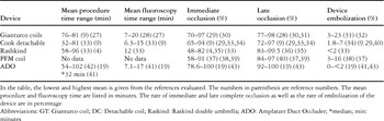
Since its first description by Porstmann et al.5 hundreds of manuscripts have been produced. For our review, we elected to choose patient outcome evidence that matters, as far as it was described in the published manuscripts.22 We consider such evidence in terms of pain, safety of the procedure, procedural time, efficacy, length of stay in hospital, and problems at long-term follow-up. We reviewed a total of 21 manuscripts, which described more than 2796 patients. Of the reports, 18 described experience with one device. The remaining three reports concentrated on experience with multiple devices. Over all, the Rashkind double umbrella device was used in 399 patients, Gianturco coils in 599, detachable coils in 669, PFM coils in 765, and the Amplatzer occluder in 763 patients.7, 11, 12, 18, 26–41 The results and complications for the five most commonly used devices are summarized in Table 2.
Table 2.

Pain
None of the reports examined this important issue. But generally, during the transcatheter approach, patients receive only one or two needle punctures in the groin that is already anesthetized with 1% Xylocaine. The associated pain is minimal, and lasts for a maximum of one or two days. This has to be weighed against either a 10 centimetre long scar of left thoracotomy, which is even more painful compared to sternotomy. The pain may last for a week or more. Intuitively, closure by the transcatheter closure should have a major advantage over surgical closure in this regard.
Safety of the procedure
Mode of sedation during the procedure
Almost always, patients underwent transcatheter closure of the patent duct under conscious sedation. Only rarely did infants in an unstable condition receive general anaesthesia. The risk of general anaesthesia, therefore, though very low, does not need to be added to the risk of the procedure.
Device related complications
Complications related to the device do occur, and even if it is not an error of the operator, it goes on her or his account, and the patient has to pay the bill. Complications occur because of the inability to release the device, or to premature or spontaneous disconnection of the device from the delivery system. Also, the long delivery sheaths are not kink-resistant, and can cause frustrating problems during delivery or retrieval of the device. Such complications are probably underreported. These kinds of problems underline the need for monitoring by institutions such as the Food and Drug Administration in the United States of America, or CE in Europe. They also show the significance of quality checks by the manufacturers.
Embolization {Table 2}.
Apart from death, embolization of the device is generally the most serious complication that can occur during transcatheter closure. It prolongs the time needed for both fluoroscopy and the overall procedure. Embolization of a device into the pulmonary artery is less dramatic than in the descending aorta. As has been reported in the literature, as well as in our experience, if retrieval is not possible, small embolized coils can be left in peripheral pulmonary arteries without causing any symptoms. If needed, a long Mullins sheath should be used to facilitate safe removal of the coil. Attempting short cuts can lead to entangling the coils within the tension apparatus of the tricuspid valve. In our experience, this led to open heart surgery in one patient to close the duct, retrieve the coil, and repair the tricuspid valve.
Embolization, at between 328 and 23%,31 is reported at greatest levels for the Gianturco coil, which is now rarely used. Embolization with the steerable coils or devices is rare (Table 2). The Amplatzer device has an extremely low rate of embolization. No embolization has occurred thus far in our experience with this device.18 We expect that, with the advent of more steerable devices, and the ability to select the correct size and type of device for the specific duct, the likelihood of embolization will be reduced to a minimum. Embolization is not an issue when reporting on surgical outcome.
Problems with the site of vascular access.
Due to the ongoing miniaturization of the devices, femoral arterial or venous injury is becoming very unlikely. Femoral arterial thrombosis was in excess of 20% when a 20 French sheath was used for the Porstmann device. Today, when only 4 French sheaths are warranted for the retrograde approach, the complication rate in most studies is zero. Haematoma at the site of access can still be a rare complication.
Loss of blood.
Loss of blood while using coils was not reported in any of the studies reviewed. Significant loss can occur when using the Amplatzer occluder without the back-bleed device on the sheath. In one of our patients, he lost almost 5% of volume because we did not use the back-bleed device. This potential complication can be eliminated by taking the necessary precautions.
Left pulmonary arterial stenosis or coarctation of the aorta.
While the use of a coil or device that is too small promotes an increased probability of embolization, using one that is too large carries the potential for left pulmonary arterial stenosis or coarctation or the aorta, or at least increased velocity of flow at these sites. As the potential increase in velocity of flow can be anticipated before deploying the device, a careful examiner should, in this situation, retrieve the device and choose either a different type of device, another size of the same device, or send the patient for surgery.
The reports examined show that real obstruction to any vessel is extremely rare, but increase in velocity in the left pulmonary artery of up to 2.5 metres per second is occasionally seen. In a multicentric study, this was seen in only 2 of 484 patients.39 Normal velocities immediately after the procedure do not guarantee that they will remain at follow up. Beck et al.42 reported a patient who developed aortic narrowing a few years after successful deployment of a Rashkind double umbrella device, which necessitated deployment of a stent.
Haemolysis.
Haemolysis, along with embolization, has been identified by the National Institute for Excellence as the most common complication of transcatheter closure of the arterial duct.21 This seems to occur more commonly subsequent to occlusion using coils. The incidence in most series, however, is zero. Uzun et al.,43 nonetheless, reported haemolysis in 3.6% of cases, whilst another group reported 7%.44 Haemolysis is almost always seen only in the presence of a significant residual shunt after closure. In our experience, it rarely occurs when there is only a tiny or small residual shunt, albeit that the assessment of the magnitude of the shunt can be very subjective. Immediate residual shunting, especially after deployment, is a very common occurrence11 (Table 2). Abolishing any residual shunt will prevent this complication. Most patients with small residual shunts achieve complete occlusion on short-term follow-up without ever developing haemolysis. It is appropriate, in our opinion, to accept a small residual leak across the arterial duct, providing that the patient is followed regularly.
Death.
In the large multicentric study for transcatheter closure coordinated by Pass et al.,39 there was but a solitary death, probably unrelated to closure, in 484 patients collected from 25 centres. Another death was mentioned by the National Centre for Clinical Excellence, representing one out of 316 patients reported.21
Left ventricular depression.
We have reported development of left ventricular systolic dysfunction few hours after closure of large patent arterial duct, in a series of 43 patients who underwent either transcatheter or surgical closure.45 Left ventricular systolic dysfunction was common in those having ducts with a narrowest diameter of over 3 millimetres, while it did not occur in the patients with smaller ducts. We believe that patients with large ducts who undergo either surgical or transcatheter closure should have an echocardiographic study before their discharge. Afterload reduction is occasionally warranted.
Procedural and fluoroscopic times
Fluoroscopic time for most of the available devices is in the range of 6 to 20 minutes. The longest fluoroscopic times are seen while occluding large ducts, especially when it is difficult to delineate the anatomy. In these cases, multiple angiograms are performed in different angulations to clarify the size and anatomy of the duct.18 Inadvertent embolization and retrieval of the device also increase the fluoroscopy time. For the PFM coil, in two multicentric studies with a total of 406 patients,36, 37 the fluoroscopic and procedural times were not reported. In our limited experience with 7 patients, the mean time required for fluoroscopy was 14 minutes.
The procedural time is important from the point of view of the patient, their parents, and the catheterization laboratory itself. Longer stay mostly translates into higher total doses of sedatives and analgesics administered to the patient, the risk of hypothermia, longer waiting times for very anxious parents, and a reduced output for the catheterization laboratory. It is reassuring to see that most procedural times range from a mean of 32 minutes32 for detachable coils, to a mean of 102 minutes18 when inserting the Amplatzer occluder. This can be compared to videoscopic surgery, where the mean procedural time was quoted to be 20 minutes.24
Efficacy
The rate of immediate complete occlusion, taken 10 minutes after implantation as seen angiographically, is lowest for the Rashkind double umbrella,34 and highest for the Amplatzer occluder.41 At mid-term follow-up, most devices provide at least 95% complete occlusion (Table 2). Residual shunts are seen most frequently in those with large ducts. Surgical ligation has clear advantages over transcatheter closure in this regard, albeit that success with the Amplatzer device rivals that achieved after surgical closure.39, 41
Length of hospital stay
Transcatheter closure is typically performed as a day-case procedure, and usually requires a stay in hospital of less than 24 hours.46 This is a clear and important advantage over surgical ligation, which required a mean stay in hospital of 3 days.2 Thoracoscopic clipping, however, can also be performed as a day case procedure.4
Long term follow up
The physician is responsible not only for safety and efficacy on a short-term basis, but especially in a growing patient should also feel responsible for the long-term effect of the device implanted. The young patients deserve critical follow-up to understand the long-term result. Long-term follow-up, of over 10 years, is available only for the Ivalon plug device and the Rashkind double umbrella. Only on rare occasions have problems been described in case reports.
The case reported by Beck et al.42 is a typical example for need for long-term follow-up after any kind of interventional procedure. In their patient, the protrusion of the struts of an implanted Rashkind double umbrella device in the descending aorta led a few years later to the development of coarctation at that site, which was relieved by implanting a stent. Yu et al.47 showed a potential relation between closure using a coil and the development of aortic regurgitation. Reopening of the duct after initial total occlusion is another possibility.48
Prophylaxis against endarteritis
Endarteritis has been reported as a rare complication of patency of the arterial duct. Its risk in the modern era approaches zero. Prior to the era of antibiotics, Campbell reported the annual risk of endarteritis in those with patent ducts to be 0.45%. Already in 1993, however, Schrader et al.49 estimated an annual risk of 0.14% in 101 adults with patent ducts. This risk, nonetheless, is still considered in most centres as an indication to close the duct, even in patients who are asymptomatic. A large study from Sweden15 seems to contradict this universal practice. In 270 patients with patent ducts, none developed endarteritis.
In the absence of larger studies, we recommend closure of any patent arterial duct as prophylaxis against endarteritis. We consider, however, that the silent duct is an exception to this rule, though it has been also seen in association with endarteritis. After closure with a device, antibiotic prophylaxis should be maintained for a period of at least six months, during which it is expected that the device will be covered by endothelium. The American Heart Association does not recommend prophylaxis for longer than 6 months after closure of the duct.50 In case of residual shunting, nonetheless, prophylaxis should be continued until complete occlusion is achieved.
Summary
The data from the reports we have reviewed shows that transcatheter closure of the persistently patent arterial duct causes minimal pain, and has a very low rate of complications. Embolization is becoming very rare. The procedural and fluoroscopic times are acceptable. If all the devices available for clinical use are accessible, with rare exceptions, almost every size and type of patent duct can be closed successfully using the interventional transcatheter approach. Most devices produce a very high rate of complete occlusion. The patients can be sent home less than 24 hours after intervention. Regular follow-up is still needed to assess long-term safety and efficacy of the devices.
Place in clinical management and future outlook
Studies dealing with the patent arterial duct are rarely prospective, and almost never randomized. With such a malformation, a lower level of evidence than randomized studies is accepted to alter management. Hence, it is acceptable to rely on consensus meetings, clinical recommendations of scientific societies, and national registries. Recommendations for management made by respected societies, however, rarely take economic factors into considerations.51 Hence, many of these recommendations might not be practicable from an economic point of view, for example in developing countries. Our discussion of the potential changes in the management of patent arterial duct would also not be complete without acknowledging that, even in the face of solid scientific evidence-based medicine, it is unwise to ignore, or negate, the importance of the experiential background of the good clinician.
The advisory committee of the National Institute for Clinical Excellence in Great Britain, on 22 June 2004, issued its provisional recommendations for the treatment of patients with persistent patency of the arterial duct. The relevant paragraphs upon which these recommendations are based include the following: “current evidence on the safety and efficacy of endovascular closure of patent ductus arteriosus ... support the use of this procedure ”
The committee goes further in placing the following conditions under which this is accepted: “The procedure should be … performed … using different devices”.21 (www.nice.org.uk accessed on 20th December 2004). With regard to the place of the transcatheter approach in clinical management, the document issued on 22 June 2004, states in its provisional report under Section 2.1.2: “Open surgery is the standard treatment”.
In contrast to the “conservative” conclusion of this institutional review of over 1000 adult patients with patent arterial duct, the consensus of Canadian Adult Congenital Heart Network proposed that transcatheter closure is today the preferred mode of treatment for adults with small ducts. Surgery is recommended for those with huge ducts not amenable to transcatheter approach, those with aneurysms, or in post- endarteritic situations. On the other hand, calcification of the duct favours transcatheter closure.20
In concert with the latter view, we believe that worldwide there is a general, albeit unofficial, consensus among the community of physicians dealing with congenital cardiac disease that transcatheter closure is today the treatment of choice for patency of the arterial duct, with cardiac surgery being indicated only rarely.
This trend towards depending more on transcatheter closure of patent arterial duct, can be shown clearly from our own experience. In a study from our previous institution in 1997, when only the Rashkind double umbrella device was available, out of 354 patients, excluding premature babies, almost half needed surgical ligation.7 In contrast, four years later, and when coils were also used, out of 341 patients treated, only 29% needed surgery.11 Today, in 2005, and reviewing 136 patients seen over the last four years, only 14% have undergone surgery (Fig. 14).

Figure 14. In the study published in 1997,4 46% of all patients who needed treatment for their patent duct underwent surgery. Three years later, in 2001, this number had dropped to 29%. Again four years later, in 2005, the need for surgery dropped further down to 14%. Abbreviations: TCC: transcatheter closure; KFSH: King Faisal Specialist Hospital and Research Centre.
Future outlook
The significance of the advances in the field of interventional paediatric cardiology could not have been predicted a decade or more ago. Today, we have more than four devices for transcatheter closure of the patent arterial duct. Most ducts can be closed using one of these devices. We are currently able to review immediate and mid-term results, but rarely results for periods of over 10 years. When significant long-term studies are published, the reported findings will almost certainly establish the basis of our decision-making and will help modify our techniques.
We expect that the trend on miniaturization will continue, as more sophisticated delivery systems and deployment techniques are developed. Our dream of the perfect device is one that is made of a biodegradable material, which dissolves over time, and which leaves the duct permanently closed. It could be also a device made of intelligent material, which adapts itself to the morphological configuration of the duct.
We have envisioned the possibility of a patch material, which could be positioned in the duct, moulding completely to its morphology. The material should be soft, accommodating, and able spontaneously to mould to the intrinsic form of the duct and not the reverse. We believe that it would less than prudent to expect one device to be suitable for all types and sizes of ducts.