Introduction
Cranial pneumatocoele may be defined as a collection of air located within the skull, either epidurally, subdurally or extradurally. Intracranial subdural pneumatocoele is also known as pneumocephalus, even though this definition is also inappropriately associated with the epidural form. A pneumatocoele may be caused by head or facial trauma, infection, tumours, or surgical intervention, or on rare occasions may be spontaneous. It often originates from the nasosinusal region, and less frequently from the temporal bone.Reference Stavas, McGeachie, Turner and Nelson1, Reference Rebol, Munda and Tos2
The development of otogenic pneumatocoele is favoured by mastoid hyperpneumatisation, which predisposes to the escape of air following rupture of the thin, bony walls. Even minor head trauma, or an abrupt change in middle ear pressure, may be the precipitating cause.Reference Stavas, McGeachie, Turner and Nelson1 The potential risks of the lesion include infection of the cavity, with possible intracranial complications, and a mass effect.Reference Mendelsohn, Hertzanu and Friedman3, Reference Park, Chandler, Telian and Doran4
We present an unusual case of chronic intracranial–epidural pneumatocoele of traumatic origin in a young man with mastoid hyperpneumatisation, followed 18 months later by the development of signs of intracranial hypertension.
Case report
A 19-year-old man was referred to the emergency department of a hospital in northern Italy due to head trauma caused by a car accident. The patient did not lose consciousness.
Neurological examination showed no focal signs or evidence of cranial nerve impairment. No cerebrospinal fluid rhinorrhoea or otorrhagia were observed.
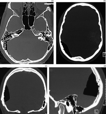
Computed tomography (CT) showed slight diastasis of the right temporal-occipital suture, but no fractures. However, a significant collection of intracranial air (measuring approximately 5 cm in depth and 2.5 × 4.5 cm at the base) was found in the right parieto-occipital area, in contiguity with the posterior mastoid cells, moderately compressing the brain (Figure 1). The temporal bones were fluid-free and showed well developed pneumatisation, with a large, partially fluid-filled cell posterior to the sigmoid sinus bilaterally, communicating on the right side with the pneumatocoele. The ventricular complex was in the midline and undilated. No haemorrhagic areas were noted.
Axial (a & b), coronal (c) and parasagittal (d) computed tomography scans, performed on the same day as the patient's head trauma, showing a right parieto-occipital pneumatocoele communicating with a large retrosigmoid pneumatic cell.

The patient was admitted to the neurological department of the same hospital and treated conservatively with absolute bed rest with the head slightly elevated, intravenous antibiotics (ceftriaxone) and dexamethasone.
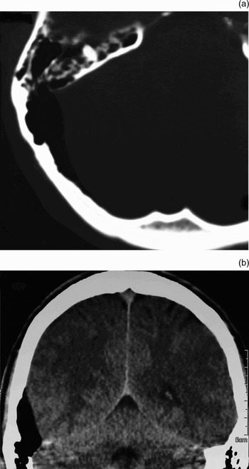
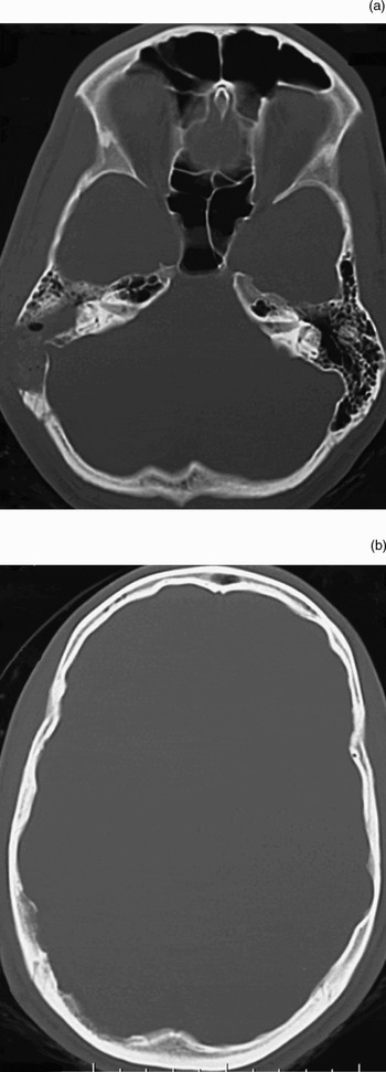
Cerebral and petrous bone CT scans, performed the next day, confirmed the earlier observations (Figure 2).
(a) Axial and (b) coronal cranial computed tomography scans performed the day after trauma, showing evidence of communication between the mastoid cells and the intracranial–epidural air collection.

Routine ENT examination, including pure tone audiometry, was normal.
Magnetic resonance imaging (MRI), performed two days later, better defined the extradural location of the pneumatocoele, and confirmed the absence of any other lesions involving the brain.
Nine days after the patient's trauma, a new cranial CT scan yielded unmodified results, and the patient was discharged with tapered corticosteroid treatment and the recommendation to avoid physical effort.
The patient remained asymptomatic and gradually resumed regular physical activity. He underwent periodic radiological follow-up examinations to verify the status of the pneumatocoele. Cranial CT scans at one, two and six months, a brain MRI scan at 12 months, and a temporal bone CT scan at 13 months all showed a persistent and substantially unmodified intracranial air collection in the parieto-occipital region. Notably, the pneumatocoele was interpreted by the neuroradiologist (at the 13-month CT scan) as a ‘giant pneumatic retrosinusal cell extending to the occipital bone’. Based on this reassuring report, the patient increased his sporting activity, including diving.
Eighteen months after the initial head trauma, the patient suffered a common cold, with nasal obstruction, rhinorrhoea and headache. During this episode, he repeatedly blew his nose, and his headache became continuous and progressive, localised in the fronto-occipital region, and unresponsive to non-steroidal anti-inflammatory drugs.
Five days after the onset of headache, clear signs of intracranial hypertension developed, with projectile vomiting, agitation and irritability, leading to immediate admission to our hospital's neurology department.
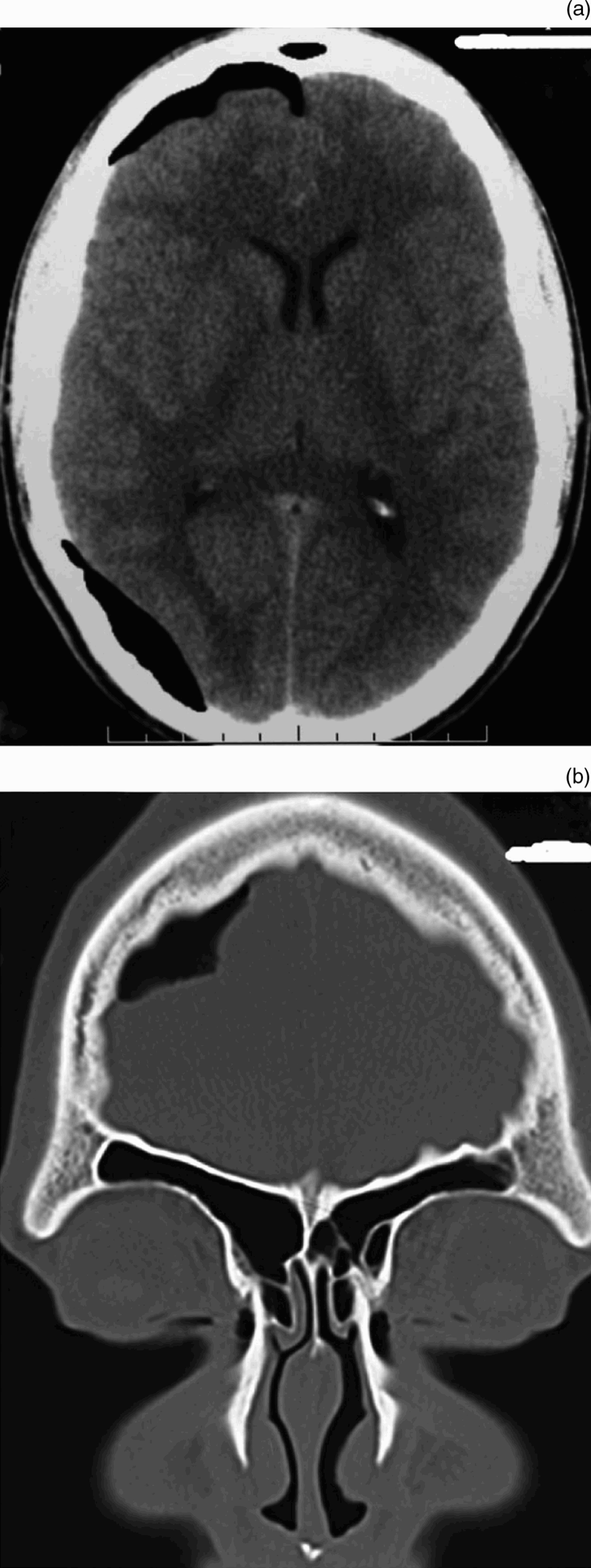
A new cranial CT scan showed spread of the pneumatocoele along a parasagittal pathway adjacent to the falx cerebri, as far as the right epidural anterior fossa, with moderate compression of the frontal lobe (Figure 3). Neurological examination showed no deficit, papilloedema or nuchal rigidity. Blood count and temperature were normal.
(a) Axial and (b) coronal computed tomography scans at 18 months, showing extension of the pneumatocoele to the anterior cranial fossa, with compression of the frontal lobe.

Bed rest with absolute prohibition of nose-blowing, together with administration of intravenous mannitol and dexamethasone, produced a rapid improvement in the patient's symptomatology. Reduction of the frontal pneumatocoele was observed on a CT scan performed on day seven of hospitalisation.
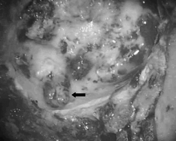
The patient was then admitted to our ENT department, and underwent surgical treatment to seal the communication between the right mastoid bone and the endocranium. A large right mastoidectomy extended to the retrosigmoid region was performed under general anaesthesia. A large retrosigmoid cell extending toward the occipital bone was detected (Figure 4) and sealed so as to be watertight, using bovine pericardium (Tutopatch; Tutogen Medical, Alachua, Florida, USA) and a mixture of bone powder and fibrin glue (Tissucol; Immuno G, Vienna, Austria). The latter mixture was also used to obliterate the mastoid cavity.
Intra-operative view showing a large retrosigmoid cell (arrow) communicating with the pneumatocoele.

The post-operative course was uneventful, and the patient was discharged symptom-free on day three.
Ten days after surgery, CT scan revealed complete reabsorption of the frontal and parieto-occipital pneumatocoele, and obliteration of the mastoid (Figure 5).
Post-operative axial computed tomography scans at (a) mastoid and (b) parietal level, showing complete reabsorption of intracranial air and obliteration of the right mastoid and retrosigmoid cell.

At six months post-operatively, the patient was asymptomatic with a normal auditory threshold, as before surgery. A further CT scan showed no change.
Discussion
Pneumatocoele is a frequent complication of cranial trauma, with an incidence ranging from 7 to 9 per cent in patients investigated by CT scan.Reference Stendel and Heckel5 Traumatic mechanisms account for more than one-third of otogenic pneumatocoeles; other frequent causes are otitis media and otological surgery.Reference Andrews and Canalis6
Hyperpneumatisation of the temporal bone, and in particular the mastoid, predisposes to pneumatocoele, since thinning of the intercellular septa and outer lamina makes them susceptible to rupture following an increase in intratympanic pressure or minor head trauma, with consequent escape of air either intra- or extracranially. In extreme, rare cases, a ‘spontaneous’ pneumatocoele may occur for which no apparent cause can be found.Reference Stavas, McGeachie, Turner and Nelson1, Reference Maier, Fradis and Scheremet7
Whatever the aetiology, any fracture in the temporal bone must be associated with a pressure increase such that the middle-ear pressure is greater than the intracranial pressure, in order for air to be forced into the cranial cavity. This may occur during nose-blowing, coughing, sneezing or performing the Valsalva manoeuvre.Reference Stavas, McGeachie, Turner and Nelson1, Reference Maier, Fradis and Scheremet7–Reference Vallejo, Gil-Carcedo, Borrás and De Campos10 When intracranial pressure increases above the level of the pneumatic cavity pressure, the air remains trapped between the bone and the dura, and no further spread occurs (i.e. a ball valve mechanism is created).Reference Dandy11 The substantial adherence between bone and dura helps prevent excessive diffusion of escaped air, which remains confined to the parieto-occipital region, with negligible brain compression. If the fistula is limited in size, spontaneous repair mechanisms are usually able to obliterate the communication with the cranial content and to resolve the pneumatocoele. Less frequently, a dynamic equilibrium is established, leading to persistence of the pneumatocoele, which may remain substantially unmodified for a lengthy period of time. In this case, the patent fistula allows repeated replacement of the portion of air which is reabsorbed, when a critical reduction of intracranial pressure occurs.
• Mastoid hyperpneumatisation predisposes to formation of intracranial pneumatocoele in response to minor trauma
• In this case report, a mastoid to cranial fistula was repaired via a transmastoid approach
• Following this repair, intracranial hypertension resolved
The diagnosis of otogenic pneumatocoele is usually straightforward, although some misinterpretation may occur, as shown in the reported case. The presence of fibrous bands connecting the dura to the bone may be mistaken for intercellular bony septae, and the pneumatocoele may be interpreted as a hyperpneumatisation extending to bones contiguous with the temporal bone.
When a pneumatocoele is limited in extent, clinical features are usually few, with headache being the main symptom.Reference Andrews and Canalis6, Reference Vallejo, Gil-Carcedo, Borrás and De Campos10 However, forceful spread of air into intracranial spaces may have serious consequences. In such a case, the resistance afforded by the adherence between the dura and the bone may be overcome by the increasing air pressure. A rapid increase in intracranial pressure may be hard to compensate for, giving rise to intracranial hypertension, with florid symptoms such as agitation, delirium, loss of consciousness, meningism, lethargy, disorientation and papilloedema.Reference Maier, Fradis and Scheremet7
Our patient developed signs of intracranial hypertension 18 months after onset of an asymptomatic, traumatic pneumatocoele. An abrupt increase in pneumatocoele air volume occurred following forceful nose-blowing during a bout of common cold, causing an intracranial mass effect. The development of such a serious complication a considerable length of time after pneumatocoele onset suggests the need for early repair of the fistula if conservative treatment, such as bed rest, is not successful. In our patient, repair was performed a considerable period after diagnosis of the pneumatocoele. In spite of clear radiological evidence of persistent air entrapment, the patient was discharged nine days after sustaining trauma. Surgical treatment at this stage would have been advisable in order to prevent possible complications. Absence of symptoms and misinterpretation on the part of the neuroradiologist (who described the lesion as a ‘giant pneumatic retrosinusal cell extending to the occipital bone’ on a CT scan performed at 13 months) also contributed to further therapeutic delay. Closing the existing fistula, by using muscle, fascia, cartilage or bone to seal the communication between the extracranial and intracranial compartments, is usually sufficient to resolve otogenic pneumatocoele.Reference Stavas, McGeachie, Turner and Nelson1, Reference Park, Chandler, Telian and Doran4, Reference Maier, Fradis and Scheremet7, Reference Madeira and Summers8 In our patient, obliteration of the fistula was completely successful, with rapid reabsorption of intracranial air.