Introduction
Working memory impairments have been documented in subjects with bipolar I disorder during euthymic and depressed phases (Murphy et al. Reference Murphy, Sahakian, Rubinsztein, Michael, Rogers, Robbins and Paykel1999; Malhi et al. Reference Malhi, Ivanovski, Hadzi-Pavlovic, Mitchell, Vieta and Sachdev2007; Godard et al. Reference Godard, Grondin, Baruch and Lafleur2011; Yates et al. Reference Yates, Dittmann, Kapczinski and Trentini2011; Xu et al. Reference Xu, Lin, Rao, Dang, Ouyang, Guo, Ma and Chen2012; Volkert et al. Reference Volkert, Kopf, Kazmaier, Glaser, Zierhut, Schiele, Kittel-Schneider and Reif2014). Impairment of working memory in this population has been associated with occupational deficits (Bearden et al. Reference Bearden, Shih, Green, Gitlin, Sokolski, Levander, Marusak, Hammen, Sugar and Altshuler2011) and suicidal behavior (Keilp et al. Reference Keilp, Gorlyn, Russell, Oquendo, Burke, Harkavy-Friedman and Mann2013). Though there are two primary subtypes of bipolar disorder, investigations of the neuropathology of working memory impairments have focused on bipolar I disorder. For example, a recent meta-analysis (Cremaschi et al. Reference Cremaschi, Penzo, Palazzo, Dobrea, Cristoffanini, Dell'Osso and Altamura2013) of functional magnetic resonance (fMRI) studies assessing working memory in euthymic bipolar I patients points to abnormal activation patterns in the dorsolateral prefrontal cortex (DLPFC), ventrolateral prefrontal cortex, other prefrontal regions, as well as the parietal and temporal cortices.
Bipolar II disorder is also associated with neurocognitive impairments, including working memory (Torrent et al. Reference Torrent, Martinez-Aran, Daban, Sanchez-Moreno, Comes, Goikolea, Salamero and Vieta2006; Dittmann et al. Reference Dittmann, Hennig-Fast, Gerber, Seemuller, Riedel, Emanuel Severus, Langosch, Engel, Moller and Grunze2008; Hsiao et al. Reference Hsiao, Wu, Wu, Hsu, Chen, Lee, Lee, Yeh, Yang, Ko and Lu2009; Sole et al. Reference Sole, Martinez-Aran, Torrent, Bonnin, Reinares, Popovic, Sanchez-Moreno and Vieta2011, Reference Sole, Bonnin, Torrent, Balanza-Martinez, Tabares-Seisdedos, Popovic, Martinez-Aran and Vieta2012; Pålsson et al. Reference Pålsson, Figueras, Johansson, Ekman, Hultman, Östlind and Landén2013), but the functional correlates of the deficits in bipolar II disorder remain largely unexplored. Recent evidence further suggests that a major part of the cognitive impairment observed in euthymic bipolar I and II disorder may be due to subthreshold depressive symptoms rather than disease severity (Volkert et al. Reference Volkert, Kopf, Kazmaier, Glaser, Zierhut, Schiele, Kittel-Schneider and Reif2014), highlighting a need for bipolar studies during the depressed phase of the illness. Even so, the underlying neural correlates of working memory performance in bipolar II depression have yet to be identified.
With respect to bipolar I disorder, previous research has suggested, though with some disparate findings, that DLPFC dysregulation is a core feature associated with working memory performance. An fMRI study of bipolar I depression explored blood oxygenation level-dependent (BOLD) response during a 2-back working memory task and found that medicated depressed subjects with bipolar I disorder exhibited increased left DLPFC activation compared with healthy subjects (Deckersbach et al. Reference Deckersbach, Rauch, Buhlmann, Ostacher, Beucke, Nierenberg, Sachs and Dougherty2008). An increased activation pattern has also been found in studies in predominantly medicated unipolar depressed subjects (Fitzgerald et al. Reference Fitzgerald, Srithiran, Benitez, Daskalakis, Oxley, Kulkarni and Egan2008; Bertocci et al. Reference Bertocci, Bebko, Mullin, Langenecker, Ladouceur, Almeida and Phillips2012) in which performance on n-back tasks is associated with increased anterior cingulate response, which could represent compensatory activation. However, Townsend et al. (Reference Townsend, Bookheimer, Foland-Ross, Sugar and Altshuler2010), found that, relative to healthy comparison subjects, medicated subjects with bipolar I depression exhibited decreased activation in the right DLPFC and the posterior parietal cortex [Brodmann area (BA) 40] during a working memory task. These findings also parallel those reported in other work in bipolar depression of unspecified subtype (Fernandez-Corcuera et al. Reference Fernandez-Corcuera, Salvador, Monte, Salvador Sarro, Goikolea, Amann, Moro, Sans-Sansa, Ortiz-Gil, Vieta, Maristany, McKenna and Pomarol-Clotet2013) where an inverse relationship between left DLPFC activation and depression severity scores in medicated subjects was found.
The demonstrated, though somewhat unclear, differences in neural activation in bipolar I depression raise the question of whether bipolar II depression is associated with similar or different patterns of neural activity. To our knowledge, there have been no functional neuroimaging studies of working memory in bipolar II depression. Additionally, in many fMRI studies of bipolar disorder, subjects are taking various medications that have unclear effects on fMRI activation. However, one recent working memory study in euthymic subjects with bipolar I disorder demonstrated that those with greater medication load exhibited the greatest brain response within the prefrontal cortex, including in the DLPFC (McKenna et al. Reference McKenna, Sutherland, Legenkaya and Eyler2014).
Here we report an initial study of neurobiological changes associated with bipolar II depression in the absence of potentially confounding medication effects. Our primary hypothesis was that unmedicated depressed subjects with bipolar II disorder would exhibit hypoactivation of the DLPFC compared with healthy controls. We also predicted that, given our prior results in depressed subjects with bipolar I disorder (Townsend et al. Reference Townsend, Bookheimer, Foland-Ross, Sugar and Altshuler2010), there would be additional areas of hypoactivation in bipolar II subjects including in the inferior frontal cortex and parietal cortex during working memory tasks.
Method
Participants
Twenty-four depressed subjects with bipolar II disorder and 21 healthy comparison subjects were enrolled and scanned. Subjects with bipolar disorder were recruited through the UCLA Mood Disorders Clinic and through local advertising. Comparison subjects were recruited by advertisement in local newspapers and campus flyers. Participants provided written informed consent in accordance with the Institutional Review Boards at the University of California, Los Angeles.
All participants completed the Structured Clinical Interview for DSM-IV Axis I Disorders, Research Version (SCID; First et al. Reference First, Spitzer, Gibbon and Williams2002). Subjects who met DSM-IV criteria for bipolar II disorder in a current major depressive episode, and scored ⩾22 on the 30-item Inventory of Depressive Symptomatology-Clinician Rated (Rush et al. Reference Rush, Gullion, Basco, Jarrett and Trivedi1996), were eligible to participate. Bipolar subjects with a history of an alcohol or drug use disorder could participate if they had been sober for at least 3 months, as confirmed by self-report and a urine toxicology screen before scanning. Course of illness information (i.e. bipolar illness duration, prior number of hypomanic and depressive episodes) was obtained by self-report and confirmed by reference to psychiatric care records when available. Comparison participants were required to have no current or past psychiatric diagnosis (including substance abuse) as assessed by the SCID.
Exclusion criteria for all subjects included current use of psychotropic medications, left-handedness, head injury with loss of consciousness >5 min, unstable medical illness, ferrous metal implants, neurologic illness, pregnancy, and a diagnosis of borderline personality disorder assessed using the Personality Diagnostic Questionnaire (Hyler et al. Reference Hyler, Skodol, Kellman, Oldham and Rosnick1990) and confirmed with a clinical interview.
On the day of the scan, severity of hypomania and depression in bipolar II subjects was assessed using the Young Mania Rating Scale (Young et al. Reference Young, Biggs, Ziegler and Meyer1978) and the 21-item Hamilton Rating Scale for Depression (HAMD-21; Hamilton, Reference Hamilton1960). A seven-item extension of the HAMD was used to assess atypical depressive symptoms common in bipolar depression (Rosenthal & Hefferman, Reference Rosenthal and Hefferman1987).
Four bipolar II depressed participants and three control participants were excluded from the analyses because of excessive movement in the scanner (movement beyond 3 mm translation peak-to-peak over 117 functional images) or excessive magnetic susceptibility dropout. An additional bipolar subject was excluded because of poor behavioral performance. The final MRI analysis was conducted on 19 bipolar II depressed subjects and 19 comparison subjects.
Of the bipolar II depressed subjects, seven had never received psychotropic medication and the remaining 12 had been unmedicated for an average of 3.5 years (s.d. = 5.5 years, range 22 days to 20 years) at the time of the study. Two bipolar II depressed subjects had current co-morbid post-traumatic stress disorder, one had panic disorder with agoraphobia, and another had social phobia. Four bipolar subjects had past substance/alcohol use disorders.
Ethics
All procedures contributing to this work comply with the ethical standards of the relevant national and institutional committees on human experimentation and with the Helsinki Declaration of 1975, as revised in 2008.
Imaging task
The n-back paradigm included three memory-load conditions (0-back, 1-back and 2-back). For the 0-back condition, participants pressed a button whenever the letter ‘X’ appeared. For the 1-back condition, participants were instructed to press the button whenever a letter flashed upon the screen that was identical to the letter presented one position back. For the 2-back condition, participants pressed the button when the current letter appeared two letters back. The task included eight blocks [four blocks of the 0-back (control) condition, two blocks of each experimental condition] with 12 trials in each 30-s block. Instructions were presented on the computer screen for 6 s at the beginning of every block and subjects were asked to respond as accurately and rapidly as possible. Stimulus presentation time was 500 ms with an inter-trial interval of 1500 ms. The order of the experimental blocks was counterbalanced and interleaved with blocks of the 0-back condition. A rest period was presented at the beginning and end of the task (total duration 4 min 58 s). Before scanning, subjects completed a practice session outside the scanner to familiarize themselves with the task and to confirm performance accuracy.
Image acquisition
Functional imaging data were acquired on a Siemens 3T Magnetom Allegra scanner (Germany). T2*-weighted images were acquired with a gradient echo planar imaging (EPI) sequence as follows: repetition time (TR) = 2500 ms, echo time (TE) = 35 ms, field of view (FOV) = 200 × 200 mm, matrix size = 64 × 64 mm, voxel size = 3.1 × 3.1 × 3 mm, slice thickness = 3 mm with a 1-mm slice gap, number of interleaved acquired slices = 28, flip angle = 90°. High-resolution structural images aligned to the anterior and posterior commissure were acquired with the following parameters: TR = 5000 ms, TE = 33 ms, FOV = 200 × 200 mm, matrix = 128 × 128 mm, in-plane resolution = 1.56, slice thickness = 3 mm, gap = 1 mm, number of slices = 28, flip angle = 90°.
Data analysis
Demographic variables
Group differences in categorical and continuous demographic variables were computed using two-tailed Fisher's exact and independent-sample t tests, respectively. Statistical significance was defined as α = 0.05.
Behavioral data
Mean accuracy and mean correct response times were computed for each participant for all three memory-load conditions. Data were analysed using a repeated-measures analysis of variance (ANOVA) with group as a between-subjects factor, and memory load (i.e. 0-back, 1-back or 2-back) as the within-subject factor. Accuracy and reaction times were analysed separately. Behavioral data were missing for one healthy control participant.
Image preprocessing
fMRI data were analysed using the fMRI Expert Analysis Tool (FEAT) version 5.98, part of FSL [Oxford Centre for Functional Magnetic Resonance Imaging of the Brain (FMRIB) Software Library, www.fmrib.ox.ac.uk/fsl]. Structural images were skull stripped using the Brain Extraction Tool (Smith, Reference Smith2002) and used for intra-subject registration. The first two volumes of each subject's functional scans were discarded to allow for T1 equilibrium effects. Motion correction was performed using MCFLIRT (Motion Correction using FMRIB's Linear Image Registration Tool) (Jenkinson et al. Reference Jenkinson, Bannister, Brady and Smith2002). Images were smoothed using a Gaussian kernel of 5 mm full width between half maximum values of a function. All volumes underwent grand-mean intensity normalization by a single multiplicative factor and high-pass temporal filtering using a Gaussian-weighted least-squares straight line fitting, with σ = 60.0 s. A high-pass filter of 120 s was used to remove low-frequency artifact signals. Time-series statistical analysis was performed using FMRIB's Improved Linear Model (FILM) with local autocorrelation correction (Woolrich et al. Reference Woolrich, Ripley, Brady and Smith2001). Using a seven-parameter affine registration, functional images were registered to high-resolution structural images using FMRIB's Linear Image Registration Tool (FLIRT) (Jenkinson & Smith, Reference Jenkinson and Smith2001; Jenkinson et al. Reference Jenkinson, Bannister, Brady and Smith2002) then aligned to the MNI-152 atlas (Montreal Neurological Institute, Canada) using a 12-parameter affine registration.
Image analysis
For first-, or subject-level, analysis, each of the three memory-load conditions, and the instructional cue at the beginning of each block condition, were modeled separately for each subject. Functional imaging data were analysed using a general linear model, and six motion-correction parameter estimates were incorporated as covariates of no interest to control motion-related signal change. Prior research using the n-back working memory task has shown that increasing working memory load produces increased prefrontal cortex activation (Braver et al. Reference Braver, Cohen, Nystrom, Jonides, Smith and Noll1997; Cohen et al. Reference Cohen, Perlstein, Braver, Nystrom, Noll, Jonides and Smith1997; Kammer et al. Reference Kammer, Bellemann, Guckel, Brix, Gass, Schlemmer and Spitzer1997). Therefore, to model our parametric design and further reduce the likelihood of a type I error, we conducted a single analysis that considered all memory-load conditions (0-back, 1-back and 2-back) simultaneously. To do so, we assigned weights (−0.5, 0.7, 1.2) to each condition (0-back, 1-back and 2-back), respectively, and obtained a statistical map for each subject.
Higher-level statistical analyses for within- and between-group analyses were carried out using FLAME 1 + 2 (FMRIB's Local Analysis of Mixed Effects) (Beckmann et al. Reference Beckmann, Jenkinson and Smith2003). For the whole-brain analysis of within-group and between-group effects on activation during these contrasts, we report brain regions with a height threshold of Z > 2.0 and a cluster probability of p < 0.05, corrected for whole-brain multiple comparisons using Gaussian random field theory (Worsley, Reference Worsley, Jezzard, Matthews and Smith2001). For reporting purposes, Montreal Neurological Institute (MNI) coordinates were transformed to Talairach space using the MNI to Talairach Conversion Applet (www.bioimagesuite.org). Anatomical localization was performed using stereotaxic atlases (Talairach & Tournoux, Reference Talairach and Tournoux1988; Oishi et al. Reference Oishi, Faria, van Zijl and Mori2011).
Region-of-interest (ROI) analysis in the DLPFC
Given our a priori hypothesis of differential DLPFC activation, we supplemented our whole-brain analysis with a ROI analysis. To avoid bias in ROI selection (Kriegeskorte et al. Reference Kriegeskorte, Simmons, Bellgowan and Baker2009), we functionally defined the left and right DLPFC using coordinates derived from a meta-analysis of normative n-back working memory fMRI studies (Owen et al. Reference Owen, McMillan, Laird and Bullmore2005). Specifically, a 10-mm sphere was placed at the peak voxel corresponding to activation in the left and right DLPFC centered at MNI coordinate (−41, 31, 31) and (41, 31, 31), respectively. The time course from these DLPFC ROI masks during the parametric analysis of 0-back > 1-back > 2-back trials was extracted separately for each subject and used for the calculation of mean percentage signal change using FEATQuery. Correlational analyses were performed to determine if mean activity in these regions was related to illness characteristics (p < 0.05), which included: current depression severity (using the HAMD); age at onset of bipolar illness; duration of bipolar illness; and number of weeks in current major depressive episode.
Results
Participant characteristics
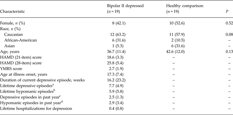
Demographic data are provided in Table 1. There was no significant difference in age between patient and comparison groups. The comparison group included more female participants, but not significantly so (χ2 = 0.42, p = 0.51). Among patients diagnosed with bipolar II disorder, there were no significant correlations between medication-free period and age at illness onset, duration of illness, current episode duration, current depression severity or lifetime number of mood episodes (all p's > 0.11).
Demographic and clinical characteristics of bipolar II depressed and healthy comparison subjects

Data are given as mean (standard deviation) unless otherwise indicated.
HAMD, Hamilton Depression Rating Scale; YMRS, Young Mania Rating Scale.
a Bipolar subjects had a range of lifetime depressive episodes from 2 to 15; patients who had a number marked as too many to count were not included in the mean calculation.
b Bipolar subjects had a range of lifetime hypomanic episodes from 1 to 20; eight patients who had a number of lifetime episodes that were marked as too many to count were not included in the mean calculation.
c Bipolar subjects had a range of depressive episodes in the past 12 months from 1 to 6.
d Bipolar subjects had a range of hypomanic episodes in the past 12 months from 0 to 12.
Behavioral data
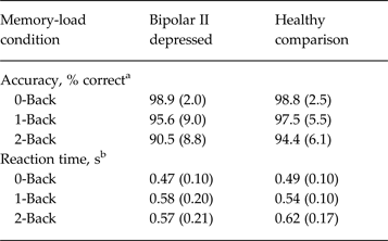
Behavioral data collected from the n-back task are shown for each group in Table 2. A repeated-measures ANOVA of accuracy data did not reveal a significant main effect of group (F 1,35 = 1.58, p = 0.22), but there was a significant main effect of memory load (F 2,70 = 13.49, p < 0.001), indicating that both bipolar II depressed and healthy comparison participants were more accurate on both the 0-back and 1-back conditions than on the 2-back memory-load condition. However, the group × memory load interaction was not statistically significant (F 2,70 = 1.32, p = 0.27).
Accuracy and reaction time by group

Data are given as mean (standard deviation).
a Accuracy data represent percentage of correct trials.
b Reaction time data represent correct response reaction times.
For reaction time, the group × memory load interaction was not statistically significant (F 2,70 = 1.20, p= 0.31). There was no significant main effect of group (F 1,35 = 0.14, p = 0.71), but there was a main effect of memory load (F 2,70 = 7.19, p = 0.001), indicating that reaction times varied across the three memory-load conditions.
fMRI results
Analysis of motion artifacts
Analysis of the three rotational (roll, pitch, yaw) and three translational (anterior to posterior, superior to inferior, left to right) parameters yielded no significant differences in motion correction between subjects with bipolar II depression and healthy comparison subjects (all p's > 0.25). Additionally, the relative and absolute motion values for each participant were examined to confirm that our patient and comparison groups did not differ significantly, which they did not (t 36 = 0.22, p = 0.82 and t 36 = 0.45, p = 0.66, respectively).
Whole-brain results: within-group findings for parametric analysis of memory load
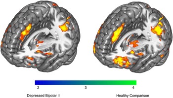
The contrast across the three memory-load conditions for whole-brain analyses of each subject group is illustrated in Fig. 1. Control subjects demonstrated significant activation in the bilateral inferior frontal gyrus (BA 44 extending to BA 9), right medial frontal gyrus (BA 8), bilateral inferior parietal lobule (BA 40), bilateral thalamus and right temporal cortices (BA 22, 37) (Z > 2.0, p < 0.05, corrected). Bipolar subjects significantly activated similar regions including the bilateral middle frontal gyrus (BA 8), bilateral superior frontal gyrus (BA 8 and BA 6), left precentral gyrus (BA 6), bilateral inferior parietal lobule (BA 40) and bilateral striatal regions (Z > 2.0, p < 0.05, corrected).
Regions of significant within-group activation in healthy comparison subjects and depressed subjects with bipolar II disorder across 0-, 1- and 2-back conditions. Maps are thresholded at Z > 2.0, p < 0.05 with correction for multiple comparisons.

Whole-brain results: between-group findings for parametric analysis of memory load
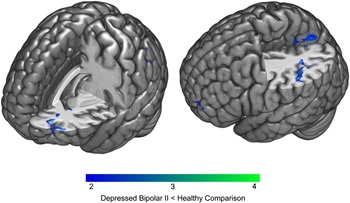
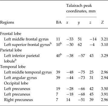
Table 3 provides the local peak maxima of the activation differences for the random-effects analysis (bipolar<control). There were no significant group differences observed in the DLPFC. In the parametric analysis of memory load, bipolar depressed subjects showed significantly reduced activity in the left middle frontal gyrus (BA 11) and left superior frontal gyrus (BA 10) relative to healthy comparison subjects (Z > 2.0, p < 0.05, corrected). Bipolar depressed subjects showed hypoactivation in additional regions including the left inferior parietal lobule (BA 40), left middle temporal gyrus and angular gyrus (both corresponding to BA 39), and occipital regions. The bipolar II depressed group did not exhibit significantly greater activation in any brain regions relative to comparison subjects across memory-load conditions. A graphic illustration of activation differences is provided in Fig. 2.
Regions of significantly decreased activation in bipolar II disorder patients relative to healthy comparison subjects across 0-, 1- and 2-back conditions. Maps are thresholded at Z > 2.0, p < 0.05 with correction for multiple comparisons.

Significant between-group differences (bipolar II depressed<comparison) in regional functional activation for the parametric analysis of memory load a

BA, Brodmann area.
a Anatomical labels and BAs were assigned according to Talairach & Tournoux (Reference Talairach and Tournoux1988) after non-linear coordinate conversion x, y and z are Talairach peak coordinates of local maxima significant at Z > 2.0, p < 0.05, corrected for multiple comparisons.
b More than one local maxima within 10 mm corresponds to this anatomical label and BA region.
ROI results: DLPFC BOLD response in bipolar II depressed versus comparison subjects
Both groups demonstrated significant bilateral DLPFC activation. Our a priori ROI analysis did not reveal statistically significant differences in BOLD response across memory-load conditions between the bipolar and comparison groups in either the left or right DLPFC, nor did we find evidence of statistically significant correlations with HAMD score, illness duration, age at illness onset, or number of weeks in current depression (for all, p > 0.05).
Discussion
Our study is an initial report of differences in neural activation across memory-load conditions in an n-back working memory task involving unmedicated, acutely depressed subjects with bipolar II disorder. Accuracy and reaction time were matched in both bipolar disorder and healthy comparison groups, and, relative to healthy subjects, we observed significantly decreased activation in the medial orbital, temporal, parietal, and occipital cortices in the bipolar group.
Our within-group findings in subjects with bipolar II depression demonstrated that our task activated regions as expected during a working memory task. Studies of healthy subjects have revealed that, across memory-load conditions, working memory tasks activate DLPFC as well as the posterior parietal cortex (Smith & Jonides, Reference Smith and Jonides1998; Curtis, Reference Curtis2006). We found similar activation patterns in both bipolar II depressed and healthy subjects.
We did not find significant differences in DLPFC activation in unmedicated subjects with bipolar II depression versus control subjects during a working memory task. A prior study by our group in medicated subjects with bipolar I disorder found reduced activation in the DLPFC and parietal cortex across mood states (Townsend et al. Reference Townsend, Bookheimer, Foland-Ross, Sugar and Altshuler2010) during a 2-back versus 0-back memory condition. Whereas some regions of hypoactivation may represent shared features of both bipolar I and II depression, DLPFC hypoactivation may be more specific to bipolar I depression. Neurobiological differences in brain activation during working memory tasks in bipolar I disorder may be trait characteristics and are thus present outside of depressive episodes. A recent review (Cremaschi et al. Reference Cremaschi, Penzo, Palazzo, Dobrea, Cristoffanini, Dell'Osso and Altamura2013) of n-back fMRI studies of medicated euthymic subjects diagnosed with bipolar I disorder revealed altered activation and connectivity within the ventrolateral circuit and parieto-temporal circuit dysfunction rather than dorsolateral. Although we did not include euthymic subjects with bipolar II disorder in this study, it may be that bipolar II disorder is associated more with ventrolateral circuit rather than dorsolateral circuit dysfunction. To the extent that this observation is true, it could account for the lack of differences in DLPFC activation in our study. Thus, our study of unmedicated depressed subjects with only the bipolar II subtype suggests that differential neural activation during working memory tasks in bipolar II depression shares some, but not all, the characteristics of activation patterns seen in subjects with bipolar I depression.
As in our previous work with subjects with bipolar I depression, we observed significant differences in BA 10 activation in bipolar II subjects compared with healthy controls. However, unlike medicated subjects with bipolar I disorder, who exhibited hyperactivation of BA 10 (Altshuler et al. Reference Altshuler, Bookheimer, Townsend, Proenza, Sabb, Mintz and Cohen2008), the present study of unmedicated depressed subjects with the bipolar II subtype found hypoactivation of BA 10. Although BA 10 is generally considered with respect to its roles in coordinating tasks (Cabeza & Nyberg, Reference Cabeza and Nyberg2000) or regulating emotion (Ochsner et al. Reference Ochsner, Bunge, Gross and Gabrieli2002), it may also be involved in circuits activated by working memory tasks (Thermenos et al. Reference Thermenos, Goldstein, Milanovic, Whitfield-Gabrieli, Makris, Laviolette, Koch, Faraone, Tsuang, Buka and Seidman2010). Previously, researchers have suggested that BA 10 aberrations could be a trait marker for bipolar disorders (Drevets, Reference Drevets1999). Thus, the region may be involved in several critical circuits that could be differentially activated across bipolar subtypes.
In the current study, depressed subjects with bipolar II disorder also exhibited significantly decreased activation relative to healthy subjects in the inferior parietal cortex (BA 40). The posterior parietal cortex (encompassing BA 40) is a component of the working memory circuit, which probably plays a role in short-term storage of verbal information (Smith & Jonides, Reference Smith and Jonides1998; Naghavi & Nyberg, Reference Naghavi and Nyberg2005; Owen et al. Reference Owen, McMillan, Laird and Bullmore2005). Decreased parietal activation may represent a more general trait of bipolar disorders, as medicated euthymic patients with bipolar I disorder also exhibit parietal hypoactivation when performing a working memory task (Monks et al. Reference Monks, Thompson, Bullmore, Suckling, Brammer, Williams, Simmons, Giles, Lloyd, Harrison, Seal, Murray, Ferrier, Young and Curtis2004; Townsend et al. Reference Townsend, Bookheimer, Foland-Ross, Sugar and Altshuler2010). Though we found decreased parietal activation among patients with bipolar II depression, performance levels did not significantly differ from comparison subjects. Our analyses did not detect the nature of a compensatory mechanism, but such a mechanism may become evident with increased levels of task difficulty.
Some researchers have suggested that differential brain activation during working memory tasks may be a distinguishing characteristic between subjects with bipolar and unipolar depression (Schöning et al. Reference Schöning, Zwitserlood, Engelien, Behnken, Kugel, Schiffbauer, Lipina, Pachur, Kersting, Dannlowski, Baune, Zwanzger, Reker, Heindel, Arolt and Konrad2009; Bertocci et al. Reference Bertocci, Bebko, Mullin, Langenecker, Ladouceur, Almeida and Phillips2012). Specifically, in a study of predominantly medicated female patients diagnosed with either bipolar I disorder or unipolar depression, Bertocci et al. (Reference Bertocci, Bebko, Mullin, Langenecker, Ladouceur, Almeida and Phillips2012) reported that patients with unipolar depression exhibited greater anterior cingulate activation during an emotional working memory task than did either patients with bipolar I depression or healthy comparison subjects. However, the emotion-regulation component of the task may have played a substantial role in this finding.
As with studies in bipolar disorder, the fMRI activation findings in subjects with unipolar depression during working memory tasks are not consistent. One study found that medicated patients with non-psychotic unipolar depression exhibit significantly less right DLPFC activation during a working memory task than do healthy subjects (Garrett et al. Reference Garrett, Kelly, Gomez, Keller, Schatzberg and Reiss2011). Other work, however, suggests that medicated patients with unipolar depression exhibit increased left dorsolateral prefrontal activation during a verbal working memory task relative to healthy subjects (Fitzgerald et al. Reference Fitzgerald, Srithiran, Benitez, Daskalakis, Oxley, Kulkarni and Egan2008), and this pattern of results is also observed in unmedicated unipolar depressed subjects (Harvey et al. Reference Harvey, Fossati, Pochon, Levy, Lebastard, Lehéricy, Allilaire and Dubois2005; Matsuo et al. Reference Matsuo, Glahn, Peluso, Hatch, Monkul, Najt, Sanches, Zamarripa, Li, Lancaster, Fox, Gao and Soares2007; Fitzgerald et al. Reference Fitzgerald, Srithiran, Benitez, Daskalakis, Oxley, Kulkarni and Egan2008).
There are several limitations in the present study. While all of the bipolar subjects were unmedicated at the time of the scan, we were not able to control for effects of past medication. The 2-back condition may not have been sufficient memory load to demonstrate DLPFC differences between bipolar II disorder subjects and healthy ones. Thus, had we used a 3-back condition we may have observed even greater activation differences along with performance deficits as well. However, in their review of n-back fMRI studies in bipolar disorder, Cremaschi et al. (Reference Cremaschi, Penzo, Palazzo, Dobrea, Cristoffanini, Dell'Osso and Altamura2013) noted the importance of having similar task performance between groups to reduce the confounds of performance-related differences on fMRI activation. Lastly, additional work with euthymic, as well as depressed, bipolar II subjects would be necessary to determine whether it is current mood or trait-like features of bipolar II that are most responsible for the effects observed.
Conclusions
Our study provides the first evidence of neural deficits associated with working memory performance in unmedicated depressed subjects with bipolar II disorder. These findings highlight the need for further study of the neural abnormalities associated with bipolar II disorder and their interactions across mood states. Such studies could provide valuable insight into the underlying neural mechanisms of bipolar disorders.
Acknowledgements
Support for this research was provided by: the National Institute of Mental Health (NIMH) (to L.L.A., K24MH01848; and to N.V., T32MH17140); a fellowship from the UCLA Integrative Study Center in Mood Disorders; the National Center for Research Resources (NCRR) (RR12169, RR13642 and RR00865); the Brain Mapping Medical Research Organization; Brain Mapping Support Foundation; Pierson-Lovelace Foundation; The Ahmanson Foundation; William M. and Linda R. Dietel Philanthropic Fund at the Northern Piedmont Community Foundation; Tamkin Foundation; Jennifer Jones-Simon Foundation; Capital Group Companies Charitable Foundation; Robson Family and Northstar Fund. The contents of this report are solely the responsibility of the authors and do not necessarily represent the official views of NIMH, NCRR or the National Institutes of Health.
The authors thank: Ana R. Aquino, Jeffrey Fischer and the UCLA Deutsch Mood Disorders Fellows for diagnostic interviewing and assessment; Jacquelyn Dang for assistance with data entry; and the research subjects for their participation.
Declaration of Interest
J.O.B. is on the speakers’ bureau for Sunovion and has received research funding from Pfizer. L.L.A. has received past funding from Takeda Pharmaceuticals North America, Inc., and H. Lundbeck A/S (advisory board honoraria, October 2012), and past and potential future funding from Sunovion Pharmaceuticals Inc. (advisory board honoraria, January 2013). The remaining authors report no financial relationships with commercial interests.