Transcatheter occlusion of secundum atrial septal defects using the Amplatzer septal occluder (AGA Medical, Golden Valley, Minnesota, Unites States of America) has become an accepted first-line treatment of this disease with a low complication rate.Reference Du, Hijazi, Kleinman, Silverman and Larntz1
However, the procedure requires placement of a guide wire and a long sheath, opening the left-sided disc in the left atrium, and carries risk of microembolism. There have been two studies on the formation of microemboli during this procedure evaluated by transcranial doppler.Reference Ferrari, Baumgartner and Tentschert2, Reference Morandi, Anzola, Cailli and Onorato3 Ferrari et al demonstrated microembolic signals in 33 of 35 patients who underwent percutaneous closure of interatrial communications, including 29 patients with a history of cryptogenic ischaemic stroke, with the highest rate during the time when the septum was crossed with the guide wire and when the left atrial disc was deployed.Reference Ferrari, Baumgartner and Tentschert2 In addition, Morandi et al reported the highest rates of microembolic signals observed during left disc opening and less during transeptal crossing, with an average count of 31 with a range from 3 to 135 and 3 ranging from 1 to 18, respectively, in 29 patients who had had a stroke or a transient ischaemic attack.Reference Morandi, Anzola, Cailli and Onorato3 Although both studies showed the highest rates of microembolic signals during left disc opening, there was no information on the factors associated with these increased microembolic signals.
Therefore, the aim of this study was to determine the frequency and factors associated with increase in microembolic signals measured by transcranial Doppler during transcatheter occlusion of secundum atrial septal defect using the Amplatzer septal occluder.
Materials and methods
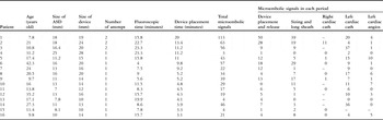
Among 21 consecutive patients who underwent successful transcatheter occlusion of atrial septal defect between February 28, 2008 and October 8, 2008, five patients were excluded because of signal noise or failure to detect reliable pulse waves. The remaining 16 patients were the subject of this study, and the patients’ demographics are presented in Table 1. The indication for transcatheter occlusion of atrial septal defect was a hemodynamically significant left-to-right shunt; no patient had had cryptogenic ischaemic stroke before the procedure. None of the patients had an atrial septal aneurysm. The study protocol was approved by the institutional review board and all patients gave written informed consent to participate in the study.
Patient profile and results of transcranial Doppler study.

Angios = angiocardiography; cath = catheterization; ASD = atrial septal defect
During the entire procedure, patients were placed supine under general anaesthesia and were continuously monitored with a 2.0 megahertz pulsed Doppler probe that was securely placed on the left temporal area and connected to a Pioneer TC-8080 (CareFusion, San Diego, California, United States of America). Sampling volume was set at a depth from 38 to 56 millimetres to detect pulse waves of the middle cerebral artery. Differentiation of microembolic signals from artefact was mainly made by automated embolic signal detection software (FS1) and occasionally by visual analysis of the waveform.
Catheterisation procedure
Aspirin (3 to 5 milligrams per kilogram) was started 2 days before transcatheter occlusion of atrial septal defect and continued for 6 months. After the introduction of sheaths, 100 Unit per kilogram of heparin was injected intravenously. Of the 16 patients, eight underwent diagnostic right and left cardiac catheterisation, as well as right upper pulmonary venography and left ventriculography, and seven underwent only right upper pulmonary venography before the sizing of the atrial septal defect. The remaining patient underwent transcatheter occlusion of atrial septal defect without any diagnostic catheterisation or angiography. In both right and left cardiac diagnostic catheterisation, we used a six French balloon catheter with side-hole (Angiographic Berman, Reading, Pennsylvania, United States of America) and left cardiac catheterisation was performed through the atrial septal defect.
Following the diagnostic catheterisation, a stiff guide wire was placed into the left pulmonary vein and a sizing balloon was advanced over the wire through the defect and was inflated to measure balloon-dilated defect size. The sizing balloon was withdrawn and an optimum-sized long sheath was placed into the left atrium over the guide wire, and the selected device was then placed through this long sheath. We checked activated clotting time of more than 200 seconds before device placement.
Procedure time was divided into five periods: right cardiac catheterisation; left cardiac catheterisation; left cardiac angiocardiography; sizing and long sheath placement; and device placement and release. In addition, total fluoroscopic time and device placement time, from mounting to releasing device, were recorded. We compared the number of microembolic signals among the five periods. In addition to identifying factors that correlated with increase in microembolic signals, we determined correlations between total number of microembolic signals and device placement time, fluoroscopic time, age, and device size.
Statistical analyses
Data were presented as median and range. Friedman's test was performed for multiple independent samples and Mann–Whitney's U-test was performed for non-parametric samples. We used Pearson's correlation coefficient to determine correlation between microembolic signals and individual variables. All data analyses were performed with a commercially available statistical analysis software package (Statview 5.0, SAS Institute Inc., Cary, North Carolina, United States of America and PASW 17.0, SPSS Inc., Chicago, Illinois, United States of America). A p-value of less than 0.05 was considered significant.
Results
In three patients, patients one to three, including two patients in whom we had to replace the device because the initial device was too small, we retrieved and deployed the device several times to conform and securely place the device. No patient developed any neurological signs within 72 hours after the procedure.
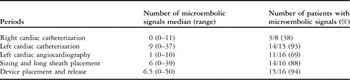
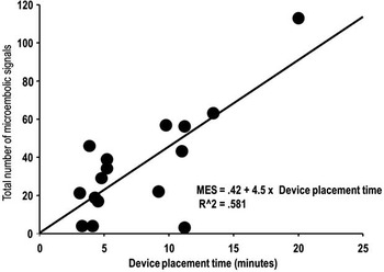
The median number of microembolic signals was 31.5, ranging from 3 to 113 during the entire procedure. Microembolic signals in three periods, left cardiac catheterisation; sizing and long sheath placement; and device placement and release, were not significantly different from one another but were significantly higher than those in the remaining two periods, right cardiac catheterisation and left cardiac angiocardiography (Table 2). This significant difference was preserved even when we looked at the eight patients who underwent all the diagnostic catheterisation as well as angiocardiography (left cardiac catheterisation, median of 6.5; sizing and long sheath placement, median of 7.5; device placement and release, median of 9.0; right cardiac catheterisation, median of 0; left cardiac angiography, median of 1, p less than 0.001, respectively). Beside left cardiac catheterisation, cumulative number of microembolic signals in the specific time, from sizing and long sheath placement to device placement and release, was a median of 11.5 and comprised mean of 58% of total number of microembolic signals. As expected, three patients, patients one to three, who required several attempts at device placement or replacement showed significantly more total microembolic signals (median microembolic signals was 63 (56 to 113) versus 22 (3 to 57)) and longer device placement time (median of 13.4 (11.4 to 20) versus 4.8 (3.1 to 11.2) minutes, p less than 0.01) than the remaining 13 patients. Importantly, device placement time significantly positively correlated with total number of microembolic signals (r equals 0.77, p less than 0.001; Fig 1), although fluoroscopic time, age, and size of atrial septal defect did not correlate with total number of microembolic signals. In addition, this positive correlation (r equals 0.84, p less than 0.0002) exists even when we excluded two patients who required device replacement.
Microembolic signals during procedures.

Total number of microembolic signals significantly positively correlated with time for device manipulation (r equals 0.77, p less than 0.001).

Discussion
This study indicates that longer time spent on device manipulation and placement can lead to more microembolic signals during transcatheter occlusion of atrial septal defect using the Amplatzer septal occluder.
In transcatheter occlusion of atrial septal defect using the Amplatzer septal occluder, microemboli occur mainly in the sequence of balloon sizing to device placement, although it occurs mainly during angiocardiography in diagnostic left catheterisation in adult coronary artery diseaseReference Leclercq, Kassnasrallah and Cesari4 or percutaneous transluminal coronary angioplasty.Reference Bladin, Bingham, Grigg, Yapanis, Gerraty and Davis5 This difference can probably be explained by the different frequency of contrast injection, because patients required only one or two angiographies in this study, but multiple coronary angiographies with different projection are required to delineate the stenosis of coronary arteries in adult coronary artery disease or coronary intervention. Although the number of microembolic signals during this procedure may range widely depending on the patient, indication of procedure, machine setting, or procedure protocol, our result was compatible with the finding that microembolic signals were observed mainly during the time specifically related to device manipulation and placement.Reference Ferrari, Baumgartner and Tentschert2, Reference Morandi, Anzola, Cailli and Onorato3
We found time spent on device manipulation to be the most significant factor associated with increase in these microembolic signals. In this study, device placement time significantly positively correlated with total microembolic signals, and patients who required multiple manipulations of the devices, including replacement, showed more microembolic signals. Although device replacement should not occur so frequently, device replacement cannot be avoided as long as an oversized device is not recommended.Reference Amin, Hijazi, Bass, Cheatham, Hellenbrand and Kleinman6 Furthermore, there was positive correlation between device placement time and total microembolic signals even when we excluded the two patients who required device replacement. Therefore, we must continue to choose device size carefully, to improve our techniques,Reference Wahab, Bairam, Cao and Hijazi7–Reference Dalvi, Pinto and Gupta10 and to plan device placement procedures to decrease the total number of attempts and shorten the procedure time. Further studies are required to clarify the incidence, mechanisms, and the relationship between microembolic signals and systemic microemboli in transcatheter occlusion of atrial septal defect.
Study limitations
As we had no multi-frequency transcranial Doppler equipment available, it was virtually impossible to more accurately distinguish gaseous microembolic signals from solid microembolic signals. However, most of the microembolic signals detected in this study showed bidirectional signals, and therefore were thought to be mainly gaseous microemboli rather than solid ones.
We found no clinically detectable consequences of cerebral microemboli during or after procedure. Clinically silent cerebral ischaemic events cannot be excluded because we could not offer sophisticated neuroradiological assessment such as diffusion-weighted magnetic resonance imaging.Reference Kloska, Wintermark, Engelhorn and Fiebach11
Conclusions
Microembolic signals observed during transcatheter occlusion of atrial septal defect using the Amplatzer septal occluder can be decreased by shortening device placement time.
Acknowledgement
We thank Dr Julien I. E. Hoffman, professor of paediatrics, University of California, San Francisco for his kind assistance in statistical analysis and editing of the manuscript.