The intricate three-dimensional arrangements of cardiac muscle fibres are important for an adequate cardiac function. Complex left ventricular deformation patterns lead to the simultaneous ejection of similar stroke volumes of the right and left ventricle despite different afterload. However, in healthy individuals, these deformation patterns are influenced by changes in loading conditions.Reference Weiner, Weyman and Khan 1
Interestingly, left ventricular deformation includes a rotational component. During systole, the normal cardiac base rotates clockwise, whereas the apex rotates counterclockwise.Reference Buchalter, Weiss and Rogers 2 , Reference Notomi, Setser and Shiota 3 This deformation is called “twist” or “torsion”. Whereas the term “twist” is often simply used to express “wringing”, torsion is defined as the base-to-apex gradient in rotation angle along the long-axis of the left ventricle.
Two-dimensional speckle-tracking echocardiography – 2D strain imaging – is a new, strain-derived and angle-independent method for the quantification of regional myocardial function.Reference Amundsen, Helle-Valle and Edvardsen 4 Several studies demonstrated that torsion can be measured with high accuracy by using two-dimensional speckle-tracking echocardiography.Reference Notomi, Lysyansky and Setser 5 – Reference Wang, Khoury, Yue, Torre-Amione and Nagueh 7
Hypertrophic cardiomyopathy causes ventricular hypertrophy in the absence of any other identifiable haemodynamic cause. In childhood, this phenotype is represented by a heterogeneous group of disorders leading to a high diversity in this patient group.Reference Braunwald, Lambrew, Rockoff, Ross and Morrow 8 – Reference Sachdev, Takenaka and Teraguchi 12
The aim of this study was to evaluate the impact of left ventricular hypertrophy on torsion by using speckle-tracking echocardiography in children with idiopathic hypertrophic cardiomyopathy in comparison to children with concentric hypertrophy of the left ventricle. Each group was compared with an age- and gender-matched control group of healthy individuals.
Methods
Study design
A single-centre, prospective, case–control study design was used. The study conforms to the principles of the Helsinki Declaration and German law. Informed consent was obtained from all subjects, and the institutional review board approved the study. All authors have read and agreed to the manuscript as written.
Patients
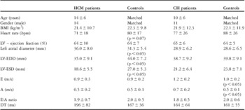
We enrolled 88 individuals in our study. A total of 24 echogenic children and young adults in sinus rhythm with idiopathic hypertrophic cardiomyopathy were compared with 24 healthy age- and gender-matched control subjects. The diagnosis of hypertrophic cardiomyopathy was based on typical clinical, electrocardiography, and echocardiographic findings with unexplained global or segmental ventricular myocardial hypertrophy occurring in the absence of any other cardiac or systemic disease that could have been responsible. In particular, significant aortic stenosis or systemic arterial hypertension was excluded, as well as storage disorders such as Fabry's disease or cardiac amyloidosis. Details are given in Table 1. Owing to the fact that the pattern of hypertrophy has been reported to affect left ventricular rotation and torsion,Reference van Dalen, Kauer and Soliman 13 patients with apical hypertrophy patterns were excluded. All patients received optimal and individualised medical therapy including β-blockers or calcium antagonists. However, no patient with left ventricular outflow tract obstruction was treated by myectomy or septal alcohol ablation therapy.
Baseline echocardiographic data of the study patients with HCM and CH, and of the gender- and age-matched controls (LV; LV-EDD; LV-ESD; DT; LVOT-G; SAM).

BMI = body mass index; CH = concentric hypertrophy; DT = deceleration time; HCM = hypertrophic cardiomyopathy; LV = left ventricle; LV-EDD = left ventricular end-diastolic diameter; LV-ESD = left ventricular end-systolic diameter; LVOT-G = left ventricular outflow tract gradient; SAM = systolic anterior movement
In order to find differences between rotational behaviour of localised – hypertrophic cardiomyopathy – and concentric hypertrophy, another patient group of 20 individuals was investigated. All of these patients had increased afterload because of aortic stenosis and/or coarctation before interventional treatment. They were compared with 20 healthy age- and gender-matched control subjects as described above. Details are given in Table 1.
Patients with disorders of metabolism, neuromuscular disease, or congenital malformation syndromes were excluded from this study. In healthy control children, cardiac disease had been ruled out by clinical examination and detailed echocardiographic study.
Echocardiography
In a university echo lab (Heart and Diabetes Center NRW, Bad Oeynhausen, Germany), we scanned all 88 individuals with a modern high-end scanner (Vivid 7, GE Vingmed Ultrasound, Horten, Norway; probe: M3S (GE Vingmed Ultrasound), 1.5–4.0 MHz). Echocardiographic examinations were performed in partial left decubitus. Harmonic imaging was used in these scanners by default.
For all left ventricular four-chamber, two-chamber, and long-axis apical views, as well as all parasternal views, image settings and frame rates (between 55 and 100 fps) were kept similar. The whole echocardiographic evaluation was performed following European Association of Echocardiography and American Society of Echocardiography recommendations.Reference Popescu, Andrade and Badano 14 For measurement of torsion, we performed parasternal short-axis views at the basal level (at mitral valve), papillary muscle level, and apical level – distal left ventricular cavity where papillary muscle was not visible.Reference Notomi, Setser and Shiota 3 In order to obtain a short-axis image at the left ventricular apical level, the transducer was positioned one or two intercostal spaces more caudal.Reference van Dalen, Vletter, Soliman, ten Cate and Geleijense 15
In hypertrophic cardiomyopathy patients, the magnitude of hypertrophy and its maximum expression was assessed by M-mode and two-dimensional transthoracic echocardiography using standard projections and off-axis views, if necessary. The presence and severity of left ventricular outflow tract obstruction and the occurrence of a systolic anterior movement of the mitral valve was recorded. Outflow gradients were assessed by continuous wave Doppler echocardiography at rest and during provocation, usually by a Valsalva manoeuvre.
Left ventricular ejection fraction was evaluated using standard software based on modified Simpson's rule in two- and four-chamber views.Reference Lang, Bierig and Devereux 16 Left ventricular mass was assessed using the two-dimensional area-length method.Reference Devereux, Alonso and Lutas 17 , Reference Wigle, Heimbecker and Gunton 18 All volumes and left ventricular mass were normalised to body surface area. Grey scale and colour Doppler recordings were digitally stored for further off-line assessment. Pulsed-wave Doppler-derived transmitral inflow velocities were obtained in the apical four-chamber view, with the sample volume placed at the mitral valve leaflet tips. Measurements included the transmitral early diastolic rapid filling (E-wave) and atrial contraction late filling (A-wave) velocities to calculate E/A ratio and deceleration time. For tissue Doppler imaging, the mitral annulus velocity was obtained with a 2-mm sample volume placed at the lateral side and septal side of the mitral annulus. Diastolic dysfunction was labelled according to the standard guidelines.
During off-line analysis, images were read from a regular computer display using the dedicated software (EchoPAc PC version BT08; GE Vingmed Ultrasound). A single echocardiographer reviewed off-line examinations of all subjects separately and in changing order, completely blinded to results of previous readings.
Speckle-tracking echocardiography or two-dimensional strain (ε) analysis
For speckle-tracking echocardiography, we used the dedicated software (EchoPAc PC version BT08; GE Vingmed Ultrasound), which provided an automated tracking score comparable to statistical standard deviation. The score ranged from 1.0 to 3.0 arbitrary units. All tracking scores with values below 2.5 were determined as acceptable for further analysis. The conversion to Lagrangian strain was performed off-line.
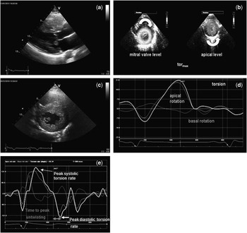
After manual tracing of the endocardial contour in a parasternal short-axis image on an end-systolic frame, the dedicated software automatically tracked the contour on the other frames of that cineloop and defined six myocardial segments for each view. After manual correction of the region of interest, adequate tracking was verified in real time. The program performed a least squares global affine transformation. The rotational component of this affine transformation was then used by the program to generate rotational profiles (Fig 1).
( a, c ) Parasternal long-axis view of massive left ventricular septal hypertrophy in HCM, systolic anterior movement of mitral valve. Short-axis view of massive left ventricular septal hypertrophy in HCM. ( b, d ) Basal clockwise and apical counterclockwise rotation during systole creating left ventricular torsion. Global rotational curves of the apex and bases; the white line represents the net difference = torsion. ( e ) Global rotation–rate curves of the apex and bases; the white line represents the net difference = torsion–rate curve (figure from Notomi et al).Reference Notomi, Srinath and Shiota 29 HCM = hypertrophic cardiomyopathy.

Statistics
Continuous data are expressed as mean value ±standard deviation. Statistical analysis was performed using the PASW™ software (SPSS Inc., Chicago, Illinois, United States of America). For continuous and normally distributed data, paired t-tests were used, and in case of non-normality of distribution Wilcoxon signed-rank tests were used. A two-tailed p-value < 0.05 was considered significant. Continuous measurements were compared by Pearson's correlation.
Results
Hypertrophic cardiomyopathy patient and control group
The mean age of the patients and the age-matched controls was 14.1 ± 5.5 years. We included 14 male patients in the hypertrophic cardiomyopathy patient group (58%) and the same number of boys in the gender-matched control group. The mean body surface area of patients and controls was comparable (1.5 ± 0.5 versus 1.5 ± 0.5 m2, p = 0.9). Out of 24 hypertrophic cardiomyopathy patients, 13 (54%) had a significant obstruction of the left ventricular outflow tract at rest (hypertrophic obstructive cardiomyopathy, peak gradient 67.3 ± 49.8 mmHg). E/E′ ratio of patients was 13.3 ± 5.9. Further baseline echocardiographic data of all patients and controls are given in Table 1.
Torsion/twist in hypertrophic cardiomyopathy patients and control group
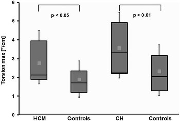
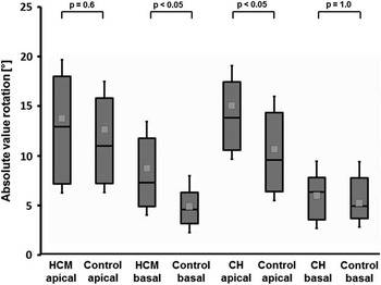
In hypertrophic cardiomyopathy patients, we found a significant increase in torsion (2.8 ± 1.6 versus 1.9 ± 1.0 °/cm in control group, p = 0.03) (Fig 2) with an increase in clockwise basal rotation (−8.7 ± 4.3° versus −4.9 ± 2.5° in control group, p < 0.001) (Fig 3) with prolonged time to peak diastolic untwisting (3.7 ± 2.4% versus 1.7 ± 0.6% of cardiac cycle in control group, p < 0.01). We found no significant differences between peak untwisting velocities in normal subjects and hypertrophic cardiomyopathy patients.
Difference between maximal torsion in HCM patients and healthy controls, and difference between maximal torsion in CH patients and matched controls. CH = concentric hypertrophy; HCM =hypertrophic cardiomyopathy.

Absolute values for rotation in HCM patients and healthy controls. Note the significant differences in basal rotation between HCM patients and healthy controls. Apical rotation values are comparable. Absolute values for rotation in CH patients and healthy controls. Note the significant differences in apical rotation between CH and controls. Basal rotation values are comparable. CH = concentric hypertrophy; HCM = hypertrophic cardiomyopathy.

Association between left ventricular outflow tract obstruction in hypertrophic cardiomyopathy and torsion
We found no significant differences for torsion in patients with versus without left ventricular outflow tract obstruction in our cohort. We found also no significant differences for peak untwisting velocities or time to peak diastolic untwisting.
Association between left ventricular muscle mass and torsion in hypertrophic cardiomyopathy
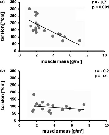
Patients with hypertrophic cardiomyopathy had a higher left ventricular muscle mass in comparison to the individuals of the control group (160.8 ± 63.9 versus 84.0 ± 19.3 g/m2, p < 0.001). We found a significant negative correlation between left ventricular muscle mass and torsion in hypertrophic cardiomyopathy (Fig 4) (r = −0.7, p < 0.001).
( a ) Negative correlation between left ventricular muscle mass in hypertrophic cardiomyopathy and torsion, ( b ) no significant correlation between left ventricular muscle mass in concentric hypertrophy and torsion.

Concentric hypertrophy patients and control group
We included seven patients with aortic stenosis and 13 patients with coarctation with a mean age of 9.9 ± 5.5 years (11 male), comparing their rotational data to gender- and age-matched controls as described above. Invasively measured mean gradient across the aortic valve was 75.6 ± 13.6 mmHg and across the coarctation 51.3 ± 22.0 mmHg.
Torsion/twist in concentric hypertrophy patients and control group
In the concentric hypertrophy group, we found an increase in torsion (3.7 ± 1.8 versus 2.2 ± 1.3°/cm in control group, p < 0.01). This increase was exclusively due to enhanced apical rotation (15.1 ± 6.3° versus 10.5 ± 5.5° in control group, p < 0.05), whereas basal rotation remained stable (−5.3 ± 4.1° versus −5.4 ± 2.7° in control group, p = 0.96).
Association between left ventricular muscle mass and torsion in concentric hypertrophy
Muscle mass was elevated in the concentric hypertrophy group (94.5 ± 31 versus 71.5 ± 17 g/m2 [controls], p < 0.05), and diastolic peak torsion rate was higher (−202 ± 88 versus −146 ± 67°/s [controls], p < 0.05) with delayed time to peak diastolic untwisting (1.8 ± 0.8% versus 1.2 ± 0.6% [controls], p < 0.05). There was no significant correlation between muscle mass and torsion in concentric hypertrophy (Fig 4).
Discussion
The contraction of a healthy ventricle is caused by different layers of myocardial fibres, which are arranged in a variety of directions resulting in distinct regional contraction patterns and an efficient pump mechanism. Subepicardial fibres run in a left-handed direction, mid-layer fibres run circumferentially, and fibres in the subendocardium run in a right-handed direction.Reference Hansen, Daughters, Alderman, Ingels and Miller 19 , Reference Gorman, Gupta and Streicher 20 Deformation of subepicardial oblique fibres results in basal clockwise and apical counterclockwise torque in systole. Vice versa, subendocardial fibres counteract with clockwise torque in the apex and counterclockwise torque in the base. Owing to the fact that the radius of rotation of the subepicardium is greater than that of the subendocardium, the subepicardium produces a greater torque than the subendocardium.Reference Nakatani 21 – Reference Dorri, Niederer, Lunkenheimer and Anderson 23
Owing to the heterogeneous patient groups during childhood that demonstrate left ventricular hypertrophy, we focused on children with idiopathic hypertrophic cardiomyopathy. The reported incidence for idiopathic hypertrophic cardiomyopathy in childhood is rare, with about 3.6 per 1 million children. In children with idiopathic hypertrophic cardiomyopathy who survive beyond age 1, the mortality rate is 1%/year and therefore comparable with that found in population-based studies in adults.Reference Colan, Lipshultz and Lowe 24
As early as 1962, Wigle et alReference Wigle, Heimbecker and Gunton 25 recognised that myocardial hypertrophy in hypertrophic cardiomyopathy could lead to a poor ventricular filling in diastole and disable a patient more than outflow tract obstruction in the systole. Almost all patients with hypertrophic cardiomyopathy demonstrate some degree of left ventricular diastolic dysfunction.Reference Nagueh, Lakkis, Middleton, Spencer, Zoghbi and Quinones 26 Well-established non-invasive measurements such as evaluation of mitral valve flow patterns by pulse wave Doppler or mitral annular velocity measurements by Doppler tissue imaging analyse later stages of diastole after mitral valve opening. However, the measurement of ventricular untwisting enables to evaluate early left ventricular filling during left ventricular diastolic suction by a non-invasive approach.Reference van Dalen, Kauer and Michels 27 , Reference Kim, Sohn and Lee 28
In children, torsion can be assessed easily because of adequate ultrasound windows. There is still little information about the amount of torsion in newborns and during early infancy. If torsion is normalised to left ventricular length, there are differences between small children with high torsion, which drops down during maturation with a secondary increase and gradual adaptation to values found in adulthood.Reference Notomi, Srinath and Shiota 29 Children with ventricular hypertrophy demonstrate a decrease of longitudinal and radial deformation. However, previous studies showed an elevation of torsion in those patients.Reference Laser, Haas and Jansen 30 – Reference Young, Kramer, Ferrari, Axel and Reichek 33
Major findings
The major findings of this study are as follows:
-
• Children with hypertrophic cardiomyopathy and patients with concentric hypertrophy had a significant higher maximal torsion than comparable healthy individuals, which is consistent with previous findings.
-
• Increase of torsion in hypertrophic cardiomyopathy was due to increased basal clockwise rotation in the localised hypertrophied regions, whereas in patients with concentric hypertrophy apical counterclockwise rotation was enhanced.
-
• Patients with hypertrophic cardiomyopathy and concentric hypertrophy had prolonged time to peak diastolic untwisting.
-
• A negative correlation between left ventricular mass and torsion was detected in hypertrophic cardiomyopathy. In patients with concentric hypertrophy, we found no correlation between torsion and muscle mass.
-
• Our observations in the hypertrophic cardiomyopathy group seem to be related to hypertrophic cardiomyopathy per se and not just to left ventricular outflow tract obstruction.
Systolic rotation, hypertrophic cardiomyopathy, and concentric hypertrophy
In children with hypertrophic cardiomyopathy, a decrease of longitudinal and radial deformation can be detected in early disease stages.Reference Ganame, Mertens and Eidem 32 Our results demonstrate, on the other hand, that circumferential deformation and rotation of hypertrophic segments is increased. This may be a compensatory mechanism in order to maintain cardiac performance. These results are comparable to previous findings.Reference Young, Kramer, Ferrari, Axel and Reichek 33 , Reference Saito, Okayama and Yoshii 34
We observed comparable results in our patients with concentric hypertrophy. It is known that a pressure overload might result in an impaired longitudinal and radial deformation.Reference Saltijeral, Perez de Isla and Veras 35 A concomitant decrease of subendocardial perfusion and remodelling processes may be responsible for a vector change in deformation resulting in enhanced torsion. The reason why highest torsion in concentric hypertrophy can be found in the apical region but is restricted to hypertrophic regions in hypertrophic cardiomyopathy remains unclear. Further research is warranted.
Association between left ventricular mass and torsion
To our knowledge, this is the first study that demonstrates a negative correlation between left ventricular mass and left ventricular torsion in hypertrophic cardiomyopathy. In patients with concentric hypertrophy, we found no comparable association. Probably this might be explained by different remodelling processes. In patients with concentric hypertrophy and elevated afterload, muscle fibre architecture is preserved. However, in hypertrophic cardiomyopathy, myocardial fibre disarray is a common finding. We speculate that hypertrophic cardiomyopathy develops from inner to the outer muscle layers. In combination with a reduction of chamber compliance and an increase of chamber stiffness due to an increased left ventricular mass and the development of myocardial fibrosis, diastolic dysfunction might occur.Reference Nagueh, Lakkis, Middleton, Spencer, Zoghbi and Quinones 26 This is also reflected by an impairment of left ventricular torsion during early diastole.
Untwisting, hypertrophic cardiomyopathy, and concentric hypertrophy
Notomi et alReference Notomi, Martin-Miklovic and Oryszak 36 demonstrated that the peak untwisting velocities in hypertrophic cardiomyopathy patients were similar to those of normal subjects. Our results support these findings. Surprisingly, we found enhanced diastolic untwisting rates in the concentric hypertrophy group, which might support the theory of a compensatory mechanism in order to increase left ventricular filling in the situation of an increased end-diastolic pressure. Our population showed a prolonged time to peak diastolic untwisting, which resembles the findings of van Dalen et al.Reference van Dalen, Kauer and Michels 27 Probably, subendocardial ischaemia in hypertrophic cardiomyopathy might be the crucial link between delayed untwisting and pathophysiology of early diastolic dysfunction.Reference Cecchi, Olivotto, Gistri, Lorenzoni, Chiriatti and Camici 37 – Reference Soliman, Knaapen and Geleijnse 39
Limitations
Owing to the fact that this was a single-centre study involving a relatively small number of patients, the results should be interpreted with caution. We did not have any invasive pressure measurements and did not perform changes of preload or afterload. Left ventricular mass was calculated using an estimated formula, which may lead to some inaccuracy of the data, particularly because of well-known heterogeneous hypertrophy patterns in hypertrophic cardiomyopathy. Therefore, patients with apical hypertrophy were excluded. Further, we included only patients with good echocardiographic image quality, which may affect the implementation of this technique in clinical practice. The fact that values of the different control groups differ from each other might be due to the age differences of the groups. There was a considerable difference in the left ventricular mass between the two groups. A potential influence of this difference on the results might be possible. Data about rotational parameters during paediatric growth are still scarce.
Clinical perspective
In hypertrophic cardiomyopathy patients with massive hypertrophy and high-normal ejection fraction, future efforts should be undertaken to promote a diastolic elastic rebound. Whether intramural implantable recoil devices – as suggested by PasipoularidesReference Pasipoularides 40 – are a possible approach must be evaluated in the future. Owing to the negative correlation between muscle mass and torsion, this parameter could be an interesting marker for the detection of early diastolic abnormalities in patients with hypertrophic cardiomyopathy, for example in order to detect a disease progression when torsion begins to decrease. The influence of medical, interventional, and surgical therapy on torsion should be the focus of further research.
Conclusions
Rotational behaviour in children with hypertrophic cardiomyopathy is characterised by predominantly enhanced systolic basal clockwise rotation and increased rotation rates resulting in increased torsion. Diastolic untwisting is delayed. Owing to the negative correlation with muscle mass, torsion could be an interesting marker to guide patients with hypertrophic cardiomyopathy.
Acknowledgements
Funding: This research was supported by an unrestricted grant from the German Heart Foundation (to Dr Prinz).