Introduction
Agouti paca (Linnaeus, 1766), commonly called paca, is a Neotropical hystricomorphic rodent found in a variety of habitats ranging from Mexico to Argentina (Costa et al., Reference Costa, Leal, Ferreira, Guimarães and França2010). This rodent is intensively hunted by people for sustenance because its meat is considered to be a good protein source (Pachaly & Werner, Reference Pachaly and Werner1998). In addition, illegal commerce of this game meat is on the rise in the Brazilian Amazon (Baía Júnior et al., Reference Baía Júnior, Guimarães and Pendu2011).
Wild rodents live in the natural ecosystem and often acquire infra-population infections of different parasites over their lifetime (Behnke, Reference Behnke2008). Indeed, parasitic co-infections are well known in the veterinary literature (Telfer et al., Reference Telfer, Birtles, Bennett, Lambin, Paterson and Begon2008); however, the helminth fauna of A. paca (Linnaeus, 1766) has seldom been studied (Ramírez-Herrera et al., Reference Ramírez-Herrera, Rodríguez-Vivas, Montes-Pérez and Torres-Acosta2001). Previous studies have reported on A. paca liver infections caused by the Echinococcus vogeli Rausch & Bernstein, 1972 metacestode in South American countries (Morales et al., Reference Morales, Guzman, Wells and Angel1979; D'Alessandro et al., Reference D'Alessandro, Rausch, Morales, Collet and Angel1981; Rausch et al., Reference Rausch, D'Alessandro and Rausch1981; Gardner et al., Reference Gardner, Rausch and Camacho1988; Meneghelli et al., Reference Meneghelli, Martinelli and Velludo1990).
Echinococcus vogeli is a parasitic tapeworm with an indirectly transmitted life cycle involving the obligatory alternation of generations between sexual reproduction within a carnivorous host and asexual reproduction within a herbivorous host. The bush dog (Speothos venaticus Lund, 1842) and the domestic dog (Canis familiaris Linnaeus, 1758) may become infected by eating an A. paca liver infected with metacestodes, whereas herbivorous hosts may become infected by eating infectious eggs released in the faeces of carnivores. Humans may also become infected through accidentally ingesting eggs that are present in contaminated soil (D'Alessandro & Rausch, Reference D'Alessandro and Rausch2008), water and vegetables (Siqueira et al., Reference Siqueira, Almeida, Chalub, Machado-Silva and Rodrigues-Silva2007).
Capillariosis is a zoonotic helminthosis of worldwide veterinary medical concern (McCarthy & Moore, Reference McCarthy and Moore2000). Ecological studies have shown that the aetiological agent Calodium hepaticum (syn. Capillaria hepatica) parasitizes the liver of numerous mammals throughout the world, especially rodents (Farhang-Azad, Reference Farhang-Azad1977; Matamoros et al., Reference Matamoros, Velázquez and Pashov1991; Reperant & Deplazes, Reference Reperant and Deplazes2005; Fuehrer et al., Reference Fuehrer, Igel and Auer2011).
The biology of C. hepaticum is unusual in that females deposit unembryonated eggs in the parenchyma of the host's liver, which is subsequently eaten by a predator (Fuehrer et al., Reference Fuehrer, Igel and Auer2011) and dispersed in the environment. Previously reported human cases suggest that humans may become infected after ingesting the eggs from contaminated soil (Fuehrer et al., Reference Fuehrer, Igel and Auer2011). After hatching in the small intestine, juveniles migrate to and mature in the liver. In addition, humans may become infected after the ingestion of wild rodent meat (Carvalho-Costa et al., Reference Carvalho-Costa, Silva, Souza, Moreira, Souza, Valverde, Jaeger, Martins, Meneses, Araújo and Bóia2009; Camargo et al., Reference Camargo, Camargo, Vera, Barreto, Tourinho and Souza2010). The presence of adult worms in the liver leads to a loss of liver cells. Large areas of parenchyma may be replaced by a cluster of eggs, which may lead to the formation of a granuloma and the development of focal necrosis (Gomes et al., Reference Gomes, Cunha, Bastos, Medrado, Assis and Andrade2006).
In Brazil, documented hosts of Capillaria spp. include synanthropic rats (Chieffi et al., Reference Chieffi, Dias, Mangini, Grispino and Pacheco1981; Galvão, Reference Galvão1981), opossum (Quintão e Silva & Costa, Reference Quintão e Silva and Costa1999), capybaras (Costa & Catto, Reference Costa and Catto1994) and wild canids (Ruas et al., Reference Ruas, Muller, Farias, Gallina, Lucas, Pappen, Sinkoc and Brum2008). To the best of our knowledge, there is currently no record of C. hepaticum infections among the paca in Brazil. This paper discusses the characteristics of a concurrent E. vogeli and C. hepaticum infection found in A. paca; these paca were captured in an endemic area where polycystic echinococcosis transmission occurs. In addition, A. paca is a novel host for C. hepaticum in Brazil, thereby expanding the known range of C. hepaticum hosts in South America.
Materials and methods
As part of an ongoing research project concerning the epidemiology of the zoonotic cestode E. vogeli, five free-ranging A. paca were captured in a permanent preservation reserve of Bujari (09°49′50″S, 67°57′08″W) located in the state of Acre (Brazil). The rodents were transported to the laboratory, anaesthetized (ketamine hydrochloride and xylazine, 10:1) and euthanized. The liver of each animal was examined macroscopically for parasitic lesions.
For the histopathological examinations, the liver tissues were fixed in 10% buffered formalin and processed according to the standard histological techniques for paraffin embedding. Tissue sections of 5 μm thickness were prepared and used for conventional haematoxylin–eosin staining.
A digital camera (Nikon Eclipse E200) connected to a microscope (Olympus BX41 microscope, Tokyo, Japan) was used to document images of the hooks of E. vogeli and the eggs of C. hepaticum. The camera output was processed and analysed with Image-Pro Plus 3 image analysis software (Media Cybernetics, Silver Spring, Maryland, USA). According to previous methods by Neves et al. (Reference Neves, Pereira, de Oliveira, Gomes and Machado-Silva1998), two morphometric variables were recorded: length (hooks and eggs) and width (eggs).
The animal protocols and surgical procedures in this study were approved by the Brazilian Institute of Environment and Renewable Natural Resources (IBAMA, no. 16 909) and the local Animal Care Committee (IPEC/FIOCRUZ, no. 0002.1.000.009-04).
Results
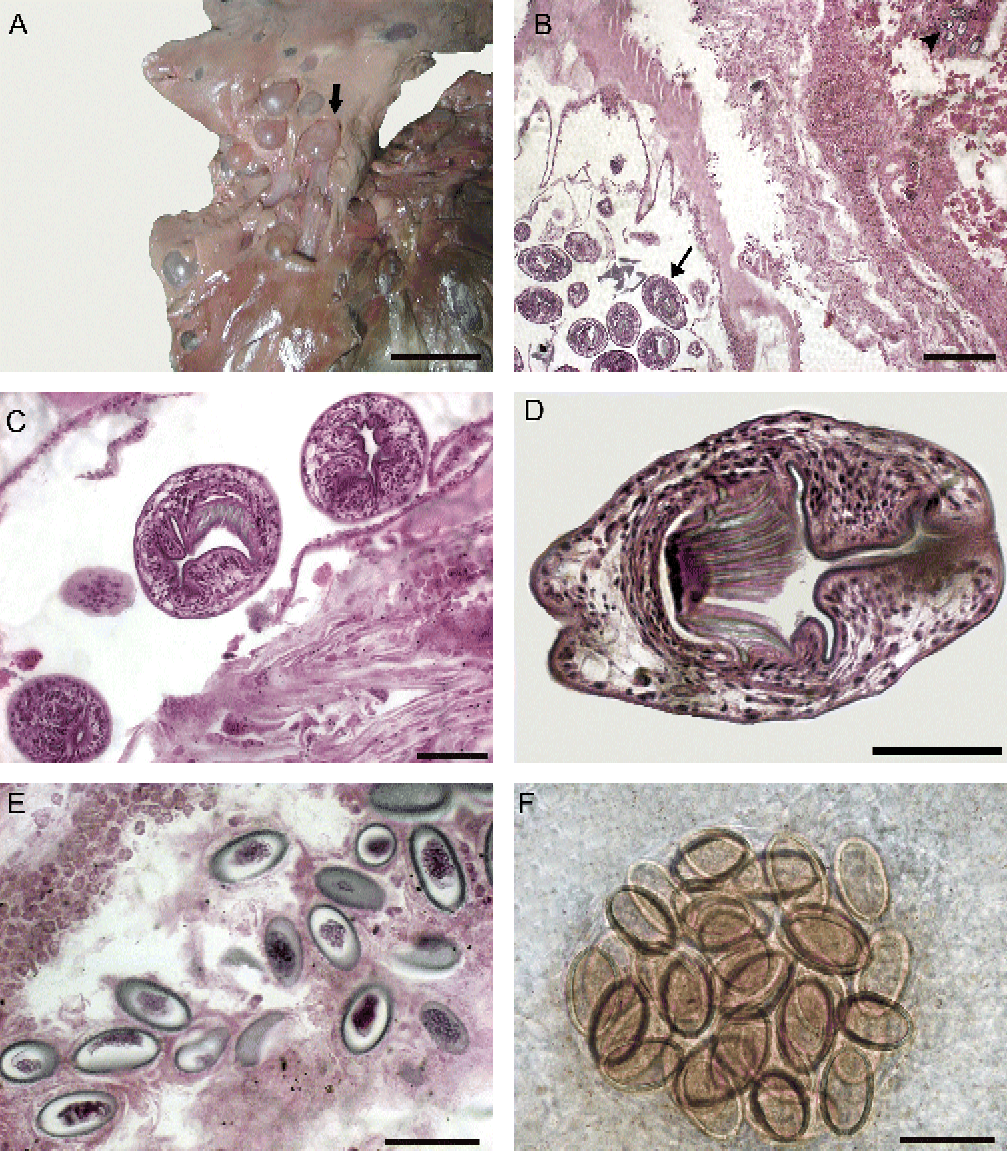
In two of the examined paca livers no gross morphological alterations were observed, whereas the three remaining livers (60%) contained multiple spherical to subspherical white or translucent lesions that were either isolated or frequently contiguous and partially covered by Glisson's capsule (fig. 1A). Hepatic histopathological examination revealed that these lesions were unilocular cystic structures that contained abundant brood capsules in which numerous protoscolices budded from the inner surface (fig. 1B). The pathological changes observed were characterized by fibrosis and a scanty inflammatory cell infiltrate of lymphocytes and macrophages around the host-derived outer adventitial layer (fig. 1B). Brood capsules containing protoscolices (fig. 1C), which had visible rostellar hooklets and calcareous corpuscles (fig. 1D), were present on the inner surface of the lesions. Echinococcus vogeli identification was based on the length of the rostellar hooks (33–41 μm) and the morphological characteristics of the blade, which was curved and longer than the handle (two-thirds of the total length). Among this infected group of paca, one rodent also had ellipsoid-shaped bipolar eggs with striated eggshells, suggestive of C. hepaticum; the eggs were located around the outer zone of fibrous tissue, with a peripheral infiltration that primarily consisted of eosinophils (fig. 1B). At higher magnification levels, wet-mounts revealed the presence of eggs in the liver parenchyma, either within a syncytium (fig. 1E) or in clusters (fig. 1F). The morphometry of the eggs (n= 30) yielded an average measurement of 44.8 ± 1.9 μm (length) × 24.4 ± 2.0 μm (width), which is consistent with C. hepaticum. No adult C. hepaticum worms were found.
(colour online) Liver with mixed infections of metacestodes of Echinococcus vogeli and eggs of Calodium hepaticum. (A) Oval lesions on the liver surface (arrowed) with a stratified cuticular membrane, scale bar = 1 cm; (B) developing protoscolices of E. vogeli (arrowed) with elliptical-shaped eggs of C. hepaticum (arrowhead), scale bar = 0.2 mm; (C) inner surface (germinal membrane) with brood capsules of E. vogeli containing two protoscoleces and rostellar hooks, scale bar = 0.05 mm; (D) prominent rostellar hooks of protoscolex of E. vogeli, scale bar = 0.03 mm; (E) liver section with eggs of C. hepaticum causing necrosis of the parenchyma, scale bar = 0.05 mm; (F) viable eggs from a wet mount of C. hepaticum, scale bar = 0.05 mm.

Discussion
We show here that free-ranging A. paca from a permanent preservation reserve in Brazil had polycystic echinococcosis and capillariosis of the liver. This finding raises several important questions: (1) How does paca become infected in the study area? (2) Are echinococcosis and capillariosis antagonists of one another because they share the same organ? (3) Is game meat such as paca the source of infection in capillariosis?
With regard to the first question, there are several factors that may account for the high prevalence observed. Carnivore definitive hosts are responsible for the contamination of the environment with E. vogeli eggs. Because pacas are free-ranging in the preservation reserve, they may inhabit environments coincident with bush dogs, which suggests a potential sylvatic route of transmission of E. vogeli. On the other hand, an urban cycle may be established when hunting dogs are fed with infected tissue of pacas (Eckert & Deplazes, Reference Eckert and Deplazes2004). Despite the fact that bush dogs have been implicated as definitive hosts of E. vogeli (D'Alessandro & Rausch, Reference D'Alessandro and Rausch2008) and domestic dogs may play a role in the domestic route (Eckert & Deplazes, Reference Eckert and Deplazes2004), there have been no previous parasitological, immunological or molecular studies for the detection of E. vogeli eggs in canids from Bujari.
The second question may be answered using the evidence in this study showing that the liver pathology was similar to that of separate cases of E. vogeli (Morales et al., Reference Morales, Guzman, Wells and Angel1979; Rausch et al., Reference Rausch, D'Alessandro and Rausch1981; Gardner et al., Reference Gardner, Rausch and Camacho1988; Meneghelli et al., Reference Meneghelli, Martinelli and Velludo1990; D'Alessandro & Rausch, Reference D'Alessandro and Rausch2008) and C. hepaticum (Gomes et al., Reference Gomes, Cunha, Bastos, Medrado, Assis and Andrade2006; Jeong et al., Reference Jeong, Do, Hong, Ji, Park, Ki, Park and Jeong2008; Mowat et al., Reference Mowat, Turton, Stewart, Lui and Pilling2009). Thus, our results suggest that there is no interplay during mixed infections.
The last question is very interesting because humans and other hosts may accidentally ingest the embryonated eggs of C. hepaticum in faecal matter that is disseminated throughout the environment (Farhang-Azad, Reference Farhang-Azad1977). Moreover, C. hepaticum eggs have been observed in faecal samples that were collected from Brazilian Indians (Coimbra-Junior & Mello, Reference Coimbra-Junior and Mello1981) and from a farmer inhabiting the northern Amazon (Carvalho-Costa et al., Reference Carvalho-Costa, Silva, Souza, Moreira, Souza, Valverde, Jaeger, Martins, Meneses, Araújo and Bóia2009). It has been hypothesized that, in the Brazilian Amazon, the ingestion of paca giblets containing unembryonated eggs could disseminate C. hepaticum eggs into the stool (Camargo et al., Reference Camargo, Camargo, Vera, Barreto, Tourinho and Souza2010). Because the eggs do not experience any biological changes when passing through the digestive tract, this phenomenon has been classified as pseudoparasitism (Carvalho-Costa et al., Reference Carvalho-Costa, Silva, Souza, Moreira, Souza, Valverde, Jaeger, Martins, Meneses, Araújo and Bóia2009) or spurious infection (Fuehrer et al., Reference Fuehrer, Igel and Auer2011). In the present study, we confirmed this classification based on our histopathological findings, morphological and morphometric data, which were consistent with previous studies of C. hepaticum (Fugassa et al., Reference Fugassa, Taglioretti, Gonçalves, Araújo, Sardella and Denegri2008). In summary, our study expands the known range of C. hepaticum hosts in South America, and, to the best of our knowledge, this is the first study to describe a mixed infection of E. vogeli and C. hepaticum. Furthermore, we confirmed that ingesting paca meat may be an alternative route of infection by C. hepaticum.
Acknowledgements
The authors would like to thank Rodrigo Méxas from the Laboratório de Produção e Processamento de Imagem Científica, IOC–FIOCRUZ for preparing the images. Financial support was received from Conselho Nacional de Desenvolvimento Científico e Tecnológico (CNPq) and Fundação Carlos Chagas Filho de Amparo á Pesquisa do Estado do Rio de Janeiro (FAPERJ).