INTRODUCTION
Human malaria is caused by species of Plasmodium, protozoan parasites transmitted by Anopheles mosquitoes. The infection induces immunity to both asexual and sexual stages of the parasite (Good and Miller, Reference Good and Miller1989; Targett et al. Reference Targett, Harte, Eida, Rogers and Ong1990; Hommel, Reference Hommel1991) and it is established that human antibodies against sexual parasite stages can reduce the infectivity of the parasite to mosquitoes, alleviating transmission pressure and hence the disease burden at the population level (Carter and Mendis, Reference Carter and Mendis1992; Shahabuddin and Costero, Reference Shahabuddin and Costero2001). When a mosquito takes a bloodmeal on an infectious host, parasite sexual stages undergo a complex developmental cycle, initially confined to the midgut (Sinden, Reference Sinden1983), and free-living parasite stages are therefore exposed to immune attack by antibody and cell-dependent effectors either ingested along with the mosquito bloodmeal (Carter et al. Reference Carter, Kumar, Quakyi, Good, Mendis, Graves and Miller1988; Lensen et al. Reference Lensen, Mulder, Tchuinkam, Willemsen, Vaidya and Sauerwein1998) or released in response to invasion (Richman et al. Reference Richman, Dimopoulos, Seeley and Kafatos1997; Sinden, Reference Sinden2002; Tahar et al. Reference Tahar, Boudin, Thiery and Bourgouin2002). Detailed analysis of the contribution of vertebrate host-related or vector-borne factors acting upon the development of malaria parasites within mosquitoes and characterization of the sexual stage(s) sensitive to each determinant is relevant to any strategy aiming at immune-mediated inhibition of transmission.
Immune-dependent inhibition of parasite infectivity by host blood factors involves a variety of sexual-stage antigens of the parasite as targets (Vermeulen et al. Reference Vermeulen, Ponnudurai, Beckers, Verhave, Smits and Meuwissen1985; Carter et al. Reference Carter, Kumar, Quakyi, Good, Mendis, Graves and Miller1988; Naotunne et al. Reference Naotunne, Karunaweera, Mendis and Carter1993; Kaslow et al. Reference Kaslow, Nussenzweig and Miller1994 ), most of which are still only poorly characterized (Kaslow et al. Reference Kaslow, Nussenzweig and Miller1994). Nonetheless, 2 major surface antigens on P. falciparum gametocytes, namely Pfs48/45 and Pfs230, are synthesized early during gametocytogenesis and expressed as a complex on gametes within the mosquito midgut (Kumar, Reference Kumar1987; Kumar and Carter, Reference Kumar and Carter1984; Rener et al. Reference Rener, Graves, Carter, Williams and Burkot1983). Antibodies to these surface proteins have been observed in humans exposed to endemic malaria (Graves et al. Reference Graves, Carter, Burkot, Quakyi and Kumar1988; Premawansa et al. Reference Premawansa, Mendis, Perera, Begarnie, Mendis and Carter1994; Roeffen et al. Reference Roeffen, Lensen, Mulder, Teelen, Sauerwein, Van Druten, Eling, Meuwissen and Beckers1995a, Reference Roeffen, Beckers, Teelen, Sauerwein, Meuwissen and Elingb) and can be induced experimentally by immunization with specific gamete antigens (Carter and Gwadz, Reference Carter, Gwadz and Kreier1980; Kaslow et al. Reference Kaslow, Isaacs, Quakyi, Gwadz, Moss and Keister1991; Carter et al. Reference Carter, Mendis, Miller, Molineaux and Saul2000). Monoclonal antibodies (MoAb) against Pfs230 and Pfs48/45 have been shown to reduce or completely block infectivity of Plasmodium to mosquitoes (Rener et al. Reference Rener, Carter, Rosenberg and Miller1980; Roeffen et al. Reference Roeffen, Beckers, Teelen, Sauerwein, Meuwissen and Eling1995b). These surface proteins provide a basis for the development of transmission-blocking vaccine (Carter et al. Reference Carter, Mendis, Miller, Molineaux and Saul2000). The mechanism whereby Plasmodium stages that are confined to the lumen of the midgut succumb to the immunity elicited by Pfs230 and Pfs48/45 has been under intense scrutiny. It is established that antibodies to sexual stage antigens are key mediators of Plasmodium killing within mosquitoes (Carter and Mendis, Reference Carter and Mendis1992) and whilst there is evidence as to where and when during the sporogonic development this suppression may occur (Shahabuddin and Costero, Reference Shahabuddin and Costero2001; Sinden et al. Reference Sinden, Butcher, Billker and Fleck1996; Gouagna et al. Reference Gouagna, Bonnet, Gounoue, Verhave, Eling, Sauerwein and Boudin2004) it is unclear as to which immune effectors are the most crucial. Contrary to the effects of anti-Pfs48/45 antibodies that appear to be based on complement-independent mechanisms, naturally acquired antibodies to Pfs230 are fundamentally of complement-fixing IgG subclasses (IgG1 and/or IgG2 antibodies) (Read et al. Reference Read, Lensen, Begarnie, Haley, Raza and Carter1994; Roeffen et al. Reference Roeffen, Beckers, Teelen, Sauerwein, Meuwissen and Eling1995b). Proof that one of the other complement pathways may effectively mediate suppression of Plasmodium within the mosquito has been achieved in experimental laboratory models, namely P. gallinaceum (Grotendorst et al. Reference Grotendorst, Carter, Rosenberg and Koontz1986; Grotendorst and Carter, Reference Grotendorst and Carter1987), P. yoelii (Tsuboi et al. Reference Tsuboi, Cao, Torii, Hitsumoto and Kanbara1995) and P. berghei (Margos et al. Reference Margos, Navarette, Butcher, Davies, Willer, Sinden and Lachmann2001). Also, the effects of the host's complement system on extracellular parasite stages have been demonstrated in several insect-transmitted haemoflagellates species namely Trypanosoma (Nogueira et al. Reference Nogueira, Bianco and Cohn1975), Leishmania (Pearson and Steigbigel, Reference Pearson and Steigbigel1980). Similar experiments on the effect of host complement on human Plasmodium (e.g. P. falciparum) transmission have rarely been reported previously (Healer et al. Reference Healer, McGuinness, Carter and Riley1999). In laboratory experiments, however, more effective complement-dependent suppression of infectivity of malaria parasites to mosquitoes has been found in several examples involving monoclonal antibodies against gametes (Kaushal et al. Reference Kaushal, Carter, Rener, Grotendorst, Miller and Howard1983; Rener et al. Reference Rener, Graves, Carter, Williams and Burkot1983; Roeffen et al. Reference Roeffen, Beckers, Teelen, Sauerwein, Meuwissen and Eling1995b; Healer et al. Reference Healer, McGuinness, Hopcroft, Haley, Carter and Riley1997). Thus, the level of parasite response to serum complement may be one of the key determinants for sporogonic development.
Quantification of the effect of host complement and/or its interaction with other factors involved in the passage of malaria parasites from man to mosquitoes is not only relevant to the understanding of natural factors affecting malaria transmission but also important for the development of effective malaria transmission-blocking vaccines. Here we used field sera from naturally infected gametocyte carriers to investigate the role of complement, alone or in association with naturally acquired transmission-reducing immunity, on parasite transmission. The contribution of specific antibodies against Pfs230, the relevant target of complement-mediated transmission-reducing mechanisms (Roeffen et al. Reference Roeffen, Beckers, Teelen, Verhave, Eling and Sauerwein1993; Healer et al. Reference Healer, McGuinness, Carter and Riley1999), was also assessed. Since a negative effect of endemic sera on P. falciparum malaria transmission to the vector has been reported in previous studies (Mulder et al. Reference Mulder, Tchuinkam, Dechering, Verhave, Carnevale, Meuwissen and Robert1994; Roeffen et al. Reference Roeffen, Lensen, Mulder, Teelen, Sauerwein, Van Druten, Eling, Meuwissen and Beckers1995a, Reference Roeffen, Beckers, Teelen, Sauerwein, Meuwissen and Elingb; Lensen et al. Reference Lensen, Van Druten, Bolmer, Van Gemert, Eling and Sauerwein1996; Drakeley et al. Reference Drakeley, Mulder, Tchuinkam, Gupta, Sauerwein and Targett1998; Mulder et al. Reference Mulder, Lensen, Tchuinkam, Roeffen, Verhave, Boudin and Sauerwein1999), we specifically looked at the association between repeated stage-specific parasite killing during early P. falciparum development and the overall reductions of oocyst prevalence and infection intensity within An. stephensi mosquitoes.
MATERIALS AND METHODS
Human sera
Field sera were collected during malaria transmission studies reported previously (Gouagna et al. Reference Gouagna, Bonnet, Gounoue, Verhave, Eling, Sauerwein and Boudin2004). Briefly, Plasmodium falciparum gametocyte carriers were recruited from cross-sectional parasitological surveys among resident villagers of Mengang (100 km from Yaoundé). Malaria is highly endemic in this area with seasonal transmission (Meunier et al. Reference Meunier, Safeukui, Fontenille and Boudin1999; Bonnet et al. Reference Bonnet, Paul, Gouagna, Safeukui, Meunier, Gounoue and Boudin2002). Thick smears were stained for 15 min with 10% Giemsa and examined with a light microscope (100× oil immersion lens). Volunteer gametocyte carriers (5–10 years old) and their parents were informed about the purpose of the study and asked to cooperate upon oral consent. The Cameroonian scientific and ethical review committees approved all recruitment and experimental procedures reported here. Each selected gametocyte-positive patient provided 6 ml of venous blood that was collected into a sterile glass tube without anticoagulant. These blood samples were allowed to clot for 20 min at room temperature and centrifuged at 2000 g for 10 min in a centrifuge set at 37°C. Sera were collected and frozen at −20°C for subsequent bioassay experiments, which took place at the Catholic University of Nijmegen, The Netherlands. A single pool of non-immune sera obtained from Dutch blood-bank donors (AB type) that were tested negative in the standard membrane feeding assay was used throughout for all infection experiments, both as the source of complement and for control assays (Van der Kolk et al. Reference Van der Kolk, deVlas and Sauerwein2006).
Serological assays for specific antibodies against Pfs230
In order to assess serological correlates of complement-dependent TRA, all field sera were analysed by capture ELISA for the presence of antibodies against P. falciparum sexual-stage antigens, mainly the Pfs230 which is the relevant target of complement-dependent transmission-reducing mechanisms. In brief, antibodies specific to Pfs230 were assayed in ‘Capture ELISA’ by coating 10 μg/ml of anti-Pfs230 mouse mAb 63F2A2 in phosphate buffered saline (PBS, pH 7·4) on Sterilin ELISA plates (PS, ref 53011, International Medical Products B.V., Zutphen, The Netherlands). Plates were blocked with 5% low-fat dry milk (Marvel, Premier International Foods Ltd, Spalding, Lines, UK) in PBS. For Pfs230 antigen capture, plates were incubated with gametocyte extract (250 000 parasite equivalents/well). Sera (1:100 dilution) were added to the wells and incubated for 2 h. Bound IgG antibodies were detected by horse-radish-peroxidase (HRPO)-labelled goat anti-human IgG (Pierce 31 412). Wells were washed 3 times with PBS and subsequently incubated with tetramethyl benzidine (TMB) substrate solution for 20 min. The colour reaction was stopped with 4 m H2SO4, and optical density (OD) read at 450 nm (Anthos 2001 microplate reader: Labtec BV). All incubations were carried out at room temperature. All serum samples were tested in duplicate with a concurrent positive control and a minimum of 3 negative (Dutch blood-bank donor) controls per plate. Sera were considered positive if the OD exceeded 3 standard deviations above the mean of negative controls.
Blood preparation for membrane feeds
We used the human malaria, P. falciparum, NF54 laboratory isolate because of its highly infectious characteristics (Ponnudurai et al. Reference Ponnudurai, Meuwissen, Leeuwemberg, Verhave and Lensen1982). All parasite batches had undergone fewer than 10 serial passages in the automated ‘tipper’ system prior to use in experiments described below. After 14 days in culture, gametocytaemia (proportion of red blood cells infected with sexual forms of parasites), and the red blood cell (RBC) density were measured prior to bloodmeal preparation. The exact number of gametocytes was estimated for the time immediately preceding the membrane feed. Consequently, gametocytes were counted per 1·25×103 RBCs using a coulter counter (Luton, UK). Gametocytaemia was estimated from the product of RBC densities and parasites per RBC. The male/female gametocyte ratio in infectious blood of the different experimental trials was approximately 1/3 (i.e. the number of female gametocytes higher than the number of males).
The culture material containing about 0·3 ml of gametocyte-positive packed cells (1%) was spun at 500 g for 2 min. The supernatant was carefully removed and the pellet containing parasitized red blood cells (RBC) was mixed with 0·75 ml of freshly washed and pre-warmed uninfected RBC from an ‘AB’ group Dutch donor (Lensen et al. Reference Lensen, Van Druten, Bolmer, Van Gemert, Eling and Sauerwein1996). A volume of 150 μl of the parasitized RBC suspension was quickly added to each field-collected serum (OWN) in the presence and absence of active human complement. All gametocyte carriers' OWN and control (AB) sera were initially heat inactivated at 56°C for 30 min, in order to disable the complement activity. A volume of 90 μl of each inactivated field and inactivated control serum sample was added to 30 μl of freshly prepared AB donor serum that was not heat treated, in order to restore complement activity. In parallel, samples of 120 μl complement-depleted samples of OWN serum and AB serum were conditioned at 37°C. In the second step, each serum preparation (with and without complement) was added to 150 μl of freshly washed human erythrocytes (50% final haematocrit) in vials prior to the feed.
The physiological activity of complement in these AB pool sera was tested in a series of preliminary experiments, which showed that bloodmeals mixed with mouse 63F2A2.2a MoAb (an IgG2a switch variant of the IgG1 isotype against Pfs230) (Roeffen et al. Reference Roeffen, Beckers, Teelen, Sauerwein, Meuwissen and Eling1995b) and active complement achieved TRA over 90% in An. stephensi. These preliminary experiments (unpublished data) were important, because technical bias due to inadequate blood preparation could theoretically alter the infection outcome. Like in previous experiments reported by Roeffen et al. (Reference Roeffen, Beckers, Teelen, Sauerwein, Meuwissen and Eling1995b) this pre-test provided evidence that the use of 30 μl of untreated normal AB donor serum is adequate to ensure a physiological-sufficient level of complement in the bloodmeal.
Mosquitoes and experimental infections
The membrane feeding assay used in this study has the advantage for experimental investigation of clear-cut TRA in which parasite responses can be monitored in standardized and controlled conditions using the highly infectious NF54 gametocyte strain. Female An. stephensi, 2 to 3 days old, were drawn from the same colony cohort and used in these experiments. Although An. stephensi is not the natural vector for P. falciparum, routine experiments with cultured NF54 isolates showed this species to become infected reliably using the membrane feeding assays (Ponnudurai et al. Reference Ponnudurai, Meuwissen, Leeuwemberg, Verhave and Lensen1982; Roeffen et al. Reference Roeffen, Lensen, Mulder, Teelen, Sauerwein, Van Druten, Eling, Meuwissen and Beckers1995a, Reference Roeffen, Beckers, Teelen, Sauerwein, Meuwissen and Elingb; Lensen et al. Reference Lensen, Van Druten, Bolmer, Van Gemert, Eling and Sauerwein1996; Mulder et al. Reference Mulder, Lensen, Tchuinkam, Roeffen, Verhave, Boudin and Sauerwein1999; Van der Kolk et al. Reference Van der Kolk, De Vlas, Van de Vegte-Bolmer, Eling, Saul and Sauerwein2005). During each trial, 4 batches of 100 mosquitoes each were randomly constituted and starved 5–6 h prior to the feed. Using previously described feeding procedures (Ponnudurai et al. Reference Ponnudurai, Lensen, Van Gemert, Bensink, Bolmer and Meuwissen1989), they were infected via mini-feeders containing mature gametocytes from cultured parasite lines re-suspended in either individual field sera under test (OWN) or control sera (AB). These pair-matched feeds were performed simultaneously, each with blood mixtures containing either a complement-rich serum or complement-deficient (heat-inactivated) serum. Mosquitoes were allowed to engorge each blood preparation for 15 min through a Parafilm® membrane, warmed to 37°C with a miniglass water jacket. After feeding, all unfed mosquitoes were removed and the remaining were kept under laboratory conditions (25–28°C, 80% RH) with daily access to 10% sucrose solution.
Mosquito dissection and detection of early parasite stages and oocysts
Contrary to previously reported transmission studies that assessed only the overall level of reducing activities at the oocyst stage (Roeffen et al. Reference Roeffen, Lensen, Mulder, Teelen, Sauerwein, Van Druten, Eling, Meuwissen and Beckers1995a, Reference Roeffen, Beckers, Teelen, Sauerwein, Meuwissen and Elingb; Lensen et al. Reference Lensen, Van Druten, Bolmer, Van Gemert, Eling and Sauerwein1996; Mulder et al. Reference Mulder, Lensen, Tchuinkam, Roeffen, Verhave, Boudin and Sauerwein1999; Van der Kolk et al. Reference Van der Kolk, De Vlas, Van de Vegte-Bolmer, Eling, Saul and Sauerwein2005, Reference Van der Kolk, deVlas and Sauerwein2006), here, we quantified the inhibiting effect of serum and complement component on the various sporogonic stages: gametes, zygotes, ookinetes and oocysts. Therefore, stage-specific parasite loads within individual mosquitoes that had been fed bloodmeals with control sera and field serum, with and without a functional host complement were assessed at 30 min, 3 and 24 h, post-infection by immuno-fluorescence staining of individual midgut homogenates (Gouagna et al. Reference Gouagna, Mulder, Noubissi, Tchuinkam, Verhave and Boudin1998, Reference Gouagna, Bonnet, Gounoue, Verhave, Eling, Sauerwein and Boudin2004). Schematically, pools of 5 mosquitoes each were dissected in phosphate-buffered saline (PBS, pH7·2) at 30 min post-feeding and their midguts were crushed and homogenized. The homogenates were incubated with fluorescein-isothiocyanate (FITC)-conjugated anti-Pfs48/45 monoclonal antibody (MoAb), in 0·05% Evans blue for 30 min at room temperature. Following incubation, preparations were washed with PBS (pH 7·2) and mounted under calibrated Bürker-Türk counting chambers covered with 24×24 mm no. 1 cover-slips and examined with an incident fluorescence light microscope (50×, immersion lens) to detect gametes.
For other pre-oocyst stages, samples of 5 mosquitoes per group were dissected at 3 h post-feeding (for zygotes and macrogametes both known as round forms) and 20–24 h post-feeding (for ookinetes). Instead of using MoAb against Pfs48/45 which is already shed on zygotes and ookinetes, midguts were homogenized and stained with FITC-labelled MoAb against Pfs25, the major antigen present on pre-oocyst stages of the malaria parasite. Midgut preparations were processed as described above. In order to assess overall TRA, the remainder of the mosquitoes (>30 mosquitoes per batch) that took ‘OWN’ or control bloodmeal were kept in an insectary (approximately 26°C and >80% relative humidity) for 7 days and subsequently dissected to determine the presence of oocysts (stained with mercurochrome 2%) on midgut. Both the presence and absence of oocysts were recorded on each occasion.
Estimation of transmission-reducing activity (TRA)
The overall transmission level was expressed as the proportion of infected mosquitoes (prevalence) and the mean oocyst number per mosquito (intensity). TRA of each field serum was obtained by expressing the level of oocyst infection in the OWN group as the percentage of that in the AB group from each paired experimental feed. Various indicators have been proposed previously to measure the magnitude of immune-dependent regulation of malaria transmission: (i) the standardized difference between the mean oocysts in control and experimental feeds (Van der Kolk et al. Reference Van der Kolk, De Vlas, Van de Vegte-Bolmer, Eling, Saul and Sauerwein2005) and (ii) the ratio of the oocyst densities in the test group relative to its pair-matched control (Lensen et al. Reference Lensen, Van Druten, Bolmer, Van Gemert, Eling and Sauerwein1996), or (iii) the comparison of prevalences of infected mosquitoes (Mulder et al. Reference Mulder, Tchuinkam, Dechering, Verhave, Carnevale, Meuwissen and Robert1994) between test serum and non-immune serum. However, it is not known which of the indicators proposed is most suited to describe the full picture of transmission-reducing patterns under field conditions. Based on previous reports (Roeffen et al. Reference Roeffen, Lensen, Mulder, Teelen, Sauerwein, Van Druten, Eling, Meuwissen and Beckers1995a, Reference Roeffen, Beckers, Teelen, Sauerwein, Meuwissen and Elingb; Mulder et al. Reference Mulder, Lensen, Tchuinkam, Roeffen, Verhave, Boudin and Sauerwein1999), the commonly used ‘R-value’ was chosen as the privileged indicator of serum-dependent regulation of transmission. The following formula was used: R/100=((Tc−c), where Tc is the geometric mean of oocysts number observed in the control group with non-immune sera and Ti is the geometric mean oocyst number observed in mosquitoes that fed on each test serum ‘i’. TRA of test sera was divided into 3 categories according to R-values: Blocking (R⩾90%), Reducing (30%⩾R<90%), and Non-blocking (R<30%) (Lensen et al. Reference Lensen, Van Druten, Bolmer, Van Gemert, Eling and Sauerwein1996).
The total variation in the level of infectivity of each parasite lot can be divided into mortality components involving all the major steps in sporogonic development. Therefore, data for macrogametocytes, macrogametes, round forms (Robert et al. Reference Robert, LeGoff, Essong, Tchuinkam, Fass and Verhave1995), ookinetes and oocysts were computed in the form of a life table, thus permitting definition of where the blockade or the reduction occurred. Interstage parasite losses were estimated at each transition by the coefficient of mortality (k-values) as described previously (Vaughan et al. Reference Vaughan, Noden and Beier1992; Gouagna et al. Reference Gouagna, Mulder, Noubissi, Tchuinkam, Verhave and Boudin1998). In brief, the conversion rate of gametocytes into gametes was the percentage of gametocytes present in the bloodmeal that escaped from the host cell and subsequently expressed the Pfs48/45 protein on their surface (see the detection method). The conversion rate of gametes into round forms (Robert et al. Reference Robert, LeGoff, Essong, Tchuinkam, Fass and Verhave1995) was the percentage of gametes that underwent exflagellation and fertilization, detected with anti-Pfs25 monoclonal antibody at 3 h post-feeding. The conversion rate of gametes/zygotes into ookinetes was the percentage of female gametes that developed within 20–24 h into ookinetes of the total of parasites (gamete and zygote) counts recorded at 3 h post-feeding.
Statistical analyses
All statistical procedures were performed using SPSS 11.0 for Windows (SPSS, Inc., Chicago, IL). Separate analyses were done both at group level (centring the interest on the group effect represented in the data) by combining data from feeding series in order to detect the source of variability in infection outcomes and on individual infection experiments to account for differences in TRA between gametocyte carriers' sera.
At group level, infection outcomes (infection rate, oocyst intensity and mean proportional parasite losses) obtained in both OWN and AB were compared with respect to complement. Therefore, the Pearson's chi square test was applied to test the association between the percentages of infection generating feeding experiments within each group. Our data consisted of 4 response variables: infection rate, parasite intensity, interstage parasite losses and categories of TRA. Each one was analysed separately using General Linear Models (GLM), which examine the contribution of each effect. Individual infection assay (coded as 1–24), infection group (OWN and AB), complement treatment (active or heat-depleted) were added as explanatory variables. Interactions between all explanatory variables were also fitted, where significant (P<0·05). Prior to analyses, mosquito infection rates were square-root arcsine-transformed while stage-specific parasite counts per infected mosquito were log10 transformed (number of parasites+1) to bring their distributions close to normal.
At individual level, the TRA of each serum was expressed as a percentage of that in the AB group from each paired experimental feed (see above, Lensen et al. Reference Lensen, Van Druten, Bolmer, Van Gemert, Eling and Sauerwein1996), and the GLM was used to test the effect of complement activity and Pf230 antibody level (with OD values for specific antibodies as continuous variable) on TRA. In addition to the main effect, interaction effects between all explanatory variables were also fitted. Pearson correlations were used (1) to determine whether parasite mortality rates obtained from the ‘within-group analyses’ were consistent across all infection experiments and (2) to correlate the presence of specific antibodies to the TRA of the sera that were tested.
RESULTS
Complement-dependent TRA of 24 serum samples from naturally infected gametocyte carriers (OWN) were evaluated using cultured NF54 P. falciparum gametocytes and laboratory-reared Anopheles stephensi. Infection trials were performed 2 or more days apart and on the day of each experiment, 2 separate serum samples were tested simultaneously, with and without complement and a single control (non-immune serum, AB group) was therefore considered for both serum samples. Thus, a total of 48 (24 field sera×2 complement treatments) and 24 (12 control serum samples×2 complement treatments) experimental feeds were effected. In addition to assessing the effect of complement on (1) the proportion of infected mosquitoes, (2) oocyst intensity and (3) stage-specific parasite losses, we also assessed the contribution of specific antibody level in each serum on the relative TRA.
Comparison of the complement effect in OWN and AB infections experiments
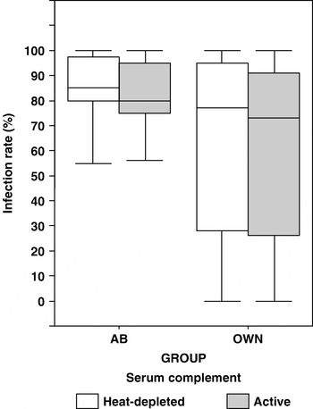
Figure 1 depicts the influence of serum-complement treatment combinations on the proportion of mosquitoes becoming infected within the infection-generating experiments. The means and medians of infection rate and oocyst intensity were not equal, suggesting a non-symmetrical distribution of these outcome variables. Therefore, only medians of infection outcomes (and interquartile ranges, IQR) are reported throughout. The median of infection rate from OWN bloodmeal was significantly lower than from AB blood, both in active complement [73 (73·5) % versus 82 (20) %)] (F1, 35=4·44, P=0·04) and complement-deficient group [77 (68) % versus 85 (20) %] (P=0·04). Although the proportion of infected mosquitoes recorded in the OWN group with active complement tended to be lower than that in the complement-deficient group, the difference was not statistically significant at the 0·05 level. (F1, 46=1·68, P=0·56). Similarly, no significant effect of complement on the proportion of mosquitoes becoming infected after ingestion of infectious bloodmeal with control sera was found (F1, 22=0·02, P=0·84), suggesting that transmission of P. falciparum to mosquitoes was not sensitive to complement alone.
Effect of host serum factors on NF54 Plasmodium falciparum infection rate in Anopheles stephensi. Data are presented separately for pairs of infection experiments in which mosquitoes were allowed to feed on a gametocyte-infected bloodmeal mixed either with individual serum samples from ‘immune’ gametocyte carriers (OWN) or control sera from naïve blood donors (AB type), assayed both in the presence of active complement and after complement activity has been heat-depleted. Boxplot displays for each of the groups the median (horizontal bar), the interquartile range (box) and 5th to 95th percentile range (error bar).

The number of oocysts per mosquito was 4 times lower in OWN [Median (IQR)=4·1 (21·2) oocysts per mosquito] than in AB group [17·6 (23·9) oocysts per mosquito] (F1, 35=4·7, P=0·03) when mosquitoes were provided with a bloodmeal with active complement. The same trend in oocyst intensities was observed between OWN and AB when sera were heated [4·6 (16·8) oocysts per mosquito in OWN versus 22·2 (32·7) oocysts per mosquito in AB group] (F1, 33=6·6, P=0·01). These results also indicate that the average numbers of oocysts per infected mosquito was not affected when complement activity was restored, both in OWN (F1, 46=0·10, P=0·74) and AB group (F1, 23=0·02, P=0·87).
Transmission-reducing activity (TRA) of immune sera against P. falciparum in mosquitoes
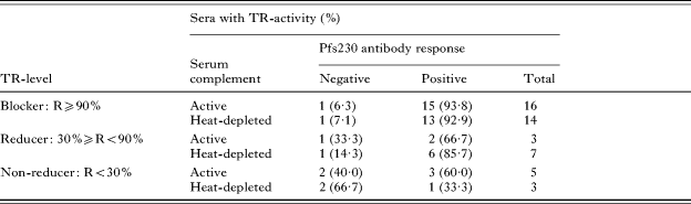
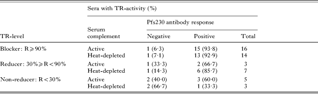
Table 1 shows the percentages of anti-Pfs230 positive ELISA titres and the distribution of the number of reducers and non-reducers with respect to complement. The relative TRA of each serum sample was expressed as a percentage reduction of oocyst numbers in the OWN group relative to that of the control with which it was performed. Regardless of the anti-Pfs230 reactivity (see last column in Table 1), the numbers of sera that significantly suppressed oocysts formation (with R⩾90%) and those that exhibited no significant suppression of transmission (0<R⩽30) were relatively greater in the presence of active complement than in the heat-treated group. TRA was highly variable among the various sera tested (F22, 24=13·18, P<0·001). Overall, the percentage of sera that significantly suppressed infectivity in the presence of active complement was not significantly higher than that in the heat-treated group (Pearson Chi-Square: P=0·35). Two serum samples that reduced parasite infectivity in the absence of active complement completely prevented oocyst formation in mosquitoes when complement activity was restored. In contrast, 4 other sera that significantly reduced transmission in the absence of complement became paradoxically less potent in generating the same level of activity in the presence of active complement.
Effects of host complement and Pfs230 antibody response on TRA against Plasmodium falciparum in Anopheles stephensi
(An infectious bloodmeal with cultured P. falciparum gametocytes (NF54 isolate) was provided to An. stephensi either in the presence of gametocyte carriers serum sample (OWN) or control sera from malaria naïve Dutch blood donors (AB group), both in the presence of active complement or heat-depleted (56°C) of complement activity. Antibodies specific to Pfs230 were assayed in ‘capture ELISA’. TRA was estimated as a ratio of the oocyst densities in the test group relative to its pair-matched control (Lensen et al. Reference Lensen, Van Druten, Bolmer, Van Gemert, Eling and Sauerwein1996).)
Relationships between Pfs230 specific antibodies and complement-dependent TRA of individual serum samples
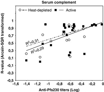
Serological data were analysed with reference to the predictive ability to induce TRA in the membrane-feeding assay. For instance, we tested whether the observed sero-reactivity to Pfs230 correlated with a reduction in transmission capacity, as defined by the R-value (Table 1). Of the sera that were positive for Pfs230 antibody in ‘Capture ELISA’ (83·3%), 2 (10%) elicited transmission reduction and 15 (75%) completely suppressed transmission in the presence of complement. Two sera which were negative for Pfs230 did, however, mediate significant TRA in the presence of complement. Overall, the magnitude of complement-dependent TRA was not significantly greater in sera that showed sero-reactivity to Pfs230 than those that did not (χ2=3·8, d.f.=2, P=0·32). Nevertheless, there was a significant linear relationship between Pfs230 antibody titres [Median OD (IQR)=0·23 (0·21)] and TRA, both in the presence (Pearson's correlation coefficient=0·41, P=0·02) and absence of active complement (Pearson's correlation coefficient=0·45, P=0·01) (Fig. 2). Restricting the analysis to the sera exhibiting significant TRA (R⩾90% and 30%⩾R<90%, n=19), the odd ratio for the relationship between anti-Pfs230 antibody titres and the percentages of infection reduction in mosquitoes that fed heat-treated bloodmeal [OR=1·29, 95% CI (0·95 – 2,42), P=0·05] increased substantially after the activity of serum complement was restored ([OR=1·4, 95% CI (1·02 – 2,44), P=0·006]). This result suggests that there could be some synergistic effect of serum complement on the ability of anti-Pfs230 antibodies to reduce transmission. Despite the observed relationships, the F statistic further indicated that the influence of complement activity alone (F1, 36=1·32, P=0·73) or combined with anti-Pfs230 antibody response (P=0·38) did not account for the variability in the TRA.
The influence of serum complement on the relationships between Pfs230 antibody titres and TRA against Plasmodium falciparum in Anopheles stephensi. Data for 24 pairs of infection experiments in which batches of An. stephensi mosquitoes were given a NF54 gametocyte-positive bloodmeal prepared either (1) with serum samples from naturally infected P. falciparum gametocyte carriers (OWN) or (2) with non-immune sera from malaria naïve blood donors (AB type), both in the presence of active complement and after complement activity has been heat-depleted. All sera were tested using ‘capture ELISA’ and positive antibody titres were calculated as the serum dilution given an optical density greater than 3 standard deviations above the mean of negative controls. In the GLM that included both complement treatment and antibody response to Pfs230 as explanatory variables, only the antibody titre to Pfs230, but not complement treatment, was significantly related to TRA, here expressed as a percentage reduction of oocyst numbers in the OWN group relative to that of the control with which it was performed (Lensen et al. Reference Lensen, Van Druten, Bolmer, Van Gemert, Eling and Sauerwein1996).

Mechanisms responsible for the differential reduction in P. falciparum transmission by individual serum samples
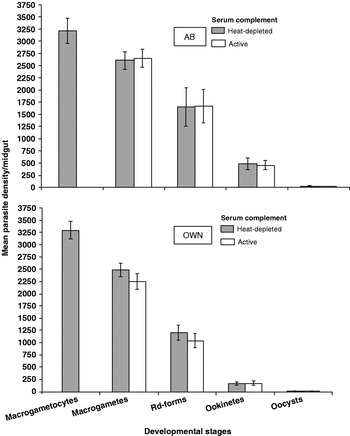
The mechanistic basis of serum-mediated or complement-dependent transmission reduction was investigated by the estimate of the percentage reduction of macrogametes, gametes/zygotes and ookinetes within mosquitoes at 30 min, 3 h and 24 h post-feeding, respectively. As shown in Fig. 3, parasite numbers declined from one stage to the next in all serum and complement treatment groups. There was no significant difference in mean numbers of macrogametes gamete/zygotes and ookinetes in mosquitoes that fed on bloodmeal with AB control sera and active complement and those from the matched-group with depleted complement activity (Fig. 3). Mosquitoes that were fed bloodmeals with OWN sera and active complement tended to exhibit lower mean numbers of macrogametes, round-forms and ookinetes than those fed bloodmeals with heat-depleted complement (Fig. 3). Throughout the early sporogonic development, however, no meaningful complement-dependent reduction of parasite numbers was evident.
Effect of serum complement on early Plasmodium falciparum sporogonic development within Anopheles stephensi fed on bloodmeals supplemented with immune and non-immune sera. Laboratory-reared An. stephensi were given infectious bloodmeals prepared either with sera from naïve Dutch donor (AB type) or pair-matched field serum samples (OWN), both with and without active complement. The number of macrogametocytes in the feed was similar for both groups. At different times following the infectious bloodmeals, mean parasite densities were estimated at each early developmental stage including macrogamete, macrogamete/zygote or round form (Rd-form), Ookinete and Oocyst. Average parasite density and standard error of the mean (error bars) from 24 independent infection experiments are shown. In both the OWN and AB groups, there was no statistically significant difference between the distribution patterns of early sporogonic parasite stages in mosquitoes fed with bloodmeals with or without active complement.

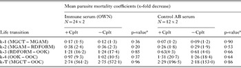
From those declining parasite numbers, we derived stage-specific mortality rates at successive developmental transitions and subsequently related the magnitude of parasite losses to complement treatments (Table 2). In both the OWN and AB group, substantial parasite losses were observed across different parasite developmental stages within mosquitoes. Consistently in all infection trials, the interstage parasite mortality rates were significantly higher in OWN than in AB, both when mosquitoes had fed on a bloodmeal with active and inactivated complement. In most cases except for 5 immune serum samples (data not shown), heat-treated field serum samples suppressed as effectively as the matched samples with active complement (Table 2). The bulk of serum-related TRA occurred mainly at 3 developmental transitions: from macrogametocytes to macrogametes (F1, 46=3·8, P=0·05), from gamete/zygotes to ookinetes (F1, 46=36·5, P<0·01) and from ookinetes to oocysts (F1, 46=12·2, P=0·001). Despite the lack of significant differences in parasite losses among infected mosquitoes that received a bloodmeal with active complement and inactivated complement, there were significant differences in cumulative parasite mortality (K-t between macrogamete and oocyst stage) associated with serum sources: 2·7 (569-fold decrease) in OWN with complement compared to 2·3 (195-fold decrease) in the AB plus active complement group (F1, 46=4·8, P=0·03). Restricting the statistical analyses to the sera exhibiting significant TRA (n=19), with total (R⩾90%) and partial reducers (30%⩾R<90%) assigned a value of 1, and 0 for those without significant TRA (R<30%), the blockade in response to field sera occurred mainly at the gamete and ookinete stages (data not shown). In the OWN group, there was a consistent correlation between accumulated parasite mortality (K-t) both with infection rate (Pearson's correlation coefficient=−0·85, P<0·01) and oocyst intensity (P coefficient=−0·98, P<0·01). In concordance with the previous analyses, there was still no significant effect of serum complement on the patterns of these relationships.
Effect of complement and host serum factors on early Plasmodium falciparum development in Anopheles stephensi infections by standardized membrane-feeding assay
(An infectious bloodmeal with cultured P. falciparum gametocytes (NF54 isolate) was provided to An. stephensi either in the presence of gametocyte carriers serum sample (OWN) or control sera from malaria naïve Dutch blood donors (AB group), both in the presence of active complement (+Cplt) or after heat (56°C) inactivation of complement activity (−Cplt). Mean parasite density was estimated at each early developmental stage including macrogametocytes (MGCT), macrogamete (MGAM), gamete/zytgote or round forms (RDFORM), Ookinete (OOK) and Oocysts (OOC). k-I, mortality coefficient indicating interstage parasite losses between two consecutive developmental stages. k-T, accumulated mortality coefficient describing the overall losses between macrogametocytes and oocysts.)

a The F test was used to compare mean mortality coefficient at each transition between infection groups with and without complement, the mean difference being significant at the 0·05 level.
DISCUSSION
The present study was undertaken to determine the role of host complement in immune-mediated TRA of NF54 Plasmodium falciparum in laboratory-infected Anopheles stephensi. Although the NF54 parasite strain was used in infection experiments (Ponnudurai et al. Reference Ponnudurai, Meuwissen, Leeuwemberg, Verhave and Lensen1982, Reference Ponnudurai, Lensen, Van Gemert, Bensink, Bolmer and Meuwissen1989), the sera tested came from 24 naturally infected P. falciparum gametocyte carriers, for whom natural immunity was expected to be stimulated by the presence of gametocytes. In view of this, it was expected that the majority of serum samples would reveal significant TRA, which possibly would be enhanced by the presence of active complement in the mosquito bloodmeal. The evidence gathered from infection experiments performed separately indicated that providing mosquitoes with bloodmeals incorporating immune sera resulted in a substantial decline of effective parasite numbers at each developmental stage in the mosquito midgut, resulting in low oocyst production. Both the proportion of infected mosquitoes and the oocyst intensity were reduced by factors present in the majority of immune sera tested, of which the Pfs230 antibody was suspected to play an important role, but in a manner uninfluenced by the presence of the heat-labile principle or serum complement. Indeed, it is established that natural transmission-reducing immunity rests mainly on the action of specific antibodies against antigens of the pre-oocyst stages (Targett et al. Reference Targett, Harte, Eida, Rogers and Ong1990; Carter and Mendis, Reference Carter and Mendis1992). Contrary to previous reports (Premawansa et al. Reference Premawansa, Mendis, Perera, Begarnie, Mendis and Carter1994; Mulder et al. Reference Mulder, Lensen, Tchuinkam, Roeffen, Verhave, Boudin and Sauerwein1999), a correlation was found between antibody titres to Pfs230 target and TRA. It was expected that the presence of active complement in the bloodmeals would increase the rate and magnitude of TRA imposed by individual immune serum. This expectation was supported by previous laboratory studies (Tsuboi et al. Reference Tsuboi, Cao, Torii, Hitsumoto and Kanbara1995; Grotendorst et al. Reference Grotendorst, Carter, Rosenberg and Koontz1986; Margos et al. Reference Margos, Navarette, Butcher, Davies, Willer, Sinden and Lachmann2001), but surprisingly our data indicated that the mechanism(s) involved in the inhibition of the sporogony is not the early complement-dependent lysis of macrogametes as previously observed in in vitro lysis experiments (Grotendorst and Carter, Reference Grotendorst and Carter1987; Healer et al. Reference Healer, McGuinness, Hopcroft, Haley, Carter and Riley1997; Williamson, Reference Williamson2003). Since adequate preparation of the bloodmeal fed to mosquitoes in this study ensured that the level of complement reached the level normally found in serum (Roeffen et al. Reference Roeffen, Beckers, Teelen, Sauerwein, Meuwissen and Eling1995b), it is not known why host complement, despite being present in the bloodmeal, only poorly contributed to TRA of our serum sample against cultured P. falciparum within An. stephensi.
Host serum has multiple components that may interfere with parasite development within the mosquito. These include white blood cells, anti-sexual antibodies, cytokines and complement. The complement system which forms one of the main effector mechanisms of antibody-mediated immunity, involves the action of 23–30 plasmatic proteins (Umotong et al. Reference Umotong, Amanor-Boadu, Okerengwo and Hedo1994; Walport, Reference Walport2001). Serum complement has often been suspected to be involved in gamete lysis or phagocytosis in vitro and in vivo (Grotendorst and Carter, Reference Grotendorst and Carter1987: Roeffen et al. Reference Roeffen, Beckers, Teelen, Sauerwein, Meuwissen and Eling1995b; Healer et al. Reference Healer, McGuinness, Hopcroft, Haley, Carter and Riley1997), thereby affecting malaria transmission from vertebrate hosts to mosquitoes but the expression of proteins by malaria parasites that inhibit complement may also be implicated (Grotendorst et al. Reference Grotendorst, Carter, Rosenberg and Koontz1986). Another possibility is that immune serum probably lacks components that can effectively initiate complement activation within the vector.
It is assumed that functions of naturally acquired anti-Pfs230 antibody may require the activation of cofactors such as serum complement to lyse the early gamete stage, decreasing the numbers of oocysts. Indeed, Quakyi et al. (Reference Quakyi, Carter, Rener, Kumar, Good and Miller1987) and Read et al. (Reference Read, Lensen, Begarnie, Haley, Raza and Carter1994) have shown that only the isotypes of Pfs230 that fixed complement can efficiently lyse gametes and block transmission in vitro. Roeffen et al. (Reference Roeffen, Beckers, Teelen, Sauerwein, Meuwissen and Eling1995b) further demonstrated that specific-Pfs230 MoAbs of isotype IgG2b and IgG2a, but not IgG1 isotype, are effective in reducing P. falciparum transmission only in the presence of complement. This means that the role of complement in Pfs230-based transmission reduction may be accomplished by an isotype-dependent effector mechanism (Healer et al. Reference Healer, McGuinness, Hopcroft, Haley, Carter and Riley1997). Our results suggested a slight trend of decreasing parasite infectivity due to serum complement which, however, was not statistically significant. The low complement-dependent inhibition of parasite infectivity observed in this study cannot be explained in the light of weak immune activity against Pfs230 in the sera tested. In fact, gametocyte carriers from whom our serum samples were collected live in an area where malaria is highly endemic (Bonnet et al. Reference Bonnet, Paul, Gouagna, Safeukui, Meunier, Gounoue and Boudin2002). Further, the frequency of anti-Pfs230 antibodies is higher in gametocyte-positive individuals than in their counterparts without gametocytes (Roeffen et al. Reference Roeffen, Beckers, Teelen, Sauerwein, Meuwissen and Eling1995b). In the present study the prevalence of anti-Pfs230 antibodies in a single-epitope capture ELISA was high indeed, and there was significant association between the antibody level within serum samples that significantly reduced parasite transmission to mosquitoes, irrespective of complement activity. Possibly, the absence of the appropriate complement-fixing anti-Pfs230 isotypes (Quakyi et al. Reference Quakyi, Carter, Rener, Kumar, Good and Miller1987; Read et al. Reference Read, Lensen, Begarnie, Haley, Raza and Carter1994; Roeffen et al. Reference Roeffen, Beckers, Teelen, Sauerwein, Meuwissen and Eling1995b), might account for the absence of an effect of complement in our serum samples. Previous studies with Cameroonian sera (Mulder et al. Reference Mulder, Lensen, Tchuinkam, Roeffen, Verhave, Boudin and Sauerwein1999) and in Sri Lanka residents (Premawansa et al. Reference Premawansa, Mendis, Perera, Begarnie, Mendis and Carter1994) showed no meaningful association between TR-activities and specific antibody response to Pfs230 antigens. Nevertheless, surveys elsewhere revealed evidence of serum positivity for anti-sexual stage antibody to Pfs230 antigen that results in significant levels of inhibition of parasite infectivity to mosquitoes (Graves et al. Reference Graves, Carter, Burkot, Quakyi and Kumar1988; Healer et al. Reference Healer, McGuinness, Hopcroft, Haley, Carter and Riley1997; Williamson, Reference Williamson2003). Extending our interest to all possible factors that could affect parasite infectivity, the TRA represented in our data would have arisen due to several physiological and immunological factors that our study design did not allow us to look at. Of the host-related immune factors, previous serological studies in samples of Cameroonian gametocyte carriers have indicated that the prevalence of an antibody-specific response against Pfs48/45 was higher in gametocyte carriers' sera (60%) (Roeffen et al. Reference Roeffen, Lensen, Mulder, Teelen, Sauerwein, Van Druten, Eling, Meuwissen and Beckers1995a). We suspect the suppressive activity of specific antibodies to Pfs48/45 on the early sporogonic development to outweigh the lytic effect of complement alone or complement-dependent agglutination of gametes by anti-Pfs230 IgG subclasses.
In all mosquitoes exposed to immune sera with active complement, the numbers of viable macrogametes, gametes/zygotes, ookinetes and oocysts were comparable to that in mosquitoes exposed to complement-depleted bloodmeal. Our findings on P. falciparum are consistent with previously reported studies with P. berghei (Ranawaka et al. Reference Ranawaka, Alejo-Blanco and Sinden1993; Grotendorst and Carter, Reference Grotendorst and Carter1987). These authors have shown that complement components are present and active within mosquito midgut, at least within 2–3 h following bloodmeal ingestion. However, emerging P. berghei gametes and early zygotes may show resistance against complement lytic activity during this period, after which parasites became increasingly sensitive to serum complement. Our analysis did not allow us to verify whether the reduction of ookinetes we observed in the group of mosquitoes fed active complement reflected the sensitivity of this stage to complement. During the development of ookinetes, however, the major gamete surface proteins namely Pfs230 and Pfs48/45 are shed and replaced by the Pfs25 coat (Carter and Mendis, Reference Carter and Mendis1992). It has been suggested that this antigen may play an important role in both protecting and promoting ookinete formation and its migration through the midgut epithelium (Meis et al. Reference Meis, Pool, Van Germert, Lensen, Ponnudurai and Meuwissen1989). In addition, ookinetes are visible 6–12 h post-infection, coinciding with the decline in complement activity as previously shown by Margos et al. (Reference Margos, Navarette, Butcher, Davies, Willer, Sinden and Lachmann2001). Thus any effector mechanism that requires host complement could not possibly be involved in the reduction of ookinetes.
The observed inhibition of post-ookinete development in this study may be the outcome of potentially synergistic interactions between mosquito and host-related factors. Indeed, the level of TRA is intrinsically determined either by aspects of the host immunity or parasite response to vector immunity that also regulate its transmissibility (Richman et al. Reference Richman, Dimopoulos, Seeley and Kafatos1997; Dimopoulos et al. Reference Dimopoulos, Seeley, Wolf and Kafatos1998; Blandin et al. Reference Blandin, Shiao, Moita, Janse, Waters, Kafatos and Levashina2004; Osta et al. Reference Osta, Christophide and Kafatos2004). When comparing parasite mortality rates from the experimental relative to the control groups, we showed that host immunity only partly explained parasite mortality, but elucidation of the influence of other mosquito-borne factors, e.g. defensive toxins, would be necessary in order to understand the mechanistic basis of TRA exhibited by each immune serum and complement interactions. Across all experiments, there was a negative association between overall variation in mortality rate and infection intensity. This correlation, however, may have emerged only because of high mortality enabled by immune factors across individual sera, rather than because of the accrued impact of serum-by-complement activity as parasites develop within mosquitoes.
In conclusion, this study confirmed that whereas immune serum (and most likely serum with a positive antibody response against Pfs230 antigen) appears to cause significant reductions of P. falciparum infectivity within the An. stephensi midgut, host complement did not play a major role in either interstage parasite mortality rate or in the reduction of infection rate and intensity. In terms of reducing P. falciparum infections in mosquito, ‘immune sera’ from naturally infected individuals do not necessarily result in complete blockage, as in previous studies (Drakeley et al. Reference Drakeley, Mulder, Tchuinkam, Gupta, Sauerwein and Targett1998; Mulder et al. Reference Mulder, Lensen, Tchuinkam, Roeffen, Verhave, Boudin and Sauerwein1999; Van der Kolk et al. Reference Van der Kolk, deVlas and Sauerwein2006). TRA against malaria parasites can be driven by a wide range of vector-borne factors (reviewed by Shahabuddin and Costero, Reference Shahabuddin and Costero2001), the precise nature of which, under natural conditions, remains elusive. An improved understanding of host-parasite-vector interactions will allow assessment of the epidemiological importance of each component of the malaria life-cycle and perhaps provide crucial insight that will facilitate the development and testing of future control strategies such as transmission-blocking vaccines (Carter et al. Reference Carter, Mendis, Miller, Molineaux and Saul2000) or genetic engineering strategies (Riehle et al. Reference Riehle, Srinivasan, Moreira and Jacobs-Lorena2003). Both field and laboratory-based studies routinely show that P. falciparum successfully infects mosquitoes despite undergoing a dramatic decrease in numbers. Therefore, a complete blockade of the parasite development process would be required to significantly reduce the impact of malaria in endemic areas.