Introduction
Thalassocarididae C.S. Bate, 1888 is generally recognized as an independent caridean family, placed within superfamily Pandaloidea Haworth, 1825 (De Grave et al., Reference De Grave, Pentcheff, Ahyong, Chan, Crandall, Dworschak, Felder, Feldmann, Fransen, Goulding, Lemaitre, Low, Martin, Ng, Schweitzer, Tan, Tshudy and Wetzer2009; De Grave & Fransen, Reference De Grave and Fransen2011). However, a recent phylogenetic analysis does not support the family status of Thalassocarididae and indicates that it should be incorporated within Pandalidae (Komai et al., Reference Komai, Chan and De Grave2019; Liao et al., Reference Liao, Ma, De Grave, Komai, Chan and Chu2019). Members of Thalassocarididae are restricted to the Indo-Pacific and represented only by four species in two genera (Chace, Reference Chace1985; De Grave & Fransen, Reference De Grave and Fransen2011): Chlorotocoides spinicauda (De Man, 1902), Thalassocaris crinita (Dana, 1852), Thalassocaris lucida (Dana, 1852) and Thalassocaris obscura Gopala Menon & Williamson (Reference Gopala Menon and Williamson1971).
Interestingly, despite the family having a wide distribution, information for all species are rather scarce (De Grave & Chan, Reference De Grave and Chan2010). Chlorotocoides spinicaudata has been reported from the Andaman and Maldives Islands, the South China Sea, Indonesia and the Philippines at depths of 15–141 m (Chace, Reference Chace1985; Li, Reference Li2006). Thalassocaris crinita is mostly a shallow-water species inhabiting coral reefs with a geographic distribution well defined in tropical-subtropical regions of the Indian Ocean and Western Pacific (De Grave & Chan, Reference De Grave and Chan2010; Marin & Chan, Reference Marin and Chan2011). Thus, it has been reported from several localities: South of China (Li & Komai, Reference Li and Komai2003; Li, Reference Li2006), Philippines (De Grave & Chan, Reference De Grave and Chan2010), Vietnam, Taiwan (Marin & Chan, Reference Marin and Chan2011), Japan (Hayashi, Reference Hayashi2007), and scattered records from the Indian Ocean including the coasts of southern India, Arabia, East Africa (Borradaile, Reference Borradaile1917; Gopala Menon & Williamson, Reference Gopala Menon and Williamson1971). The other two species, T. lucida and T. obscura are mainly pelagic and inhabit oceanic waters down to 1000 m depth (Gopala Menon & Williamson, Reference Gopala Menon and Williamson1971). Thalassocaris obscura is mainly concentrated in its distribution in the northern part of the Indian Ocean, particularly in the Arabian Sea, but also occurs in the Red Sea at smaller numbers in the south-east Indian Ocean, with the easternmost range to Indonesia (Gopala Menon & Williamson, Reference Gopala Menon and Williamson1971; Chace, Reference Chace1985). Thalassocaris lucida seems to be most common in the warmer waters of the western Pacific (Japan, Marianas, the Fiji Islands and Hawaii) and in the eastern Indian Ocean (Gopala Menon & Williamson, Reference Gopala Menon and Williamson1971; Chace, Reference Chace1985; Podeswa & Pakhomov, Reference Podeswa and Pakhomov2015). The concentration of these pelagic species in the upper 100 m can be locally abundant (Gopala Menon & Williamson, Reference Gopala Menon and Williamson1971) and has been reported as prey items in the stomach contents of tuna species (George & George, Reference George and George1964) that suggests their importance among foraging pelagic fish.
Regarding the early life history of the species in the two genera, the literature available is even more scarce. The only study dealing with the morphology and distribution of the planktonic stages of Thalassocaris species was carried out by Gopala Menon & Williamson (Reference Gopala Menon and Williamson1971). Using the abundant larval specimens collected in the plankton during the International Indian Ocean Expedition, Gopala Menon and Williamson described the morphology of T. crinita, T. lucida and T. obscura. They provided valuable illustrations and tried to re-construct the larval series, estimating a total of 10–13 zoeal stages. However, the true identity of all those larvae attributed to T. crinita and T. lucida, as well as later zoeal stages of T. obscura collected in the plankton remain obscure. Moreover, they were able to obtain and describe the first larval stage of T. obscura from females collected during that cruise.
Aside from the initial contribution by Gopala Menon & Williamson (Reference Gopala Menon and Williamson1971), no further studies have appeared concerning the larval morphology of Thalassocaris. In a recent research cruise in Japan, two shrimp larvae fitting different advanced zoeal stages of Thalassocaris according to Gopala Menon & Williamson (Reference Gopala Menon and Williamson1971) were collected. Molecular barcoding analysis confirmed that the two larvae truly belonged to Thalassocaris and showed a positive match of both larvae with T. lucida. The present work reports this finding and re-describes the two larvae in detail corresponding to the modern standard of decapod crustacean larval description by Clark et al. (Reference Clark, Calazans and Pohle1998).
Materials and methods
Sample collection
The specimens were found in a plankton sample collected during a research cruise by TV ‘Toyoshio Maru’, Hiroshima University (Cruise No. 2016-25). Collection was made with an ORI net (Rigo 5543-B) towed at a depth from 737 to 750 m (bottom depths ranging from 1235 to 1250 m) off east Tanegashima Island, Kagoshima, Japan (30′48″681–30′53″610N, 131′30″675–131′31″638E) on 20 October 2016. The net was hauled back on deck without closing the mouth. Duration of tow was 30 min. The plankton sample was sorted under a stereomicroscope (Olympus SZX7) and the two Thalassocaris larvae were preserved in 70% ethanol.
Morphological analysis
Before dissection for larval description, each larva was placed in lactic acid (about 24 h) to facilitate the observation of the appendages and setae. Specimens were dissected in lactic acid using a stereomicroscope (Olympus SZX7) and close observations were carried out using a compound microscope (Olympus BX51). Drawings were made with the aid of a camera lucida and description follows the method proposed by Clark et al. (Reference Clark, Calazans and Pohle1998). The specimens were in relatively good condition, but the loss of pereiopods and maxillipeds prevented a more detailed description. The specimen in earlier zoeal stage is described in detail, and only the main differences with the more developed specimen are noted. The stage of development was estimated following the pattern of antenna, pleopods, telson and uropods described in Gopala Menon & Williamson (Reference Gopala Menon and Williamson1971). The carapace length (CL), was measured from the posterior orbital margin to the mid-dorsal end of the posterior carapace margin, the rostrum length (RL), from the tip to the base of the rostrum, and the carapace width (CW) as the maximum distance across the carapace. The specimens examined in this study were deposited in the Natural History Museum and Institute, Chiba, Japan (CBM).
Molecular identification
One pleopod with some pleon muscles were taken from each of those two preserved larvae. Crude genomic DNA was extracted by QIAamp DNA Micro Kit (Cat. No. 56304, Qiagen, Venlo, the Netherlands) following the manufacturer's protocol except the final DNA elution was concentrated into 40 µl. Attempts in sequencing the general barcoding gene mitochondrial COI (Hajibabaei et al., Reference Hajibabaei, Singer, Hebert and Hickey2007) using the universal primer (HCO2198/LCO1490, Folmer et al., Reference Folmer, Black, Hoeh, Lutz and Vrijenhoek1994) had failed. Therefore, another commonly used barcoding gene for crustaceans, the mitochondrial 16S rRNA gene (Palero et al., Reference Palero, Guerao, Hall, Chan and Clark2014; De Grave et al., Reference De Grave, Chan, Chu, Yang and Landeira2015; Wakabayashi et al., Reference Wakabayashi, Yang, Shy, He and Chan2017) was sequenced (~550 bp, 16Sar/16S1472, Schubart, Reference Schubart, Martin, Crandall and Felder2009; Pascoal et al., Reference Pascoal, Barros-Velázquez, Ortea, Cepeda, Gallardo and Calo-Mata2011). The PCR reagent components, thermal cycling profile and further steps to get assembled sequences followed Wakabayashi et al. (Reference Wakabayashi, Yang, Shy, He and Chan2017).
The sequences obtained from the two larvae were added to the 16S rDNA dataset of the most comprehensive molecular phylogeny analysis on Pandaloidea published recently and covering 88% of the genera and about one-third of the known species (Liao et al., Reference Liao, Ma, De Grave, Komai, Chan and Chu2019). The present two larvae formed a robust clade with Thalassocaris crinita but do not have a positive match for the species. GenBank only has two 16S rDNA sequences of T. crinita (the other sequence, not included in Liao et al., Reference Liao, Ma, De Grave, Komai, Chan and Chu2019, is EU868712) for the genus Thalassocaris. Therefore, the 16S rDNA segment of a specimen of another species T. lucida from Japan (CBM-ZC 7772) was sequenced and added to the dataset for analysis. The remaining species in the genus, T. obscura, has a geographic distribution restricted to the Indian Ocean and Red Sea (Chace, Reference Chace1985). The final analytic dataset of 16S rRNA gene thus includes 66 Pandaloidea sequences and one of the 12 outgroups used in Liao et al. (Reference Liao, Ma, De Grave, Komai, Chan and Chu2019), namely Saron marmoratus, was used as an outgroup. The dataset included 560 bp nucleotide length after the sequence alignment by MAFFT v.7 (Katoh & Standley, Reference Katoh and Standley2013). An optimal model of DNA substitution was evaluated and selected by the Akaike information criterion (AIC) of jModelTest v.2.1.3 (Darriba et al., Reference Darriba, Taboada, Doallo and Posada2012), and the pairwise divergence calculation was implemented by MEGA v. 7 (Kumar et al., Reference Kumar, Stecher and Tamura2016). Maximum likelihood tree was constructed using IQ-TREE 1.5 (Nguyen et al., Reference Nguyen, von Schmidt, Haeseler and Minh2015) as implemented on the W-IQ-TREE web server (http://iqtree.cibiv.univie.ac.at/; Trifinopoulos et al., Reference Trifinopoulos, Nguyen, von Haeseler and Minh2016). Substitution model option and rate heterogeneity were determined by the optimal model selection from jModelTest, and the node support by 1000 ultrafast bootstrap replicates (UFBS, Minh et al., Reference Minh, Nguyen and von Haeseler2013).
Results
DNA barcoding identification
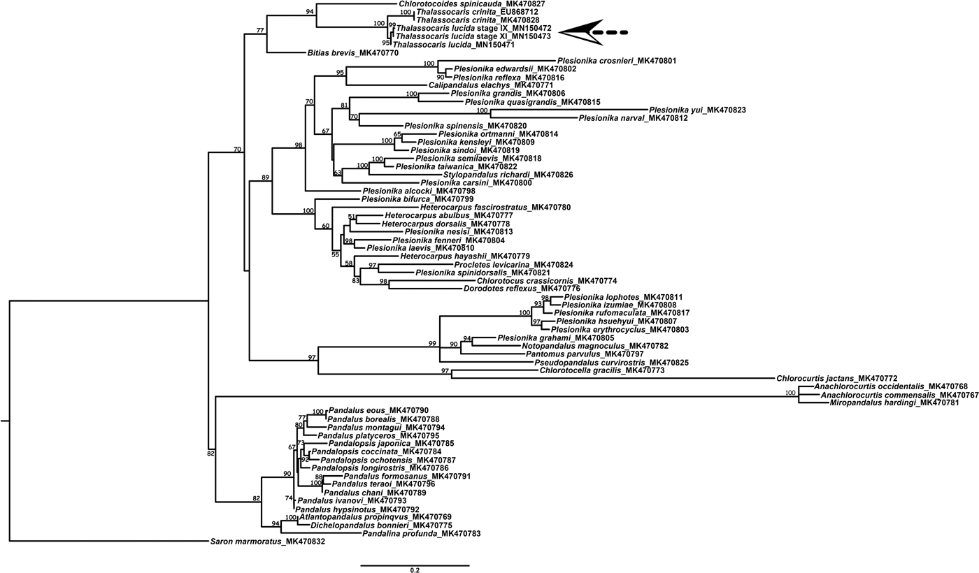
The present dataset includes the two larvae with 64 Pandaloidea species. The best model of present 16S rDNA segment dataset estimated by jModelTest (selected with corrected AIC) was the HKY + I + G evolutionary model (I = 0.339, G = 0.557). The constructed molecular phylogenetic tree (Figure 1) inferred strongly that the two larvae belong to the genus Thalassocaris (UFBS = 100). Furthermore, there is a positive barcoding match of the two larvae with T. lucida with 99.4–99.6% sequence identity (and 99.8% similarity between the two larvae). Moreover, there is no sequence divergence (0%) between the two individuals of T. crinita (EU868712 and MK470828) and they have 4.0–4.3% divergence from the T. lucida and the present two larvae. Accordingly, the present two larvae can be confirmed to be T. lucida.
Maximum likelihood phylogenetic tree constructed by IQ-TREE analysis from mitochondrial 16S rDNA segment (484–546 bp) on the present two larvae (indicated by arrows) using the most comprehensive sequence dataset for Pandaloidea molecular phylogeny (Liao et al., Reference Liao, Ma, De Grave, Komai, Chan and Chu2019), with the adding of another 16S rDNA sequence of T. crinita (EU868712) available in GenBank and a new sequence from T. lucida (MN150471). Ultrafast bootstrap support values (UFBS) are indicated at the branches.

Larval description
Order DECAPODA Latreille, 1802 Infraorder CARIDEA Dana, 1852 Superfamily PANDALOIDEA Haworth, 1825 Family PANDALIDAE Haworth, 1825 Genus Thalassocaris Stimpson, 1860 Thalassocaris lucida (Dana, 1852) (Figures 2–5) ZOEA IX (CBM-ZC15349)
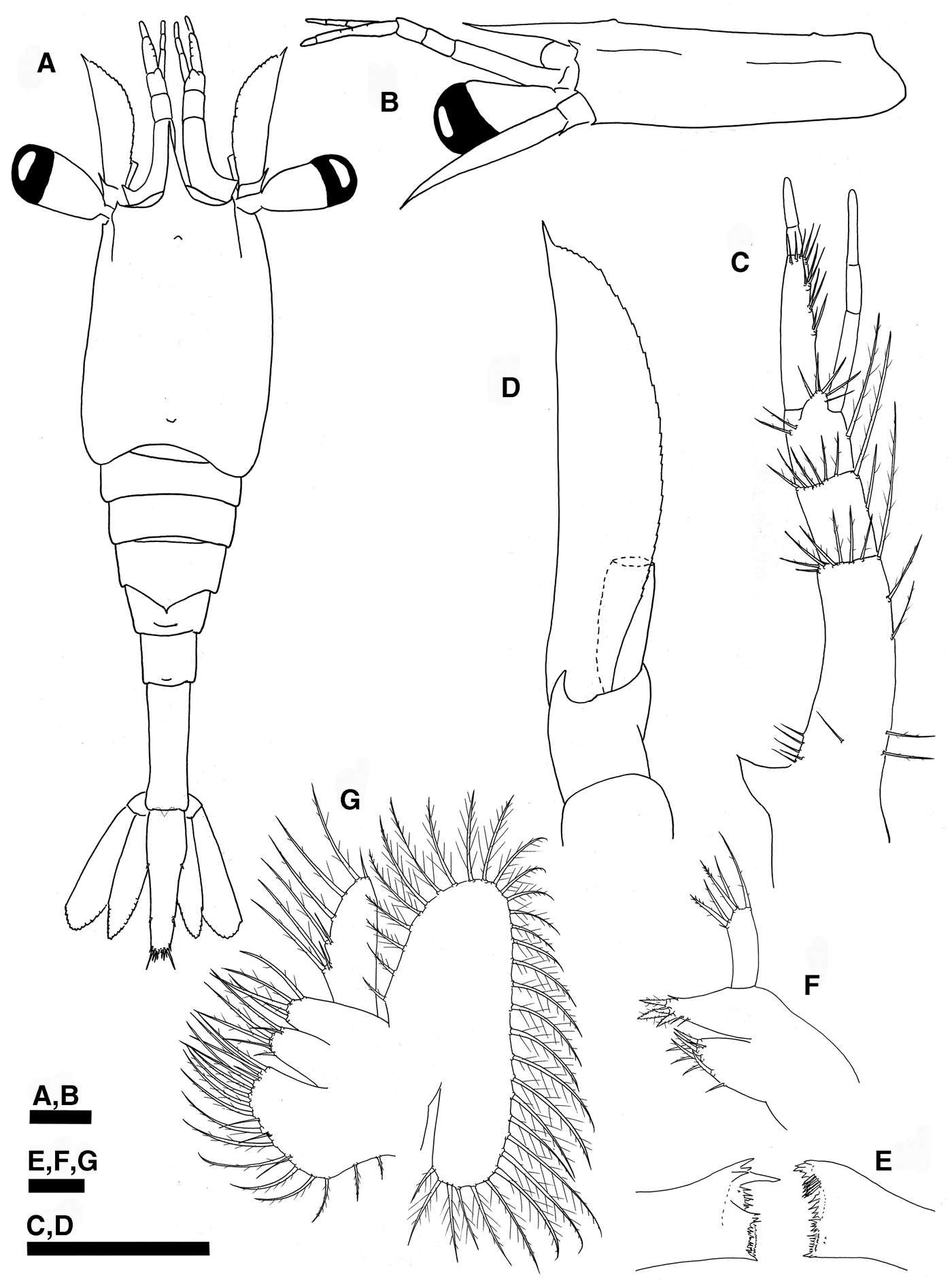
Carapace (Figure 2A, B) – Broad, smooth and rather flattened dorsoventrally (CL: 1.9 mm; CW: 1.5 mm); rostrum slender and unarmed, as long as first segment of antennular peduncle (RL: 0.67 mm); dorsal anterior and posterior processes present; stout supraorbital spine; small antennal and pterygostomial spines; absence of spines on ventral margin; eyes stalked, funnel-shaped.
Thalassocaris lucida (Dana, 1852) zoea IX, (A) habitus, dorsal view; (B) carapace and cephalic appendages, left lateral view; (C) antennule, dorsal view; (D) antenna, dorsal view; (E) mandibles, ventral view; (F) left maxillule, ventral view; (G) left maxilla, ventral view. Scale bars: A–D, 500 µm; E–G, 100 µm.

Antennule (Figure 2C) – Peduncle 3-segmented: basal segment with stylocerite and 8 proximal plumose setae, 2 plumose setae, plus 1 long, 7 short, plumose setae along distal edge; medial segment with 1 long, plumose seta, plus 1 long, and 6 short, plumose setae along distal edge; distal segment with 1 long, 2 short plumose setae plus group of 5 plumose setae; endopod 3-segmented without setae; exopod 3-segmented with 2 + 2 + 3 + 5 aesthetascs in proximal segment.
Antenna (Figure 2D) – Pointed and slender; basis with distolateral spine; scaphocerite broad and with sharp distolateral tooth clearly overreaching blade, inner margin with 30 long plumose setae; endopod segmented (missing, not described).
Mandible (Figure 2E) – Asymmetrical; incisor and molar processes developed, palp absent.
Maxillule (Figure 2F) – Coxal endite with 9 plumodenticulate setae; basial endite with 4 cuspidate and 3 simple setae; endopod unsegmented, with 1 small, simple seta, 2 sparsely plumose setae on basal lobe, 2 sparsely plumose setae on median lobe, 1 sparsely plumose seta on distal lobe; exopod absent.
Maxilla (Figure 2G) – Coxal endite unilobed with 21 plumose setae; basial endite bilobed with 6 + 4 sparsely plumose setae; endopod with 3 + 2 + 1 + 1 + 2 sparsely plumose setae; scaphognathite margin with 33 long plumose setae.
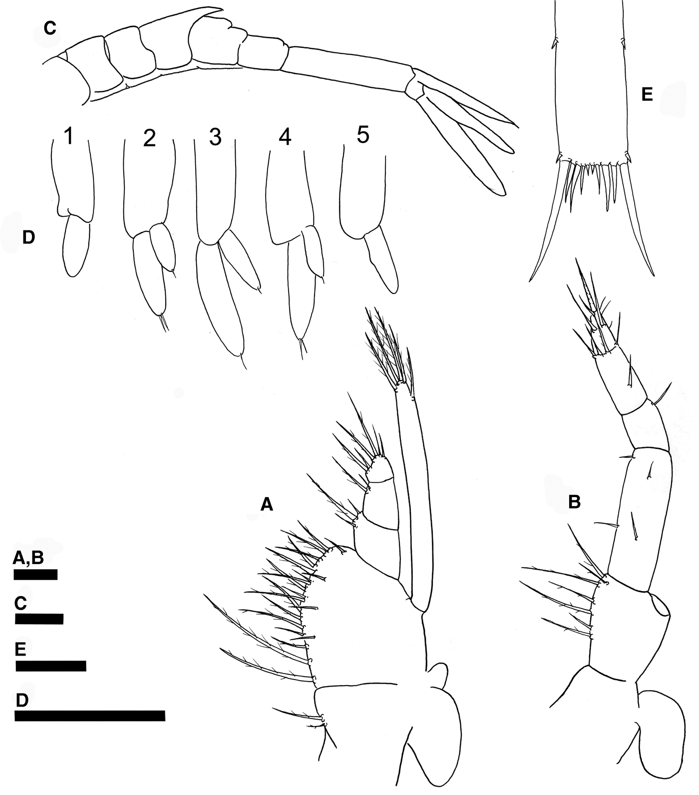
First maxilliped (Figure 3A) – Coxal endite with 2 plumose setae; basial endite with 21 plumose setae; endopod 3-segmented with 3, 2, 2 + 4 sparsely plumose setae; exopod unsegmented with 1 subterminal and 4 terminal, plumose, natatory setae; large bilobate epipod present.
Thalassocaris lucida (Dana, 1852) zoea IX, (A) left first maxilliped, ventral view; (B) left second maxilliped, ventral view; (C) pleon and telson, lateral view; (D) left pleopods 1–5; (E) mid-distal section of telson. Scale bars: A, B, E, 500 µm; C–D, 100 µm.

Second maxilliped (Figure 3B) – Coxal endite without seta, and with epipod; basial endite with 2 + 2 + 3 plumose setae; endopod 4-segmented with 2 + 2 simple setae, 1 sparsely plumose setae, 1 + 4 sparsely plumose setae, and in the distal segment with 3 simple setae and 4 serrulate setae; exopod missing, not described.
Third maxilliped – Missing, not described.
Pereiopods – Missing, not described.
Pleon (Figures 2A & 3C) – Smooth, with 6 somites; tergum of third pleomere with posterior spinose projection that overlaps half of fourth somite length; deep and shallow notch on tergum posterior to mid length of fourth and fifth pleomeres respectively; small anal spine present.
Pleopods (Figure 3D) – Present on pleomeres 1–5; endopod present with 1 simple seta on the second-fourth pleopod, absent on first and fifth pleopods; exopods without setae on first and fifth pleopod and with 2, 1, 2 simple terminal setae on second, third and fourth pleopods, respectively.
Uropods (Figure 2A) – Well differentiated; protopod without seta; endopod well developed with 25 plumose setae; exopod with 24 plumose setae plus 1 plumose seta on outer margin, 1 small simple seta at outermost apex.
Telson (Figures 2A & 3E) – From rectangular to inverted triangle in shape; 3 pairs of lateral simple setae, posterior margin with 5 pairs of plumoserrulate setae.
ZOEA XI (CBM-ZC15350)
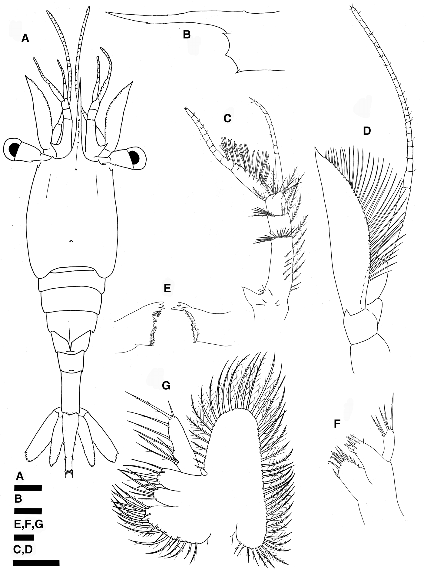
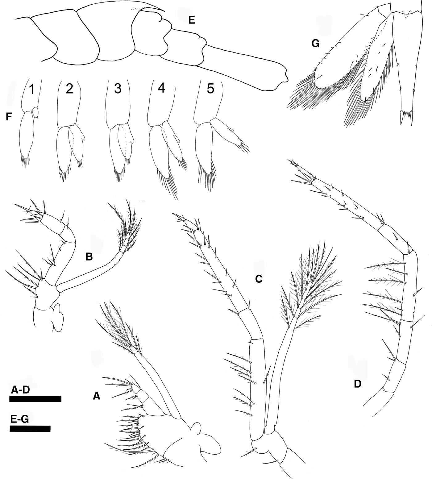
Carapace (Figure 4A, B) – (CL: 2.1 mm; CW: 1.7 mm) rostrum carinated, elongated, longer than antennular peduncle (RL: 1.5 mm), armed dorsally with 3 small spines and ventrally armed with 2 small teeth along mid-distal section.
Thalassocaris lucida (Dana, 1852) zoea XI, (A) habitus, dorsal view; (B) mid-distal section of carapace, left lateral view; (C) left antennule, dorsal view; (D) left antenna, dorsal view; (E) mandibles, ventral view; (F) left maxillule, ventral view; (G) left maxilla, ventral view. Scale bars: A, C, D, 500 µm; B, 200 µm; E–G, 100 µm.

Antennule (Figure 4C) – Peduncle 3-segmented: basal segment with stylocerite and 9 proximal plumose setae, and 12 plumose setae along inner side and arranged in 2 lines, plus 14 short plumose, plus 1 long plumose setae along distal edge; medial segment with 1 long, plumose seta, plus 1 long, and 7 short, plumose setae along distal edge; distal segment with 1 long, 4 long plumose setae plus group of 5 plumose setae; endopod 8-segmented with setation pattern 0, 0, 1, 0, 0, 1, 3, 0 simple setae; exopod 10-segmented with setation pattern 2 + 3 + 3 + 4 + 4 + 4 + 4 aesthetascs, 4 aesthetascs and 0, 2, 0, 2, 2, 2, 2, 3 simple setae.
Antenna (Figure 4D) – Basis with smaller distolateral spine; scaphocerite with inner margin with 33 long, plumose setae; endopod with 24 segments (0, 0, 1, 2, 1, 1, 0, 1, 3, 1, 2, 3, 1, 1, 2, 0, 2, 2, 1, 0, 2, 0, 1, 6 simple setae) and twice length of scaphocerite.
Mandible (Figure 4E) – Unchanged.
Maxillule (Figure 4F) – Coxal endite with 11 plumodenticulate setae; basial endite with 4 cuspidate and 4 simple setae.
Maxilla (Figure 4G) – Basial endite bilobed with 6 + 6 sparsely plumose setae; scaphognathite margin with 48 long plumose setae.
First maxilliped (Figure 5A) – Coxal endite with 3 small, plumose setae; basial endite with 23 plumose setae; endopod 3-segmented with 3, 2 + 1, 2 + 4 sparsely plumose setae; exopod unsegmented with 1 + 2 + 4 plumose, natatory setae (count of setae not accurate due to damage).
Thalassocaris lucida (Dana, 1852) zoea XI, (A) left first maxilliped, ventral view; (B) left second maxilliped, ventral view; (C) left third maxilliped, ventral view; (D) left fifth pereiopod ventral view; (E) pleon and telson, lateral view; (F) left pleopods 1–5; (G) telson and left uropods, dorsal view. Scale bars: A–G, 500 µm.

Second maxilliped (Figure 5B) – Endopod thicker and 4-segmented with 4 + 1 + 1 sparsely plumose setae, 1 sparsely plumose seta, 1 sparsely plumose seta + 4 serrulate setae, and in distal segment with 3 simple setae and 4 serrulate setae; exopod unsegmented with 2 + 2 + 2 + 3 plumose, natatory setae.
Third maxilliped (Figure 5C) – Coxal endite without seta; basial endite with 1 proximal sparsely plumose seta + 1 sparsely plumose seta on distal globose lobe; endopod 4-segmented with 8 plumose + 1 simple setae, 2, 17, 2 serrulate setae + 2 simple setae; exopod unsegmented with 2 + 2 + 2 + 2 + 2 + 4 plumose, natatory setae.
First four pereiopods – Missing, not described.
Fifth pereiopod (Figure 5D) – Uniramous; coxa without seta, basis with 1 + 2 sparsely plumose seta, ischium with 1 + 3 sparsely plumose setae, merus with 6 inner plumose + 1 simple + 2 serrulate setae, carpus with 1 + 2 serrulate setae, propodus with 2 + 2 + 3 + 1 + 4 + 4 serrulate setae + 2 simple setae, dactylus with 4 simple + 3 serrulate setae.
Pleon (Figures 4A & 5E) – Posterior spinose projection on the third pleomere, more evident, pointed and carinated.
Pleopods (Figure 5F) – Exopod with 8, 10, 10, 12, 13 plumose natatory + 2 simple setae on pleopods 1–5, respectively; endopod with 2 simple, 4, 5, 6, 6 plumose natatory setae on pleopods pleopods 1–5, respectively; appendix interna present on endopod of pleopods 2–4.
Uropods (Figure 5G) – Endopod well developed with 32 terminal, 2 subterminal and 5 dorsal plumose setae plumose setae; exopod longer than endopod with 34 terminal, 10 subterminal plumose setae plus, 1 small simple seta at outermost apex.
Telson (Figures 4A & 5G) – Inverted triangle shape; posterior margin with 4 pairs of plumoserrulate setae.
Discussion
The molecular identification of the larvae described in the present study confirms that, at least those late zoeal stages described by Gopala Menon & Williamson (Reference Gopala Menon and Williamson1971), truly belonged to Thalassocaris. Although only two of the three known species of Thalassocaris are included in the molecular genetic analysis, there is a positive match (99.4–99.6% sequence identity in 16S rDNA sequences) of the two larvae with T. lucida. Generally, less than 1% sequence divergence in the 16S rRNA gene is considered as conspecific in DNA barcoding identification for decapod crustaceans (Matzen da Silva et al., Reference Matzen da Silva, dos Santos, Cunha, Costa, Creer and Carvalho2011; Lavery et al., Reference Lavery, Farhadi, Farahmand, Chan, Azhdehakoshpour, Thakur and Jeffs2014; Wakabayashi et al., Reference Wakabayashi, Yang, Shy, He and Chan2017). Moreover, only T. crinita and T. lucida have been reported from Japan and environs (Hayashi, Reference Hayashi2007) and both of them are included in the molecular analysis. The other known species in Thalassocaris, namely T. obscura, is not included in the analysis but is not present in Japan (Hayashi, Reference Hayashi2007) and so far has only been reported from the Indian Ocean and Red Sea (Chace, Reference Chace1985).
The larval description of Gopala Menon & Williamson (Reference Gopala Menon and Williamson1971) was not detailed enough to fully understand the setation pattern of their appendages, though the information provided matches well with the present Thalassocaris larvae. We cannot compare the early zoeal stages described by Gopala Menon & Williamson (Reference Gopala Menon and Williamson1971), but late zoeal stages of Thalassocaris share distinct characters, which are easy to identify: (1) carapace broad and dorsoventrally flattened, (2) coxal endite of maxilla with only one lobe, (3) basis of third maxilliped with a distal globose lobe. According to Gopala Menon & Williamson (Reference Gopala Menon and Williamson1971), these are distinct characters that do not support the inclusion of Thalassocaris in the Pandalidae. Recent systematic positioning of Thalassocaris shrimps placed them in a separate family, Thalassocarididae, within Pandaloidea (De Grave & Fransen, Reference De Grave and Fransen2011), although the placement of this group of shrimps has been controversial in the past. Traditionally, Thalassocarididae was considered related to Pandalidae on the basis of their mouthparts (Chace, Reference Chace1985), but other studies also suggested affinities with Crangonidea and Processidae based on morphology (Christoffersen, Reference Christoffersen1988) and molecular data (Bracken et al., Reference Bracken, De Grave, Felder, Martin, Crandall and Felder2009). The latest systematic analysis of Pandaloidea, based on molecular and morphological data, showed that Thalassocarididae is deeply nested within Pandalidae (Liao et al., Reference Liao, Ma, De Grave, Komai, Chan and Chu2019). Therefore, Thalassocarididae should be synonymized with Pandalidae (Komai et al., Reference Komai, Chan and De Grave2019). The present confirmation of the larval characteristics of Thalassocaris allow a better assessment of the affinity of these shrimps with Pandalidae. Although the shape of the carapace, coxal endite of maxilla and basis of third maxilliped are distinct features of Thalassocaris larvae, other pandalid genera also show quite different larval morphologies. For instance, Dichelopandalus and Atlantopandalus are the only members in Pandalidae with one pair of dorsolateral spines on the fifth pleomere (Pike & Williamson, Reference Pike and Williamson1964). Similarly, Austropandalus is unique in bearing one exopodal plumose seta in the maxillule (Thatje & Bacardit, Reference Thatje and Bacardit2000). Even more significant is the circular-spatulate shaped telson in dorsal view of some members of Pandalus (e.g. P. gracilis, P. prensor, P. niponensis, P. coccinata and P. japonica, with Pandalopsis is now synonymized with Pandalus by Liao et al., Reference Liao, Ma, De Grave, Komai, Chan and Chu2019) (Lee et al., Reference Lee, Hong and Kim2007), or the presence of posterior margins of pleonites fringed with spines, serrulate-shaped, in Pandalus tridens (see Haynes, Reference Haynes1980).
The larval specimens of Thalassocaris lucida described in this study showed other interesting features that deserve more discussion. One of these features is the presence of two notches on the tergum of fourth and fifth pleomeres. Within Pandalidae, the presence of notches on the pleomeres is not a common character in larval stages and it has been reported only on the fourth pleomere in Heterocarpus abulbus from zoea VIII (Jiang et al., Reference Jiang, Landeira, Shih and Chan2016), and Plesionika grandis from zoea IV (Jiang et al., Reference Jiang, Chan and Shih2017). Interestingly, Gopala Menon & Williamson (Reference Gopala Menon and Williamson1971) only described the presence of one notch on the fourth pleomere. It is possible that the smaller notch on the fifth pleomere was overlooked in that description, but also it could be a character not present in the other two congeneric species, T. crinita and T. obscura. Therefore, it could be a potential character for species discrimination.
Another key character observed in T. lucida is the spiny dorsal projection on the third pleomere. According to the description of Gopala Menon & Williamson (Reference Gopala Menon and Williamson1971), this character appears for the first time in the zoea X stage and is fully developed in the following zoea, that is the last stage. Following this, the more developed specimen should be in zoea XI stage since it shows a slender carinated dorsal spine and well developed pleopods with plumose, natatory setae, that generally marks the end of the zoeal phase (Anger, Reference Anger2006). It is worthwhile to mention that Gopala Menon & Williamson (Reference Gopala Menon and Williamson1971) examined specimens in the last zoeal stage which were about to moult or in the process of moulting and found that the decapodid stage would incorporate spines on the external margin of the scaphocerite, a character of adult phase. In the other younger specimen described in our study (that shows a small dorsal spine), the pleopods are less developed than in the zoea X described by Gopala Menon & Williamson (Reference Gopala Menon and Williamson1971), suggesting that this spine can appear earlier, in zoea IX. In any case, the developmental mode of Thalassocaris, with a long series of larval stages, resembles the development of Chlorotocus (Landeira et al., Reference Landeira, Jiang, Chan, Shih and González-Gordillo2015), Heterocarpus (Jiang et al., Reference Jiang, Landeira, Shih and Chan2016), Plesionika (Landeira et al., Reference Landeira, Chan, Aguilar-Soto, Jiang and Yang2014; Jiang et al., Reference Jiang, Chan and Shih2017) and Stylopandalus (Lebour, Reference Lebour1940). Affinities with these four pandalid genera are also suggested by the shape of the eyes. Thalassocaris lucida shows the typical pedunculated funnel-shaped eyes of these genera, but less elongated than in the larvae of Stylopandalus (Lebour, Reference Lebour1940) though this genus is considered as a junior synonym of Plesionika by Liao et al. (Reference Liao, Ma, De Grave, Komai, Chan and Chu2019).
Notwithstanding that the larvae described by Gopala Menon & Williamson (Reference Gopala Menon and Williamson1971) were collected from plankton samples, they attempted to identify them at species level providing key characters. First of all, they noted that those larvae identified as T. crinita were smaller (3.5 mm of carapace + rostral length in zoea XI) than those of T. obscura and T. lucida (5.0 mm). The size of the last zoeal stage of T. lucida described in the present study is 3.6 mm, notably smaller than those larvae assigned to T. lucida by Gopala Menon & Williamson. Nowadays it is well known that the size is a plastic larval trait that is controlled by environmental factors and maternal phenotype/condition (González-Ortegón & Giménez, Reference González-Ortegón and Giménez2014). Therefore, the use of the larval size should be considered with caution in discriminating species from distant geographic areas, as has been demonstrated in other decapod larvae (e.g. Dungeness crab Metacarcinus magister, Shirley et al., Reference Shirley, Shirley and Rice1987; snow crab Chionoecetes opilio, Landeira et al., Reference Landeira, Matsuno, Yamaguchi, Hirawake and Kikuchi2017).
Gopala Menon & Williamson (Reference Gopala Menon and Williamson1971) also suggested that the length of the spines of the telson could be useful to differentiate the three species. Thus, the fourth pair of spines are the longest, and never more than twice the length of the sixth pair in T. obscura, exceeding twice the length of the sixth pair in the last two zoeal stages of T. crinita, and about four times longer than the sixth pair in the last two zoeal stages of T. lucida. The description of T. lucida provided here, shows that the fourth pair of spines are slightly more than twice the length of the sixth pair, and therefore more similar to the previous description of T. crinita than of T. lucida. Regarding the antennal scaphocerite, our description suggests that it becomes progressively more pointed in later stages with reduction of the terminal lamella, but does not disappear. According to Gopala Menon & Williamson (Reference Gopala Menon and Williamson1971) in T. obscura and T. lucida the scaphocerite tapers to a sharp point in the last zoeal stage, but in T. crinita the terminal spine and distal lamella remains distinct as in the present larvae. Therefore, there is a clear disagreement between our description of T. lucida and the previous study, indicating that new larval descriptions are necessary for T. crinita and T. obscura to realize their exact distinguishing characteristics.
Acknowledgements
The authors express their gratitude to Prof. Susumu Ohtsuka who organized the cruise where we obtained the Thalassocaris specimens. Our gratitude also goes to the captain of ‘Toyoshio Maru’ Mr Kazumitsu Nakaguchi, the chief officer Mr Shuhei Yamaguchi, and other crew members for their on-board assistance.
Financial support
This work was supported by grants to TYC from the Ministry of Science and Technology, Taiwan, R.O.C. (MOST107-2621-B-019-001-MY3), and the Center of Excellence for the Oceans (National Taiwan Ocean University), which is financially supported by The Featured Areas Research Center Program within the framework of the Higher Education Sprout Project by the Ministry of Education in Taiwan, R.O.C.