INTRODUCTION
Fasciola hepatica is a parasite that causes significant losses to livestock farmers, and is of interest from a One Health prospective as it also affects wildlife and humans (Cwiklinski et al. Reference Cwiklinski, O'Neill, Donnelly and Dalton2016). The initial stage of F. hepatica infection in the definitive host is rapid and transient and it occurs via the intestine. Although F. hepatica newly excysted juveniles (NEJ) are able to penetrate various segments of the intestine, the small intestine is the anatomical region where the highest rate of migration is observed (Hanna, Reference Hanna1980; Kawano et al. Reference Kawano, Yamamoto, Koga, Shimizu and Kimura1991), suggesting that there could be some specific features, receptors or molecules here that the parasite is able to recognize. It is well known that upregulation and secretion of members of the cysteine proteases (cathepsin L3, L4 as well as cathepsin B) occurs at the NEJ stage (Cancela et al. Reference Cancela, Ruétalo, Dell'Oca, da Silva, Smircich, Rinaldi, Roche, Carmona, Alvarez-Valín, Zaha and Tort2010; Robinson et al. Reference Robinson, Hutchinson, Donnelly and Dalton2010) and that these are important for migration through the gut wall (McGonigle et al. Reference McGonigle, Mousley, Marks, Brennan, Dalton, Spithill, Day and Maule2008). In addition, components with glycosidase activity – including β-hexaminidase, β-galactosidase and β-glucosidase – have been detected in the tegument and excretory/secretory (ES) repertoire of NEJ and adult flukes (Moore and Halton, Reference Moore and Halton1976; Irwin et al. Reference Irwin, Morrissey, Ryan, Walshe, O'Neill, Carrington, Matthews, Fitzpatrick, Mulcahy, Corfield and Dalton2004), which could potentially degrade host mucins, thus aiding migration.
Parasite glycans and their lectin receptors are often involved in determining final tropisms in the definitive host. Among protozoa with enteric tropism for example, galactose (Gal) and N-acetyl-galactosamine (GalNAc) lectin is required for mucin adhesion for Entamoeba histolytica (Chadee et al. Reference Chadee, Petri, Innes and Ravdin1987; Petropolis et al. Reference Petropolis, Faust, Deep Jhingan and Guillen2014) and Cryptosporidium parvum (Barnes et al. Reference Barnes, Bonnin, Huang, Gousset, Wu, Gut, Doyle, Dubremetz, Ward and Petersen1998; Cevallos et al. Reference Cevallos, Bhat, Verdon, Hamer, Stein, Tzipori, Pereira, Keusch and Ward2000; Hashim et al. Reference Hashim, Mulcahy, Bourke and Clyne2006). Adherence of Giardia duodenalis to enterocytes can be blocked by glucose (Glc) and mannose (Man) (Sousa et al. Reference Sousa, Gonçalves, Bairos and Poiares-Da-Silva2001). No work has yet been carried out to address these potential interactions in F. hepatica. More recently, high mannose N-glycans, monofucosylated truncated N-glycans and mucin-type core-1 (also denominated T-antigen) O-glycans on the tegument and at the oral and ventral suckers have been identified on the F. hepatica NEJ (Garcia-Campos et al. Reference Garcia-Campos, Ravidà, Nguyen, Cwiklinski, Dalton, Hokke, O'Neill and Mulcahy2016b ) and we hypothesized that interaction between these parasite glycans and lectin-receptors in the host gut might be important during initial infection of the definitive host.
The aim of this study was to assess whether the migration of F. hepatica NEJ can be disrupted by the blockage of glycans found on the parasite or those expressed in the host intestinal tract using an ex vivo method.
MATERIALS AND METHODS
Reagents
All the biotinylated-lectins, FITC-labelled streptavidin and Vectashield mounting media used were purchased from Vector Laboratories (Peterborough, UK). Unless otherwise stated the remainder of the chemicals and reagents were purchased from Sigma-Aldrich Ltd. (Ireland).
Lectin selection and histology staining
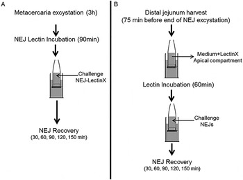
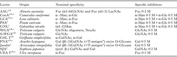
Lectins used are listed in Table 1. The lectins used in Experimental Plan A (Fig. 1A) were selected on the basis of binding to NEJ tegument and NEJ somatic antigen (Garcia-Campos et al. Reference Garcia-Campos, Ravidà, Nguyen, Cwiklinski, Dalton, Hokke, O'Neill and Mulcahy2016b ). A total of eight lectins were chosen, which covered a wide sugar specificity spectrum.
Scheme of the two approaches performed to evaluate the role of glycans in F. hepatica NEJ invasion in host intestine in vitro. (A) NEJ were excysted in vitro, separated into batches and incubated with RPMI 1640 (control) or with RPMI 1640 + 10 µg mL−1 of biotinylated-lectin for 90 min at 39 °C. NEJ were then washed with RPMI 1640 at 39 °C and inoculated into the in vitro system as described (Garcia-Campos et al. Reference Garcia-Campos, Baird and Mulcahy2016a ), and allowed to migrate through rat distal jejunum from 30 to 150 min. (B) 75 min before the end of the NEJ excystation in vitro, rat distal jejunum was used to mount the in vitro system. The apical compartments were pre-incubated with RPMI 1640 + 10 µg mL−1 of the biotinylated-lectin for 60 min at 39 °C. Medium from the apical compartments was discarded, washed, replaced by the RPMI 1640 medium at 39 °C and challenged with NEJ previously washed in RPMI 1640 medium at 39 °C. A total of nine chambers were used at each condition.

List of the nominal specificity and specific inhibitors of lectins

a Lectins employed to evaluate glycan recognition in distal jejunum.
b Lectins employed in Experimental Plan B. Fuc (Fucose), Man (Mannose), Glc (Galctose), Lac (Lactose), GlcNAc (N-Acetyl-Glucosamine), GalNAc (N-Acetyl-Galactosamine), LacNAc (N-Acetyl-Lactosamine), NeuAc (Neuraminic acid), Ser (serine)m Thr (threonine).
c Lectins employed in Experimental Plan A.
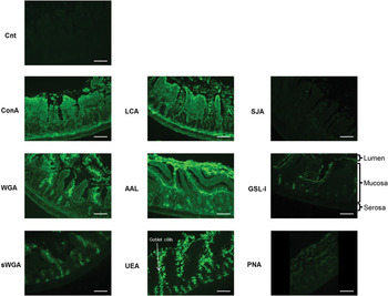
Selection of eight lectins used in Experimental Plan B (Fig. 1B) was based on their sugar specificity and their ability to bind to glycans nearest to the lumen, to goblet cells and uniformly through the intestinal mucosa in a preliminary screen of rat jejunum by histochemistry (Fig. 2). Briefly, sections were incubated with blocking solution (blocking solution: PBS–BSA – 1%) for 3 h at 4 °C. Slides were rinsed three times with washing solution (PBS–BSA – 0·1%) and incubated for 3 h with 1 mL of 10 µg mL−1 of biotinylated lectin in 0·1% BSA–PBS solution. Negative controls comprised gut sections incubated with all lectins previously mixed with their specific inhibitors for 1 h. After three washes, sections were incubated with 1 mL of 5 µg mL−1 of FITC-labelled streptavidin in 0·1% BSA–PBS solution for 1 h in the dark. At the end of the incubation period, sections were washed, mounted in glycine and PBS in a ratio 9:1, and sealed with nail polish. Slides were viewed using an Olympus BX40FA microscope (Olympus Optical Co, Ltd.) using 10× and 20× Achromatic objectives (Olympus) equipped with an epifluorescence source and a filter system for FITC fluorescence.
Recognition of glycans by a panel of lectins in rat distal jejunum. Rat distal jejunum was fixed in 10% formalin solution, embedded in paraffin and cut in 5 µ m sections. Sections were blocked with 1% BSA–PBS for 3 h and incubated for another 3 h with 10 µg mL−1 of the biotin-labelled lectins ConA, WGA, sWGA, LCA, UEA, AAL, PNA, GSL-I or SJA in 0·1% BSA–PBS. For the negative control, all the lectins were incubated together for 1 h with their specific sugar inhibitors before incubating them with sections. Incubation of the sections with 5 µg mL−1 of FITC-labelled streptavidin in 0·1% BSA–PBS for 1 h was performed in order to recognize positive lectin binding. Scale bar = 50 µ m.

Excystation of metacercariae and NEJ lectin incubation
Two sources of F. hepatica metacercariae (n = 16 000) were used: metacercariae were purchased from Baldwin Aquatics, Inc. (Monmouth, Oregon) (Oregon strain), and Ridgeway Research Ltd. (St Briavels, UK) (Italian strain). NEJ were excysted for 3 h as previously described (Carmona et al. Reference Carmona, Dowd, Smith and Dalton1993) and washed in RPMI 1640 solution (Biosciences, Ireland). NEJ from the Oregon strain (n = 10 000) were used for Experimental Plan A (Fig. 1A). Briefly, NEJ were divided into nine batches of 90 NEJ and each batch incubated for 90 min at 39 °C in 3 mL of RPMI 1640 medium supplemented with 10 µg mL−1 of one of eight lectins. A batch of NEJ incubated with RPMI 1640 medium only was used as negative control. After incubation, all NEJ were washed in RPMI for 5 min and their viability assessed microscopically. NEJ from the Italian strain (n = 6000) were directly introduced into the horizontal diffusion chambers in Experimental Plan B (Fig. 1B).
Tissue donors, chamber configuration and lectin incubation in distal jejunum
All procedures involving animal tissue were carried out following approval by the University College Dublin (UCD) Animal Research Ethics Committee (AREC approval 15-01). Sixteen 6-week-old male Wistar rats (250–300 g) were used for these experiments. Rats were housed in the Biomedical Facility in UCD in controlled environmental conditions. The animals were euthanized by stunning, then cervical dislocation. Laparotomy was performed, the caecum exposed and the portion of small intestine between 25 and 5 cm just before the caecum (distal jejunum) dissected, opened by cutting along its mesenteric border and maintained in RPMI 1640 medium at 39 °C.
The methodology employed was based on the protocol described in Garcia-Campos et al. (Reference Garcia-Campos, Baird and Mulcahy2016a ) Briefly, chambers were constructed consisting of a basolateral compartment of 9 mL capacity and an apical compartment of 200 µL capacity. The two compartments were separated by a sheet of rat distal jejunum mounted, so that the apical (lumenal) surface was uppermost and the basolateral surface was bathed in the larger volume of RPMI. Nine to twelve horizontal diffusion chambers were mounted per donor.
The chambers were then filled with RPMI 1640 medium at 39 °C and exposed to NEJ. In Experimental Plan A, mucosal sheets were exposed to NEJ pre-incubated with lectins or, in Experimental Plan B, the lumenal surface of sheets mounted in the chambers were pre-incubated with a lectin for 60 min, washed three times and replenished with fresh medium before challenge with NEJ.
NEJ challenge and assessment of NEJ migration
To examine whether lectin pre-treatment of NEJ influenced their migration, 40–45 lectin-incubated NEJ were added to the apical (lumenal) compartments. The actual number added was verified by counting the total infecting dose. Numbers of transmigrating NEJ, which sedimented at the bottom were then counted at 30 min intervals.
In Experimental Plan B, to examine whether pre-treatment of tissues by lectins influenced migration of NEJ, the apical (lumenal) surface of distal jejunal sheets was pre-incubated with a specific lectin before challenge with 35–40 fresh non-treated NEJ. The NEJ challenge number was calculated and the number of transmigrating NEJ which appeared in the basolateral chamber was determined at 30 min intervals.
Sectioning of NEJ and staining
Batches of 20 lectin-incubated NEJ were washed in RPMI 1640 at 39 °C for 5 min after excystation, immediately fixed in 10% formalin solution and maintained at 4 °C (T0). Another twenty NEJ per lectin treatment were incubated for 150 min at 39 °C in RPMI 1640, mimicking the conditions of the horizontal diffusion chamber. Viability of all NEJ was assessed at the end of the incubation period by observation of parasite coloration, integrity and body contractility. NEJ were fixed in 10% formalin solution and maintained at 4 °C (T150).
Lectin-incubated NEJ fixed at T0 and T150 were transferred to 24-well plates. As the NEJ remained at the bottom of the wells, excess supernatant was removed carefully from the top of the well. Once the supernatant was removed, 1 mL of haematoxylin was then added and after 10 min the stain was gently removed, taking care not to disturb the NEJ. In parallel, 1 mL of 1% semi-liquid agar was poured into separate wells without any parasites. Next, fixed and haematoxylin stained-NEJ were harvested with a transfer pipette and placed gently into the centre of the semi-liquid agar. After incubation at 4 °C overnight, the solidified agar plugs containing the NEJ were collected and inserted into tissue cassettes (Simport). After removal of excess water and lipids from the agar plugs (Pathcentre, Shandon), they were embedded in paraffin and sectioned using a microtome. Repeated 5 µ m-thick sections were mounted and stained with eosin for NEJ localization.
After localization of the NEJ in the blocks, sections were blocked, washed, incubated with FITC-labelled streptavidin solution and mounted with Vectashield mounting medium as described above. Slides of lectin-incubated NEJ were viewed using an Olympus BX40FA (Olympus Optical Co, Ltd.) using 10× and 20× Achromatic objectives (Olympus) equipped with epifluorescence source and filter system for FITC fluorescence.
Sample size calculation and statistical analysis
Sample size was calculated as described by Hulley et al. (Reference Hulley, Cummings, Browner, Grady and Newman2013) assuming detection of significant differences at α = 0·05, β = 0·20, power = 80% and a minimum effect desirable to detect between control and treated groups of 30%. Because all tissues used were obtained from animals of the same breed, sex, weight and age, the apical compartment was designated as the experimental unit. Nine chambers from at least three different donors were employed for each lectin. Means of NEJ migration in all control chambers were calculated and used for data normalization of the controls for each independent experiment. Lectin effects were normalized to their respective control. Counts are expressed by percentage means (±s.e.). The Kruskal–Wallis test was used to compare multiple groups and Mann–Whitney U test was used as a post-hoc test for pairwise comparisons. Differences where P < 0·05 were deemed significant (*P ⩽ 0·05, **P ⩽ 0·01, ***P ⩽ 0·001).
RESULTS
Mannose binding lectins significantly decreased NEJ migration across the distal jejunum
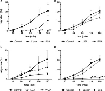
The viability of the Oregon strain-NEJ was maintained in the control and in all lectin-incubated NEJ, but migration across the distal jejunum differed between groups. For the control group, chambers from all donors showed NEJ migration at T150, with counts that varied from 1 to 25 NEJ, representing an average of 20·96% migration. Significant effects on migration were detected from 120 min post-challenge onwards (P = 0·0014) following incubation of NEJ. The Man-binding lectin ConA completely inhibited NEJ migration, with no migration observed in any of the chambers up to and including T150 (P = 0·0053) (Fig. 3A). GNL, which has a very similar glycan-binding specificity, also significantly inhibited NEJ migration, with one NEJ detected in only three of the chambers, representing a migration rate of 1·66% (P = 0·0005) compared with that seen in the control group. The lectin WGA, which has GlcNAc-binding specificity, showed a migration rate of 5·6%, although this decrease as compared with controls was not statistically significant. The Fuc-binding-lectins PSA, LCA and UEA showed similar NEJ invasion rates,11·35, 12·83 and 13·88%, respectively, which were lower than the control groups but again, not significantly different. In some cases, differences in migration kinetics could also be observed. For example, migration following incubation of NEJ with PSA was only seen after 90 min. However, LCA-incubated NEJ were able to get through the distal jejunum from the beginning of the experiment. For UEA-incubated NEJ, although the migration rate was lower at all time points, the time course followed the same pattern as seen in the control group. Lectin binding to mucin-type core-1 glycans also followed the same migratory kinetics as seen in the control group.
Kinetics of lectin-incubated NEJ invasion in rat distal jejunum in vitro. NEJ were incubated with RPMI (control) or with RPMI + 10 µg mL−1 of lectin with Man (ConA and GNL), Fuc (PSA, UEA and LCA), GlcNAc (WGA) and mucin-type core-1 (PNA and Jacalin) specificity, for 90 min before adding to the in vitro chambers. Migration of NEJ controls and NEJ incubated with PSA and ConA (A), PNA and UEA (B), LCA and WGA (C) and Jacalin and GNL (D) was assessed at 30 min intervals until 150 min post-incubation. Nine chambers were used for each condition. For each panel three independent experiments were performed. NEJ located in basolateral compartments were counted and the total percentage migration calculated. Means of NEJ migration in all control chambers were calculated and used for data normalization of the controls for each independent experiment. Lectin effects were normalized according to their respective control group. Vertical bars represent s.e. Asterisks represent significant differences in the percentage of NEJ found in the basolateral compartment between treatment and its control (**P ⩽ 0·01, ***P ⩽ 0·001).

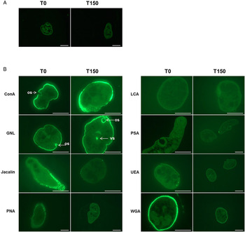
Histology showed that glycan binding by ConA and GNL on the NEJ tegument and oral sucker was still present at T150
The lectins ConA, GNL, WGA, PNA and Jacalin were able to bind to glycan structures found on the tegument at T0 (Fig. 4). ConA and GNL also bound strongly to the oral sucker at this time. At T150, positive lectin binding was still evident on the NEJ tegument for ConA and GNL, confirming that the oligomannose N-glycans were still blocked at the end of the experiment, and that they may be important for host recognition. Particularly for GNL at T150, binding to the ventral sucker was also seen, suggesting that the adhesion of the NEJ ventral sucker to the intestine could have been compromised by blocking of this glycan. Weaker binding to the tegument of WGA at T150 was also observed, which could indicate that some of the glycan population with terminal GlcNAc were also blocked during the entire migration period. The binding of PNA and Jacalin seen in NEJ incubated with PNA and Jacalin before challenge was lost by T150. None of the lectins showed visible binding to the gastrodermal epithelial cells of the NEJ – the parasite's gut – at either T0 or T150, suggesting that the secretion of ES products was not affected.
Lectin-binding recognition of glycans on NEJ from T0 to T150. NEJ were incubated for 90 min at 39 °C with RPMI 1640 (control) (A) or with RPMI + 10 µg mL−1 of biotin-labelled lectins (B) with Man (ConA and GNL), Fuc (PSA, UEA and LCA), GlcNAc (WGA) and mucin-type core-1 (PNA and Jacalin) specificity. Half of the NEJ were washed in RPMI and fixed in 10% buffered formalin (T0). In order to mimic the conditions of the migration study, the remaining NEJ were incubated in RPMI at 39 °C for 150 min. After incubation, they were fixed as previously described (T150). NEJ were loaded in 1% agarose, mounted in cassettes and embedded in wax. NEJ sections were blocked with 1% BSA–PBS for 3 h and incubated with 5 µg mL−1 of FITC-labelled streptavidin in 0·1% BSA–PBS for 3 h. Positive binding to oral sucker (os) and ventral sucker (vs) are indicated with arrows. Scale bar = 50 µ m.

Jejunal sheets incubated with lectins were equally susceptible to NEJ migration as non-treated tissue
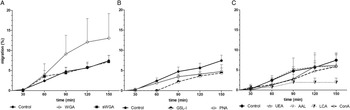
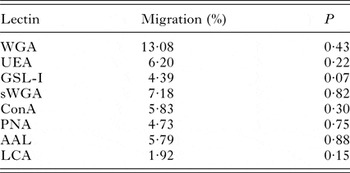
The migration curves observed for the NEJ loaded onto distal jejunum pre-incubated with the rest of the lectins showed no significant differences from those observed for the control group (Fig. 5). The migratory curve observed in the control group using the Italian strain was lower than that seen in the control group of the Oregon strain, reaching peak of 7·44% (1–9 NEJ) as average of migration rate at T150. Distal jejunum exposed to WGA showed a trend of increased NEJ migration compared with the other conditions and to the control group from 60 min onwards, showing 13·08% of migration rate at T150 (Fig. 5A). Jejunal sheets incubated with GSL-I (Fig. 5B) and LCA (Fig. 5C) showed a trend towards reductions of the NEJ migration values to 4·39 and 1·92%, respectively. However, as detailed in Table 2, the effects observed by WGA, GSL-I and LCA were not significant.
Kinetics of NEJ invasion in lectin-incubated rat distal jejunum in vitro. Jejunal sheets were mounted in the horizontal diffusion chamber and incubated with 200 µL of 39 °C RPMI 1640 (control) or with 39 °C RPMI + 10 µg mL−1 of lectin with GlcNAc/NeuAc (WGA), GlcNAc (sWGA), Gal-/GalNAc- (GSL-I), mucin-type core-1 (PNA) Fuc (UEA, LCA and AAL) and Man (ConA) specificity for 60 min and washed with 39 °C RPMI 1640 before NEJ load. NEJ were loaded and allowed to migrate through rat distal jejunum incubated with WGA and sWGA (A), GSL-I and PNA (B) and UEA, AAL, LCA and ConA (C) for 150 min. Migration was measured at 30 min intervals. A total of nine chambers were used at each condition. For each panel three independent experiments were performed. NEJ located in basolateral compartments were counted and the total percentage was calculated. Means of NEJ migration in all control chambers were calculated and used for data normalization of the controls for each independent experiment. Lectin effects were normalized according to their respective control group. Vertical bars represent s.e.

Trend of NEJ migration across rat lectin-incubated distal jejunum in vitro after 150 min post challenge

DISCUSSION
The presence of N- and O-glycans in the F. hepatica tegument has been well described (Garcia-Campos et al. Reference Garcia-Campos, Ravidà, Nguyen, Cwiklinski, Dalton, Hokke, O'Neill and Mulcahy2016b ). Nevertheless, their contribution to NEJ adhesion to the intestinal epithelial cells of the host at the earliest stages of infection has not been yet elucidated. In addition, the presence of lectin-binding receptors on the NEJ tegument has not been studied. In order to address these gaps, we have adapted an ex vivo method (Garcia-Campos et al. Reference Garcia-Campos, Baird and Mulcahy2016a ) for evaluation of host–liver fluke interactions with the distal jejunum of the rat.
When NEJ tegumental glycans were blocked by a panel of lectins with different carbohydrate specificities, ConA and GNL almost completely inhibited NEJ migration in vitro. This inhibition was maintained until the end of the experiment, reflecting continued binding by these lectins to the parasite surface, oral and ventral sucker at T150. The blockage of high and/or oligomannose N-glycans at the oral and ventral suckers could have an inhibitory effect on parasite feeding or secretion as seen with larvae of the helminth parasites Teladorsagia circumcincta, Haemonchus contortus and Trichostrongylus colubriformis when incubated with ConA in vitro (Ríos-de Álvarez et al. Reference Ríos-de Álvarez, Jackson, Greer, Bartley, Bartley, Grant and Huntley2012), and in the intimate adhesion of the NEJ to the epithelium between the ventral disc of Giardia lamblia and the host enteric epithelium (Inge et al. Reference Inge, Edson and Farthing1988). High and oligomannose-type N-glycans are the most dominant glycan population identified in the NEJ tegument (Garcia-Campos et al. Reference Garcia-Campos, Ravidà, Nguyen, Cwiklinski, Dalton, Hokke, O'Neill and Mulcahy2016b ), so it is logical that lectins binding to these had major effects on NEJ migration. Therefore, high and/or oligomannose N-glycans are likely to be involved in the establishment of the first interaction of the NEJ with intestinal epithelial host cells. There are many known examples of the importance of Man motifs in the interaction between the host and other pathogenic agents. In parasitic infections for example, Man is responsible for the invasion of Trypanosoma cruzi into heart cells (Barbosa and de Meirelles, Reference Barbosa and de Meirelles1992), and of Enteromyxum scophthalmi into turbot intestinal epithelium in vitro (Redondo and Alvarez-Pellitero, Reference Redondo and Alvarez-Pellitero2010). In other pathogenic agents with enteric tropism such as Candida albicans, it is known that oligomannosides, particularly α1-2 and β1-2 Man- N-glycans, located in the yeast wall are vital in binding to the intestinal cell line Caco-2 in vitro (Dromer et al. Reference Dromer, Chevalier, Sendid, Improvisi, Jouault, Robert, Mallet and Poulain2002; Dalle et al. Reference Dalle, Jouault, Trinel, Esnault, Mallet, D'Athis, Poulain and Bonnin2003).
Incubation of NEJ with PNA, Jacalin and WGA resulted in binding, and hence blockage of glycans, only at the beginning of the experiment. This could be due to turnover of the NEJ tegument, as is known to occur (Lammas et al. Reference Lammas, Duffus and Taylor1985; Dalton and Joyce, Reference Dalton and Joyce1987). Even though NEJ shedding may have removed some of the ConA and GNL blocked N-glycans during the experiment, binding persisted, perhaps because the mannose-type N-glycan population is more abundant and/or has a longer half-life. Tegument shedding is a selective process and it is possible that a longer period would be needed to remove all ConA and GNL attached to the surface and for synthesis of mannose-type N-glycans de novo. The mucin-type core-1 O-glycans as well as GlcNAc-capped N-glycans are not as abundant as the mannose-type N-glycan population in the tegument. When the tegument sheds, the substitution of blocked glycans by unblocked ones may occur rapidly. Therefore, there could be short O-glycan structures and N-glycans with terminal GlcNAc motifs available, which could re-establish the potential function of these glycan specificities. We observed a trend of reduction in the migration rate following WGA incubation, especially during the first 60 min of infection. This could suggest that the blockage of terminal GlcNAc N-glycans was maintained until 60 min and after that the tegument was shed, allowing new glycans to interact with the epithelium. However, as no significant differences were observed between the WGA group and the control group, confirmation of any interaction between GlcNAc residues and the host epithelium requires further studies. The lectins PNA and Jacalin did not change migration rate appreciably, meaning that either the tegument was shed very quickly, and thus the glycans were able to interact with the host epithelium from the beginning, or that mucin-type core-1 O-glycans do not play a role in invasion. In order to confirm the latter, further work with other lectins with terminal Gal- and GalNAc- binding affinities, for example GSL-I and SBA, etc. would be necessary.
When live NEJ were incubated with lectins, there was no positive lectin binding to NEJ gastrodermal epithelial cells. This could have different interpretations. The first one may be that the lectins were not taken up by the NEJ because this lifecycle stage is more involved in tissue penetration rather than nutrient intake (Robinson et al. Reference Robinson, Menon, Donnelly, Dalton and Ranganathan2009; Cwiklinski et al. Reference Cwiklinski, Dalton, Dufresne, La Course, Williams, Hodgkinson and Paterson2015). The second interpretation is that lectins were degraded by the effect of the proteases and enzymes that comprise the ES products. Finally, it could also mean that lectins were, indeed, taken up by the NEJ but the glycosylation pattern of the gastrodermal epithelial cells is not aligned with the binding specificity of the lectins used. Even though we have shown that members of the cathepsin clade contain a paucimannosidic N-glycan attached to their peptide backbone (Garcia-Campos et al. Reference Garcia-Campos, Ravidà, Nguyen, Cwiklinski, Dalton, Hokke, O'Neill and Mulcahy2016b ), and they occur not only in the tegument but also in the gastrodermal epithelial cells, they were not recognized by these lectins. No binding recognition of these short tetra-saccharides was observed for ConA in a glycoarray (Brzezicka et al. Reference Brzezicka, Echeverria, Serna, van Diepen, Hokke and Reichardt2015).
Invasion and penetration of the host intestine by NEJ may involve binding to a set of lectin receptors found on host intestinal epithelial cells such as the mannose-binding receptor (MBL) (Uemura et al. Reference Uemura, Saka, Nakagawa, Kawasaki, Thiel, Jensenius and Kawasaki2002; Kania et al. Reference Kania, Sorensen, Koch, Brandt, Kliem, Vitved, Hansen and Skjodt2010), galectins (Barondes et al. Reference Barondes, Cooper, Gitt and Leffler1994; Preston et al. Reference Preston, Beddoe, Walkden-Brown, Meeusen and Piedrafita2015), intelectins (Intls), etc. via oligomannose N-glycans. Such mechanisms have been shown for other pathogens such as the interaction of Shigella flexneri with MBL in the intestine (Zuo et al. Reference Zuo, Zhang, Lu, Liu and Chen2009), and of Leishmania sp. in extra-intestinal localization (Santos et al. Reference Santos, Costa, Krieger, Feitosa, Zurakowski, Fardin, Gomes, Weiner, Harn, Ezekowitz and Epstein2001). Even though MBL are also capable of recognizing glycans with other terminal motifs like Fuc and GlcNAc (Weis et al. Reference Weis, Drickamer and Hendrickson1992), we did not find significant differences in the NEJ migration rate when parasites were incubated with lectins specific for these glycans. Other lectin receptors that could be involved in this interaction are Intls, which are a family of Ca2+-dependent galactofuranosyl-binding lectins expressed in Paneth and goblet cells of mammalian small intestine (Nair et al. Reference Nair, Guild and Artis2006; Wrackmeyer et al. Reference Wrackmeyer, Hansen, Seya and Danielsen2006). However, lectins able to address this interaction were not included in this study. Galectins, bind most strongly to Gal and GalNAc residues, but the interaction of galectin-3 with oligomannose has also been demonstrated (Jouault et al. Reference Jouault, Fradin, Trinel, Bernigaud and Poulain1998; Fradin et al. Reference Fradin, Poulain and Jouault2000). Therefore, it is possible that members of the galectin family could bind to mannose-type N-glycans found in the NEJ tegument.
Because glycans are often important in the final tropisms of parasites in the definitive host, and the expression of mucin genes in F. hepatica NEJ (Cancela et al. Reference Cancela, Santos, Carmona, Ferreira, Tort and Zaha2015) has been described, we blocked host glycans with lectins to evaluate whether NEJ were more or less efficient in invading following lectin pre-treatment. In contrast to what has been seen in studies performed in Toxoplasma gondii (Caffaro et al. Reference Caffaro, Koshy, Liu, Zeiner, Hirschberg and Boothroyd2013), E. histolytica (Chadee et al. Reference Chadee, Petri, Innes and Ravdin1987), C. parvum (Barnes et al. Reference Barnes, Bonnin, Huang, Gousset, Wu, Gut, Doyle, Dubremetz, Ward and Petersen1998; Cevallos et al. Reference Cevallos, Bhat, Verdon, Hamer, Stein, Tzipori, Pereira, Keusch and Ward2000; Chen and LaRusso, Reference Chen and LaRusso2000; Choudhry et al. Reference Choudhry, Bajaj-Elliott and McDonald2008) and G. duodenalis (Sousa et al. Reference Sousa, Gonçalves, Bairos and Poiares-Da-Silva2001) we did not demonstrate the presence of lectin-type receptors on the NEJ capable of recognizing host glycans.
Understanding the role that glycans play in host-pathogen interaction has led to the development of therapeutic remedies that are currently in use. For instance, the blockage of sialic acid and neuraminidase interaction by zanamivir and oseltamivir, commercial antiviral drugs used for the treatment and prevention of influenza A and B. The appearance of oseltamivir-resistant viruses has stimulated the investigation of alternative remedies with similar glycan–enzyme blockage effect against influenza B virus in vitro (Charyasriwong et al. Reference Charyasriwong, Haruyama and Kobayashi2016). Similarly, research is moving towards the development of carbohydrate-based vaccines that could boost cellular immune responses or induce humoral responses with blocking properties (Astronomo and Burton, Reference Astronomo and Burton2010). Passive immunization with tyvelose-reactive antibodies against a group of glycoproteins of Trichinella spiralis found in the first larval stage provides protection as antibodies block epithelial invasion by the parasites in vitro (McVay et al. Reference McVay, Bracken, Gagliardo and Appleton2000). Our findings suggest that the development of prophylactic drugs or vaccine-induced antibodies capable of binding NEJ mannosylated N-glycans may contribute to novel strategies for the control of fasciolosis.
Difficulties with the supply of metacercariae required the use of two different metacercarial strains in these studies. Whereas metacercariae of the Oregon strain tend to hatch quickly and more uniformly, metacercariae of the Italian strain tend to hatch at a more variable rate. Although both strains transmigrated through the tissue, the use of two different strains could have contributed to variability between experiments. Even though this was not the purpose of the present study, it raises the question of whether there are some biological differences in the NEJ migratory behaviour between strains, which could be usefully explored in future studies. When comparisons between control and lectin groups were performed we assured that the same strain was used for both.
In conclusion, the current results demonstrate the inhibitory effect on NEJ migration of blocking high and oligomannose-type N-glycans on the NEJ tegument in vitro. This effect was not transient, but maintained for at least 150 min. The in-depth identification of the NEJ glycans and ligands involved in this interaction deserves further investigation, both in terms of understanding the basic biology of parasite invasion, and in the quest to develop novel methods for the control of fasciolosis.
ACKNOWLEDGEMENTS
We would like to thank Patsy Kearns, Fiona McCartney and Joanne Heade for animal care and preparation before euthanasia. We also thank Joseph Brady for his assistance with histology processing.
FINANCIAL SUPPORT
This work was supported by the EU Seventh Framework Programme (to G. M., PARAVAC – grant number 265862), EU Horizon 2020 (to G. M., PARAGONE – grant number 635408); and Science Foundation Ireland (to G. M., Project 14/14A2314).