Introduction
Rechargeable “zinc-ion” batteries (ZIBs) are a promising energy-storage technology that borrows the benefits—and some components—from well-established alkaline Zn–MnO2 batteries, but uses Zn2+-containing mild aqueous electrolytes that enable previously unattainable cycle life.[Reference Xu, Li, Du and Kang1–Reference Song, Tan, Chao and Fan3] Such cells are constructed from a zinc anode (commonly a metal foil) paired with a metal oxide-based cathode that undergoes Zn2+-supported redox reactions. Most ZIB-based research focuses on developing active materials for the cathode,[Reference Song, Tan, Chao and Fan3–Reference Kundu, Hosseini Vajargah, Wan, Adams, Prendergast and Nazar6] with manganese oxides being the most promising due to their combination of high capacity (up to the theoretical one-electron value, 308 mAh/g), high operating voltage (~1.4 V versus Zn), nominally low cost, and environmentally benign nature.[Reference Xu, Li, Du and Kang1, Reference Song, Tan, Chao and Fan3, Reference Wei, Xu, Li, Du and Kang7–Reference Ko, Sassin, Parker, Rolison and Long16]
We recently reported on the electrochemical properties in aqueous zinc-ion electrolytes of electrode architectures comprising birnessite-type MnOx affixed to carbon nanofoams (MnOx@CNF).[Reference Ko, Sassin, Parker, Rolison and Long16] These three-dimensional (3D)-wired electrodes express oxide-normalized specific capacity approaching the theoretical MnOx value, delivering that capacity at rates that meet or exceed those reported for MnOx-containing powder-composite electrodes.[Reference Song, Tan, Chao and Fan3] When cycled in mixed-salt electrolytes that also contain Na+, MnOx@CNF exhibits an additional functionality—pseudocapacitance—that can be tapped to deliver charge at time scales where Zn2+-supported redox reactions are limited. We explored the dynamics of charge storage using a suite of electroanalytical protocols (voltammetry and impedance) that revealed clear delineation between capacitor-like and battery-like response.[Reference Ko, Sassin, Parker, Rolison and Long16] Yet, uncertainties remained regarding the underlying charge-storage processes, as several distinct mechanisms have been proposed in the literature for manganese oxides in zinc-ion cells.[Reference Song, Tan, Chao and Fan3]
To address lingering mechanistic questions, MnOx@CNF electrodes conditioned at different states-of-charge are examined ex situ by: (i) scanning electron microscopy (SEM)—to visualize the electrode architecture; (ii) energy-dispersive spectroscopy (EDS) and x-ray photoelectron spectroscopy (XPS)—to examine changes in chemical composition; (iii) x-ray diffraction (XRD)—to identify the formation of new crystalline phases; and (iv) x-ray differential pair distribution function (PDF) analysis—to track the atomistic structural changes in the MnOx. With this physicochemical backdrop, the electrochemical impedance data, expressed as 3D Nyquist plots to track changes in charge-transfer resistance, cement the interpretation of the multiple charge-storage mechanisms that confer rate and capacity performance to Zn–MnOx@CNF cells.
Experimental
Synthesis of MnOx@CNF electrodes
CNF paper (“one-ply 40/500” variant) was prepared using a previously published protocol.[Reference Lytle, Wallace, Sassin, Barrow, Long, Dysart, Renninger, Saunders, Brandell and Rolison17, Reference Sassin, Hoag, Willis, Kucko, Rolison and Long18] Carbon fiber paper (Lydall) was infiltrated with a resorcinol–formaldehyde (RF) mixture comprising 40 wt% resorcinol and formaldehyde catalyzed with a molar resorcinol/sodium carbonate mixture of 500 and cured for 12 h at room temperature. These foams were then pressure cooked (Nesco 3-in-1, Target®) at ~88–94 °C for 9.5 h, then at ~80 °C for 14.5 h. The polymer nanofoam-filled carbon-fiber papers were then thoroughly rinsed with ultrapure water (>18 MΩ cm), acetone, and then pyrolyzed in a tube furnace (Thermo Scientific Lindberg Blue M) at 1000 °C for 2 h under flowing argon; the ramp rate was set at 1 °C/min. Nanoscale birnessite manganese oxide was then electrolessly deposited onto the “one-ply 40/500” CNF paper by infiltrating with 0.1 M Na2SO4 for 8 h and subsequently replacing this solution with 0.1 M NaMnO4·H2O + 0.1 M Na2SO4 for 20 h under ambient conditions.[Reference Sassin, Hoag, Willis, Kucko, Rolison and Long18–Reference Fischer, Saunders, Pettigrew, Rolison and Long20] The MnOx@CNF electrodes were then rinsed thoroughly with ultrapure water and finally dried at 50 °C overnight under flowing N2. As previously reported, this self-limiting permanganate-based deposition generates birnessite-type MnOx nanofilaments (~4-nm width and 10–40 nm length) that are homogeneously distributed across the interior and exterior surfaces of the CNF.[Reference Fischer, Pettigrew, Rolison, Stroud and Long19]
Electrochemical characterization
Electrochemical measurements (cyclic voltammetry, linear sweep voltammetry, and AC impedance) were performed with a Gamry Reference 600 potentiostat in a three-electrode configuration. The working electrode was a piece of the 0.5 × 0.5 cm2 MnOx@CNF adhered to a 1.5 cm × 0.6 cm titanium foil (Alfa Aesar, 99%) using conductive carbon glue (DAG EB-020A). The counter electrode and quasi-reference electrodes were zinc foil (1 cm × 2 cm; Alfa Aesar, 99%), and zinc wire (Alfa Aesar, 99%), respectively. The MnOx@CNF electrodes were cycled in a series of electrolytes: 0.25 M ZnSO4, 0.25 M Na2SO4 + 0.75 M ZnSO4, and 1 M ZnSO4.
For voltammetry measurements, electrodes were cycled between 0.9 and 1.9 V versus Zn/Zn2+ at 1–100 mV/s. Specific capacity was calculated from the average of the integrated charge under the anodic and cathodic regions and normalized to the MnOx mass loading (~40 wt%). Impedance measurements were performed with an AC amplitude of 10 mV while scanning the frequency between 10 mHz and 100 kHz; spectra were collected at 50 mV intervals from 1.0 to 1.9 V versus Zn/Zn2+. Preconditioning steps were applied at the desired potential for 10 min before collecting impedance. To condition the electrodes for ex situ studies, they were scanned to the desired potential, followed by holding at potential for 30 min; electrodes were then rinsed thoroughly with water and dried under vacuum.
Materials characterization
Samples of MnOx@CNF were analyzed with a Rigaku SmartLab x-ray diffractometer equipped with a Cu Kα radiation source (λ = 1.5406 Å), recording scans from 10° to 70° with a 0.01° step size and an integration time of 1 s/step. SEM and EDS of the surface (exterior) and cross-section (interior) was performed with a Leo Supra 55 at an accelerating voltage of 20 keV. Elemental analyses of the surface of the electrodes was assessed using XPS (Thermo Scientific K-Alpha x-ray) equipped with a monochromatic Al Kα source (1486.68 eV) and a 400 µm elliptical spot size. High-resolution spectra over the C1s, O1s, Mn2p, S2p, Na1s, and Zn2p were obtained. The instrument was operated with a low-energy electron flood gun and the resulting spectra were not peak-shifted prior to quantitative analysis. The spectra were analyzed with Avantage (version 5.962).
X-ray PDF analysis was performed at beamline 11-ID-B of Argonne National Laboratory. The MnOx@CNF electrodes were cycled in 0.25 M Na2SO4 + 0.75 M ZnSO4 to the desired potential, then rinsed, dried, ground with a mortar and pestle, and placed in Kapton capillary tubes; samples of native CNF paper and an uncycled MnOx@CNF were prepared using this same protocol. The PDF data were collected with a sample-to-detector distance in the RA-PDF setup of 180 mm with an x-ray energy of 60 keV.[Reference Chupas, Qui, Hanson, Lee, Grey and Billinge21] The data were integrated within Fit2D, and processed within PDFgetX3 to correct for Compton scattering, etc. and to scale the background of CNF and/Kapton.[Reference Hammersley22, Reference Farrow, Juhas, Liu, Bryndin, Boz˘in, Bloch, Profen and Billinge:23] Scaling was performed to remove the signal of the C–C peak at 1.5 Å that is present in the CNF but not in the materials of interest (manganese oxide, zinc species). The resulting data for the pristine MnOx@CNF, cycled MnOx@CNF, and standards (MnO, Mn2O3, and Mn3O4) were fit in PDFGui with models of birnessite MnOx and zinc hydroxide sulfate (Zn4(OH)6(SO4)·xH2O). Fits were performed from r = 1–20 Å with subsequent iterant refinement of (i) unit cell parameters and scale factors, (ii) atomic positions, (iii) disorder, and (iv) isotropic displacement parameters; a turbostratic disorder parameter was introduced in (iii) “disorder refinement” for the layered MnOx and Zn4(OH)6(SO4)·xH2O along the layers to simulate layer mismatch.
Results and discussion
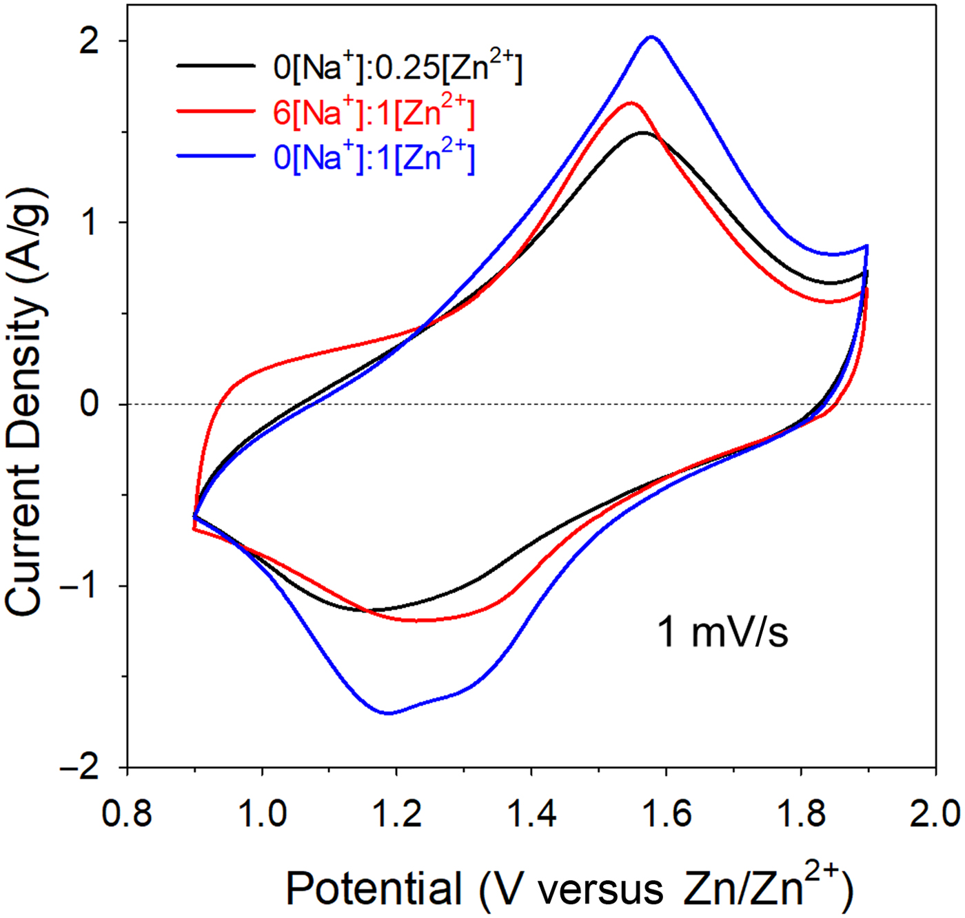
In our previous report, we identified a mixed-salt electrolyte comprising 0.75 M Na2SO4 + 0.25 M ZnSO4 (denoted as 6[Na+]:1[Zn2+]) that optimized capacity and rate capability for 3D MnOx@CNF electrodes, while also supporting greater cycling stability than that observed with 1 M ZnSO4 (“0[Na+]:1[Zn2+]”).[Reference Ko, Sassin, Parker, Rolison and Long16] For the current study, we also performed tests with 0.25 M ZnSO4 (“0[Na+]:0.25[Zn2+]”) to explore the effects of Zn2+ concentration. Representative cyclic voltammograms for MnOx@CNF cycled from 0.9 to 1.9 V versus Zn/Zn2+ in each of these three electrolytes show reversible redox peaks that are ascribed to a Zn2+-based process (Fig. 1).[Reference Song, Tan, Chao and Fan3] In the case of the Na+-containing electrolyte, redox peaks are superimposed on a pseudocapacitive background, as we reported previously.[Reference Ko, Sassin, Parker, Rolison and Long16] At 1 mV/s, the peak current is highest in 1 M ZnSO4, in keeping with the higher concentration of Zn2+ at the MnOx surface; at increasing rates, the capacity derived by integrating the redox peaks decreases by ~90% at 100 mV/s (Fig. S1). The mixed-salt electrolyte offers higher capacity retention at increasing scan rates due to Na+-supported pseudocapacitive reactions at the MnOx.
Cyclic voltammograms of MnOx@CNF electrode cycled at 1 mV/s from 0.9 to 1.9 V versus Zn/Zn2+ in 0[Na+]:0.25[Zn2+], 6[Na+]:1[Zn2+], and 0[Na+]:1[Zn2+] electrolytes.

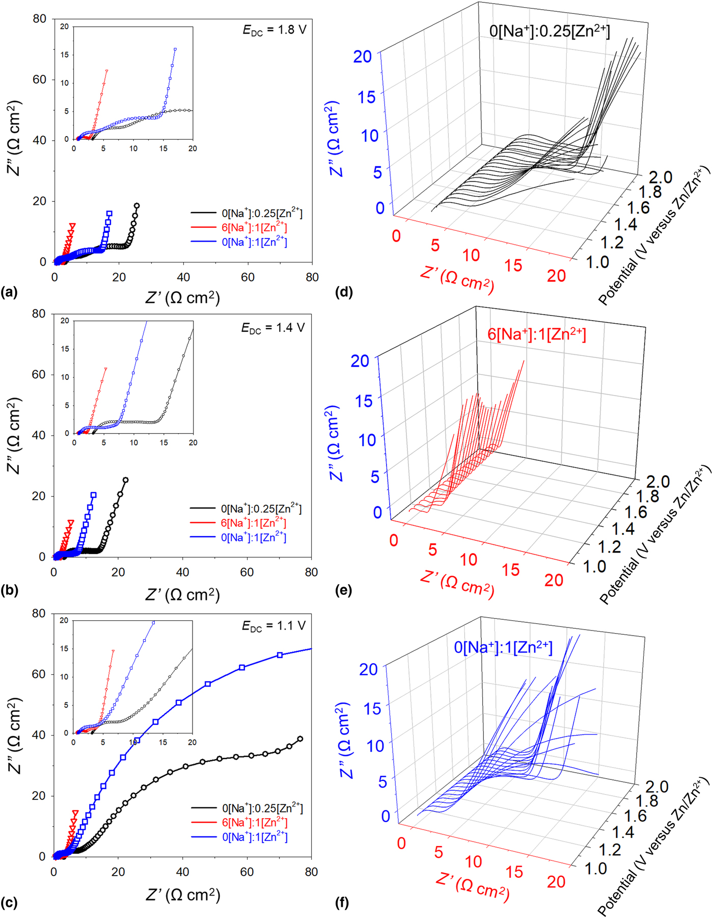
Electrochemical impedance provides valuable information on the dynamics of charge-storage processes in such complex electrode/electrolyte combinations.[Reference Ko, Sassin, Parker, Rolison and Long16] We translate impedance data collected at multiple DC potentials (E DC) from 1.0 to 1.9 V versus Zn/Zn2+ into Nyquist plots that reveal changes in the charge-transfer resistance for this series of electrolytes. At more positive potentials (E DC = 1.8 V) where the MnOx is Mn(IV)-rich (i.e., “charged”), Nyquist plots exhibit relatively simple charge-transfer arcs [Fig. 2(a)]; the highest charge-transfer resistance occurs for the two Zn2+-only electrolytes (R CT = ~15–20 Ω cm2) compared with an order of magnitude lower value for the mixed-salt electrolyte (~2 Ω cm2). In 0[Na+]:0.25[Zn2+], both the series resistance and R CT are higher because of the lower bulk ionic conductivity (14 mS/cm) of this electrolyte and a lower concentration of Zn2+ within the electrode volume.
Representative 2D Nyquist plots taken at stepped potentials from (a) 1.8 V, (b) 1.4 V, and (c) 1.1 V versus Zn/Zn2+ for MnOx@CNF electrodes and corresponding 3D Nyquist plots cycled in (d) 0[Na+]:0.25[Zn2+], (e) 6[Na+]:1[Zn2+], and (f) 0[Na+]:1[Zn2+] electrolytes.
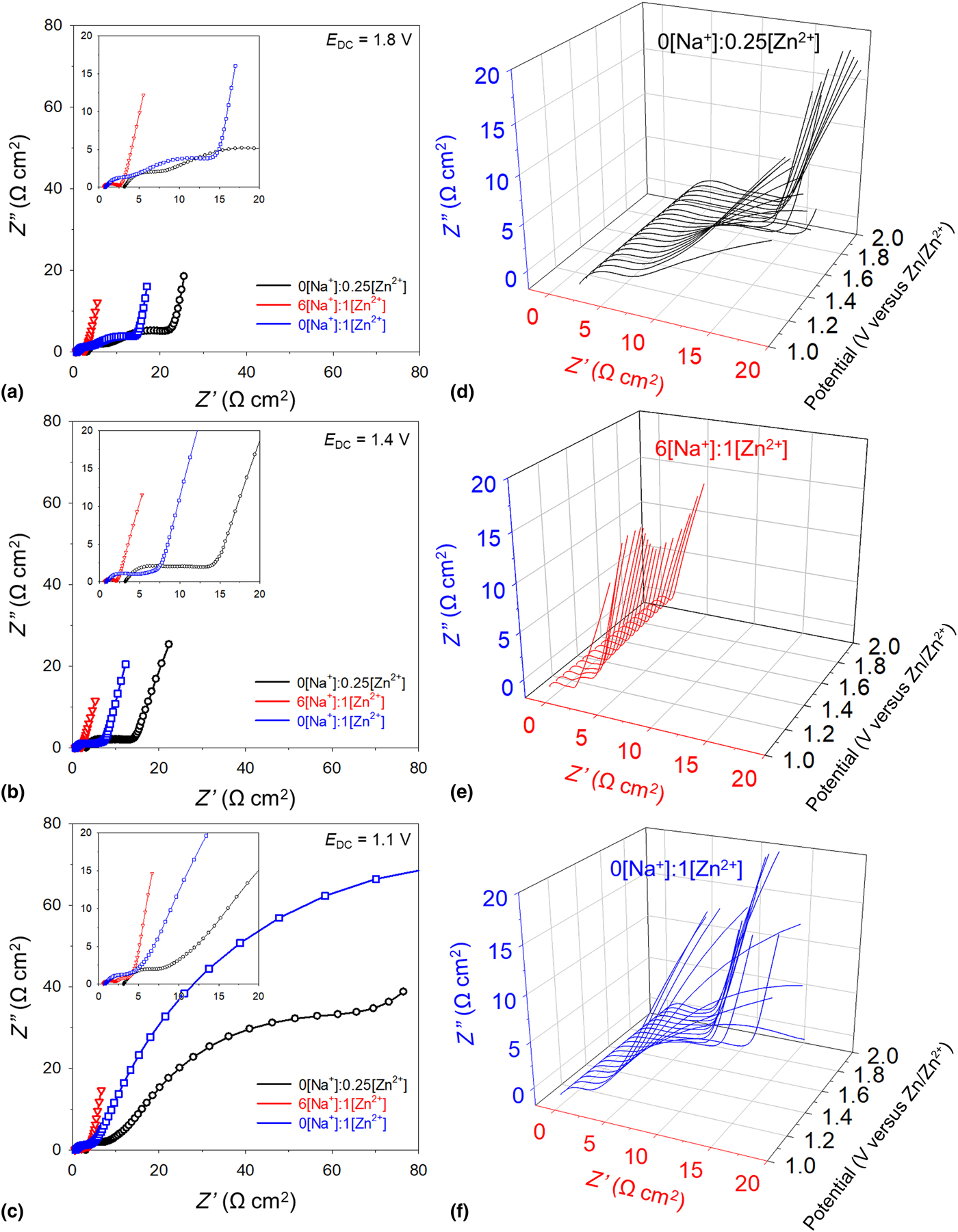
The Nyquist plots are qualitatively unchanged near the half-wave potential of the observed redox peaks (E DC = 1.4 V) with only a slight decrease in R CT for the two Zn2+-only containing electrolytes [~5–15 Ω cm2; Fig. 2(b)]. At E DC = 1.1 V, where MnOx should be in a Mn(III)-rich state (i.e., “discharged”), all three electrolytes exhibit two semicircles, but the second charge-transfer arc dominates the spectra for Zn2+-only containing electrolytes, with R CT values of ~110 and ~200 Ω cm2 for 0[Na+]:0.25[Zn2+] and 0[Na+]:1[Zn2+], respectively [Fig. 2(c); expanded scale shown in Fig. S2]. In the mixed-salt electrolyte, the second charge-transfer arc remains low (R CT < 3 Ω cm2) even at such reducing conditions, consistent with the improved rate capability obtained in this electrolyte (Fig. S1).
To better visualize changes in charge-transfer resistance as a function of charge state, we constructed 3D plots of Nyquist data (Z′ versus −Z″ versus E DC) from impedance spectra collected in 50 mV steps from 1.0 to 1.9 V [Figs. 2(d)–2(f); 3D plots at a different viewing angle are shown in Fig. S3]. Rendering impedance data in this manner complements the treatment described in our prior report, where 3D Bode representations of real and imaginary capacitance were used to interrogate charge-storage dynamics.[Reference Ko, Sassin, Rolison and Long24] In the mixed-salt electrolyte, the 3D Nyquist plot confirms that impedance is relatively unchanged across this entire potential region, with R CT < 3 Ω cm2 [Fig. 2(e)]. In the Zn2+-only electrolytes, the second charge-transfer arc observed at potentials <1.4 V is larger by an order-of-magnitude versus that observed for the mixed-salt electrolyte [Figs. 2(d) and 2(f)].
To complement this electrochemical analysis of the potential-dependent behavior of MnOx@CNF, we performed a series of ex situ measurements to determine changes in electrode structure and composition as a function of state-of-charge and electrolyte composition. Samples of MnOx@CNF were first scanned using linear-sweep voltammetry at 1 mV/s through the oxidation/reduction cycle to the targeted discharge (0.9 V) and charge (1.9 V) potentials and subsequently conditioned at the final potential for 30 min. After rinsing and drying, representative electrochemically conditioned MnOx@CNFs were analyzed by SEM coupled with EDS and the results compared to native (uncycled) MnOx@CNF.
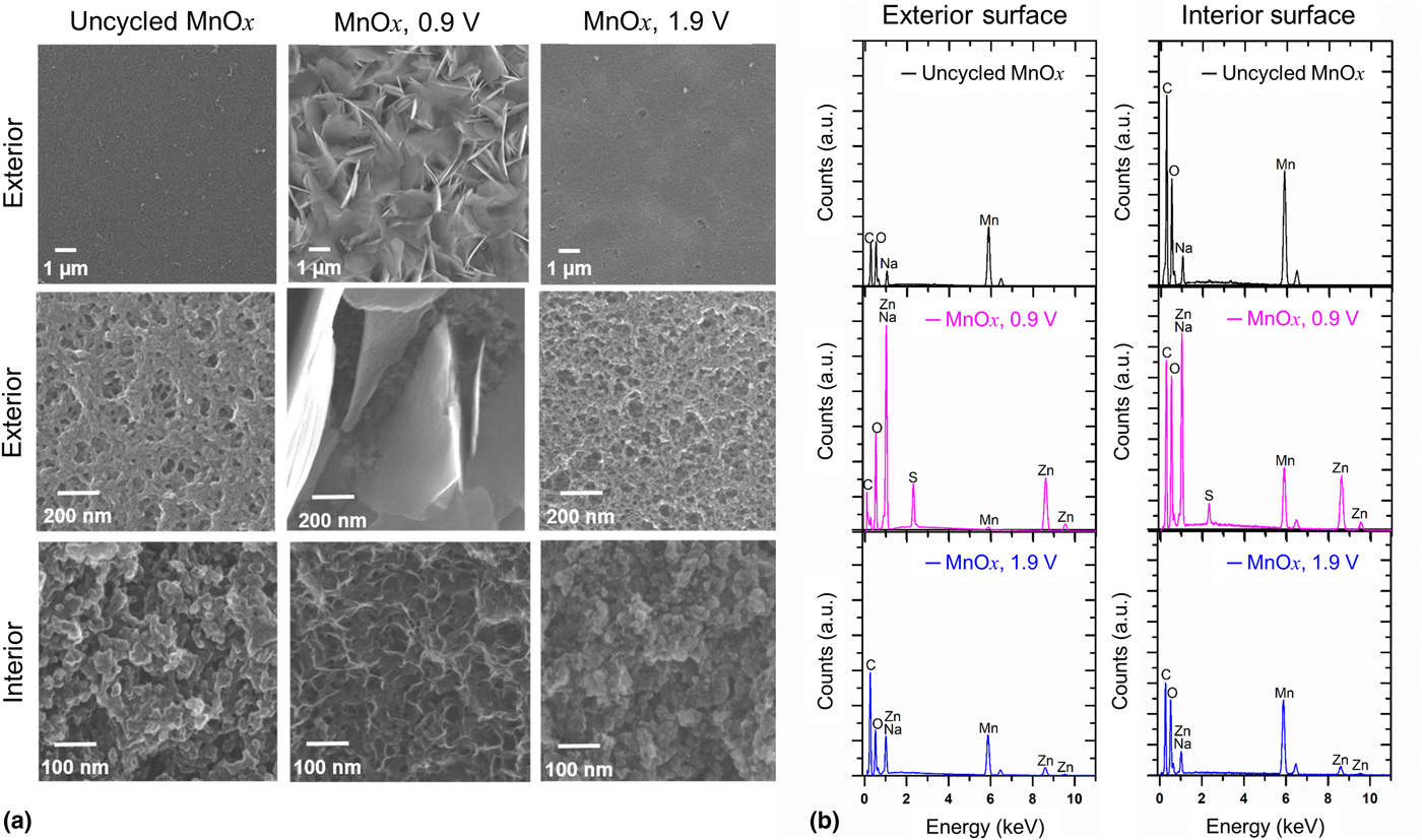
The native MnOx@CNF exhibits an aperiodic distribution of mesopores and small macropores (10–70 nm), homogeneously distributed through the ~80-μm thickness of the electrode [Fig. 3(a)].[Reference Ko, Sassin, Parker, Rolison and Long16, Reference Sassin, Hoag, Willis, Kucko, Rolison and Long18] After conditioning at 0.9 V (discharged state) in the mixed-salt electrolyte, we observe micrometer-sized platelets covering the outer boundary of the electrode. At low magnification the surface appears obstructed, but the pore structure of MnOx@CNF is still visible beneath the platelet-like deposits at higher magnification, although the interior walls exhibit some coarsening. The inner pore volume must remain accessible because the pseudocapacitive signature persists at these reducing potentials (Fig. 1). After “recharging” at 1.9 V, the exterior and interior surface of the MnOx@CNF electrode revert to the appearance of the uncycled form, with the platelet deposits no longer observable. Similar trends in exterior and interior morphology are observed in the Zn2+-only containing electrolytes; larger precipitates are present after cycling in 1 M ZnSO4 [Fig. S4(a)] with wispy sheet-like deposits in 0.25 M ZnSO4. We posit that this boundary coating is the source of the additional charge-transfer arc and overall higher resistance observed from impedance measurements after conditioning at reducing potentials (Fig. 2), particularly when 1 M ZnSO4 is used. In both Zn2+-only electrolytes, the appearance of uncycled MnOx@CNF is essentially recovered after recharge [Fig. S5(a)].
(a) Scanning electron micrographs of ex situ prepared MnOx@CNF cycled in 6[Na+]:1[Zn2+] mixed-salt electrolyte and the corresponding (b) elemental analysis performed by EDS tracking mainly Mn, Zn, and S species.

The SEM-observed precipitates are coincident with changes in the EDS spectra, namely the appearance of a prominent Zn peak after conditioning at 0.9 V, accompanied by a diminished Mn peak (attributed to MnOx being screened by overlying precipitates at the MnOx@CNF exterior). The presence of Zn is noted deep in the interior of the MnOx@CNF, albeit at a lower Zn:Mn peak ratio than observed at the exterior, suggesting that Zn-containing species are more finely dispersed at the walls that define the voids of the CNF. Concomitant with Zn detection, we observe peaks for S, arising from incorporation of electrolyte-derived sulfate. The x-ray photoelectron spectra of the same series of samples (exterior only) confirm that S is present as SO42– and is always accompanied by Zn regardless of the state-of-charge (Fig. S6). Considering recent reports of the formation of zinc–hydroxide–sulfate species at manganese oxides reduced in aqueous ZnSO4 electrolytes,[Reference Lee, Lee, Kim, Chung, Cho and Oh11, Reference Pan, Shao, Yan, Cheng, Han, Nie, Wang, Yang, Li, Bhattacharya, Mueller and Liu13, Reference Lee, Seo, Lee, Yoon, Kim, Chung, Cho and Oh14] we used these XPS results to calculate a stoichiometry of Zn4.6(OH)5.8−x(SO4)·xH2O for the precipitated film (see Supporting Information for more details). Upon reoxidation to 1.9 V, the peaks for S and Zn markedly decrease, indicating a quasi-reversible precipitation/dissolution process. We observe similar EDS trends for Zn and S detection in the Zn2+-only electrolytes [Fig. S4(b) and S5(b)].
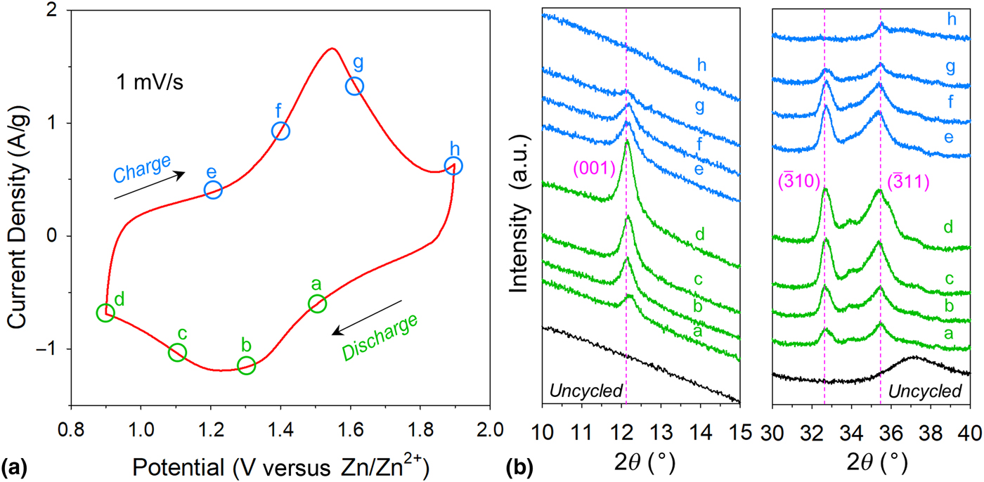
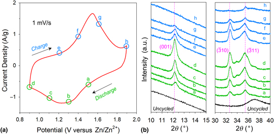
The identity of the Zn- and S-containing discharge product is further verified by XRD, a common tool used to analyze zinc-ion materials. For example, while many reports confirm crystallographic Zn2+ insertion into particular manganese oxide polymorphs via shifts in diffraction peaks,[Reference Song, Tan, Chao and Fan3] other XRD studies show that crystalline Zn4(OH)6(SO4)·xH2O forms during reduction/discharge.[Reference Song, Tan, Chao and Fan3, Reference Lee, Lee, Kim, Chung, Cho and Oh11, Reference Pan, Shao, Yan, Cheng, Han, Nie, Wang, Yang, Li, Bhattacharya, Mueller and Liu13, Reference Lee, Seo, Lee, Yoon, Kim, Chung, Cho and Oh14] We performed ex situ measurements of the MnOx@CNF electrode in the mixed-salt electrolyte as conditioned at potentials of 1.5, 1.3, 1.1, and 0.9 V for discharge and 1.2, 1.4, 1.6, and 1.9 V for charge [see the corresponding current versus potential plots and diffraction patterns of MnOx@CNF specimens conditioned at the specified potentials; Figs. 4(a) and 4(b)]. The disordered nature of the oxide in MnOx@CNF limits the utility of XRD for identifying ion-insertion reactions (e.g., for Zn2+) that would otherwise be accompanied by expansion/contraction of a crystalline lattice. Yet, the relatively featureless XRD background of MnOx@CNF also simplifies identification of any new phases that may form during the reduction/oxidation cycle.
(a) Representative cyclic voltammogram of MnOx@CNF cycled in the 6[Na+]:1[Zn2+] electrolyte showing the conditioning points (circles) for discharge (green) and charge (sky blue) where potential was held for 30 min. (b) Phase evolution of ex situ XRD patterns during this corresponding charge/discharge cycle. Dashed pink lines refer to the main Zn4(OH)6(SO4)·xH2O reflections (ICDD #00-044-0675).

New diffraction peaks are clearly observed for MnOx@CNF conditioned in the 6[Na+]:1[Zn2+] electrolyte from 1.5 to 0.9 V, with increasing intensity upon deeper reduction [Fig. 4(b); full spectra shown in Fig. S7]. Prominent peaks noted at 12°, 33°, and 35° are assigned to reflections for Zn4(OH)6(SO4)·xH2O, a layered structure comprising Zn(OH)2 sheets separated by ZnSO4 and water molecules. The XRD-confirmed presence of Zn4(OH)6(SO4)·xH2O has been attributed to increases in local pH that arise at manganese oxide surfaces upon H+-insertion, where electrochemically generated OH– reacts with Zn2+ and SO42– from the electrolyte to form this insoluble, crystalline product.[Reference Lee, Seo, Lee, Yoon, Kim, Chung, Cho and Oh14] In another study, Pan et al. performed high-resolution transmission electron microscopy to identify lattice fringes corresponding to the d-spacing from the discharged product, manganese oxyhydroxide (MnOOH), showing further evidence of H+-insertion.[Reference Pan, Shao, Yan, Cheng, Han, Nie, Wang, Yang, Li, Bhattacharya, Mueller and Liu13] Unfortunately, the reaction product, Zn4(OH)6(SO4)·xH2O, could not be imaged due to high-energy electron beam damage, though x-ray reflections of this precipitate were observed.[Reference Pan, Shao, Yan, Cheng, Han, Nie, Wang, Yang, Li, Bhattacharya, Mueller and Liu13]
Upon reversal of the conditioning potentials for MnOx@CNF toward an oxidized (charged) state, these peaks gradually diminish until the largely featureless XRD background is recovered at 1.9 V [Fig. 4(b)], coincident with the disappearance of the platelet-like features observed by SEM [Fig. 3(a)]. For the Zn2+-only electrolytes, a clear distinction between the 0[Na+]:0.25[Zn2+] versus 0[Na+]:1[Zn2+] electrolyte is observed, with the reflections at 12°, 33°, and 35° still evident upon conditioning at 1.9 V in the electrolyte with higher Zn2+ concentration (Fig. S8). The suppression of the reflections upon conditioning MnOx@CNF at 1.9 V in 0.25 M ZnSO4 are attributed to reversible precipitation/dissolution of Zn4(OH)6(SO4)·xH2O (Fig. S9).
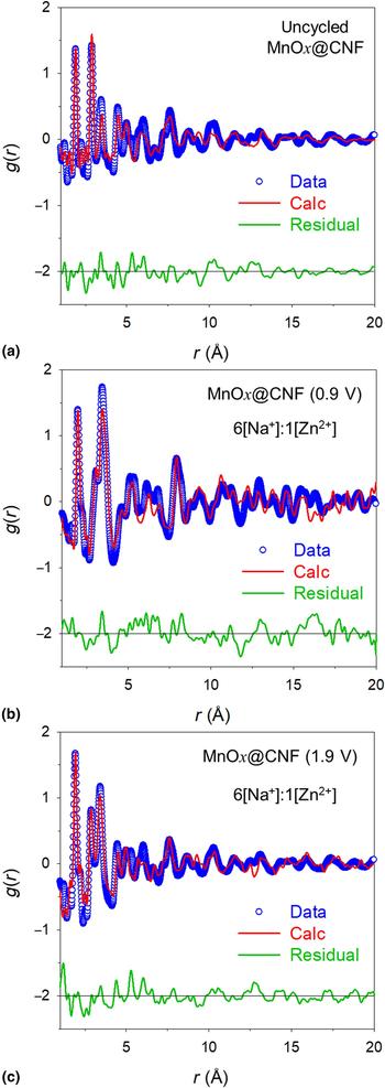
To probe possible structural changes at the nanoscale MnOx, we turned to differential PDF (dPDF) analysis. The dPDF technique allows one to subtract the signal of a substrate (in this case the CNF) to evaluate structural changes at the angstrom scale of a thin film (in this case the MnOx). For dPDF studies, we focused on three ex situ samples from the optimized 6[Na+]:1[Zn2+] electrolyte: (i) native MnOx; (ii) MnOx cycled to 0.9 V and held at potential; and (iii) MnOx cycled from 0.9 to 1.9 V and held at 1.9 V. The PDF spectrum for native MnOx has a structure analogous to that previously reported for birnessite-like MnOx electrolessly deposited within CNF paper [Fig. 5(a)].[Reference Donakowski, Wallace, Sassin, Chapman, Parker, Long and Rolison25] In the discharged state, a similar structure was exhibited but with additional correlations at 3.5 Å; these features could be well fit with a structure of Zn4(OH)6(SO4)·xH2O with a small spherical envelop approximation of ~5 Å [Fig. 5(b)]. The correlation at 3.5 Å corresponds to Zn–Zn and/or Zn–S, which suggests that these species are present on the birnessite material even after rigorous rinsing with water. Recharged MnOx has a dPDF similar to that of native MnOx [Fig. 5(c)] but with an ~0.5 Å decrease in the lamellar spacing (Table S1).
(a) The dPDF showing pair correlations for MnOx@CNF. The material exhibits turbostratic disorder of the lamellar MnOx sheets. (b) The dPDF showing pair correlations for MnOx@CNF cycled to 0.9 V, which shows that the signal is dominated by Zn4(OH)6(SO4)·xH2O that is not present in the pristine or charged material. The material contains greater correlations at 3.5 Å that were fit with a model of Zn4(OH)6(SO4)·xH2O with a spherical particle size of ~5 Å. (c) The dPDF showing pair correlations for MnOx@CNF cycled to 1.9 V (charge), which shows similar correlation to uncycled MnOx@CNF.

The nature of an intercalant can be tracked by changes in the lamellar spacing. Refining the dPDF pattern by assigning a “Na+” to the ionic site between the layers resulted in a large error for each sample (approximately ±0.5 Na+), which we attribute to the disorder present within the birnessite layers. Qualitatively, the discharged electrode shows the lowest amount of ionic content. If Zn2+ intercalation occurs, one would anticipate the higher atomic number of Zn would contribute to a greater scattering species and thus increase in apparent content. It is possible that a mix of H+ and Zn2+ is present with the low Z of H+ or H3O+ counteracting the high Z of Zn. Further element-specific studies would have to be performed to evaluate Zn2+ speciation in the material because the large amount of Zn4(OH)6(SO4)·xH2O that forms can obscure signals from interlamellar Zn2+, thus Zn2+ intercalation cannot be precluded.[Reference Sun, Wang, Hou, Yang, Fan, Ma, Gao, Han, Hu, Zhu and Wang15]
Conclusions
Our results provide insights into the charge-storage mechanisms of birnessite-like MnOx in MnOx@CNF after cycling in zinc-ion relevant electrolytes. Electron microscopy and x-ray techniques confirm that Zn4(OH)6(SO4)·xH2O reversibly precipitates and dissolves during charge/discharge in Zn2+-containing electrolytes, and that salt can only form if H+ insertion is the dominant redox process. In the mixed-salt electrolyte, although the Zn4(OH)6(SO4)·xH2O still forms but the electrode maintains low R CT throughout the cycling window, which explains the high-rate capability and may underwrite the improved cycle life, as we previously reported.[Reference Ko, Sassin, Parker, Rolison and Long16] The rigorous ex situ investigation of MnOx@CNF electrodes in this report should serve as a template for studying the complex charge-storage processes in zinc-ion cells. Future work will be directed toward in situ/operando measurements that track complex convolution of ion insertion, precipitation, and dissolution that occur in ZnSO4 electrolytes.
Supplementary material
The supplementary material for this article can be found at https://doi.org/10.1557/mrc.2019.3.
Acknowledgments
This work was supported by the U.S. Office of Naval Research. J.S.K. is an NRL-NRC Postdoctoral Associate (2016–2018). This research used resources of the Advanced Photon Source, a U.S. Department of Energy (DOE) Office of Science User Facility operated for the DOE Office of Science by Argonne National Laboratory under Contract No. DE-AC02-06CH11357. The authors would like to thank Dr. Olaf Borkiewicz of Argonne National Laboratory for data acquisition, integration, and assistance.