General concepts of sepsis
Sepsis, SIRS, and MODS
Sepsis is a complex life-threatening condition, being considered as one of the major causes of severe illness in veterinary species and human beings (Alberti et al., Reference Alberti, Brun-Buisson, Goodman, Guidici, Granton, Moreno, Smithies, Thomas, Artigas and Le Gall2003; Taylor, Reference Taylor2015). It occurs when an infectious agent triggers a systemic inflammatory response (SIRS) characterized by an imbalance between pro and anti-inflammatory elements that may cause disorders in hormonal, metabolic, cardiovascular, and coagulation systems, among others (Hotchkiss and Karl, Reference Hotchkiss and Karl2003; Singer et al., Reference Singer, Deutschman, Seymour, Shankar-Hari, Annane, Bauer, Bellomo, Bernard, Chiche, Coopersmith, Hotchkiss, Levy, Marshall, Martin, Opal, Rubenfeld, Van, Vincent and Angus2016; Smyth et al., Reference Smyth, Brace-Mcdonnell and Perkins2016). Therefore, sepsis has been defined as a SIRS caused by an infectious agent. In some cases, this inflammatory response can be deregulated and lead to multiple organ dysfunction syndrome (MODS), resulting in death (Levy et al., Reference Levy, Fink, Marshall, Abraham, Angus, Cook, Cohen, Opal, Vincent and Ramsay2003; Riedel and Carroll, Reference Riedel and Carroll2013).
Sepsis has a significant impact on human health due to its severity and high mortality rates, being a global health priority by the World Health Organization and the World Health Assembly (WHO, 2017). In addition, sepsis and its consequences also significantly impact animal health. For example, calves and foals are highly vulnerable to endotoxemia, characterized by bacteremia or septicemia due to Gram-negative organisms, presenting very high mortality rates (Fecteau et al., Reference Fecteau, Smith and George2009; Bonelli et al., Reference Bonelli, Meucci, Divers, Radcliffe, Jose-Cunilleras, Corazza, Guidi, Tognetti, Castagnetti, Intorre and Sgorbini2015a, Reference Bonelli, Meucci, Divers, Boccardo, Pravettoni, Meylan, Belloli and Sgorbini2018). Therefore, sepsis is an example of a topic that should be addressed from the ‘One-Health’ point of view (Robinson et al., Reference Robinson, Bu, Carrique-Mas, Fèvre, Gilbert, Grace, Hay, Jiwakanon, Kakkar, Kariuki, Laxminarayan, Lubroth, Magnusson, Ngoc, Van Boeckel and Woolhouse2016)
Causes
Although to the authors’ knowledge there is no specific data in veterinary medicine, it was described that more than 70% of the documented sepsis cases in human beings are attributed to bacteria, leading to pneumonia and urinary tract, intra-abdominal, skin, and other soft tissue infections (Motzkus and Luckmann, Reference Motzkus and Luckmann2017). Sepsis can also be triggered by other infectious agents like parasites, fungi, or viruses, although, for example, only around 1% of sepsis cases are classified as viral (Blanco et al., Reference Blanco, Muriel-Bombín, Sagredo, Taboada, Gandía, Tamayo, Collado, García-Labattut, Carriedo, Valledor, De Frutos, López, Caballero, Guerra, Álvarez, Mayo and Villar2008; Zahar et al., Reference Zahar, Timsit, Garrouste-Orgeas, Français, Vesim, Descorps-Declere, Dubois, Souweine, Haouache, Goldgran-Toledano, Allaouchiche, Azoulay and Adrie2011; Lin et al., Reference Lin, McGinley, Drysdale and Pollard2018). However, the prevalence of non-bacterial sepsis might be underdiagnosed because several reports indicated higher incidence. For example, one study reported that 21% of pediatric sepsis cases were attributed to viruses (Weiss et al., Reference Weiss, Fitzgerald, Pappachan, Wheeler, Jaramillo-Bustamante, Salloo, Singhi, Erickson, Roy, Bush, Nadkarni and Thomas2015).
Physiopathology
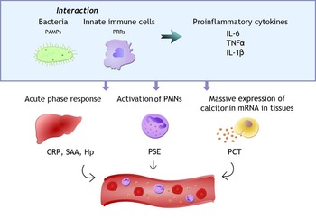
The origin of sepsis relies on the interaction between the causal agent and the ‘pattern-recognition receptors’ (PRRs) present on cells of the innate immune response (Fig. 1). These receptors can detect the ‘pathogen-associated molecular patterns’ (PAMPs) that are conserved structures of pathogens. In addition, they can detect molecules produced by the host after damage, independent of the causal agent, known as ‘damage-associated molecular patterns’. That is why sepsis and non-septic SIRS often have many similarities, making the diagnosis of sepsis challenging (Lewis et al., Reference Lewis, Chan, Pinheiro, Armitage-Chan and Garden2012; Faix, Reference Faix2013).
Proinflammatory response during sepsis. PAMPs, pathogen-associated molecular patterns; PRRs, pattern recognition receptors; IL-6, interleukin-6; TNFα, tumor necrosis factor α; IL-1β, interleukin-1β; CRP, C-reactive protein; SAA, serum amyloid A; PMN, polymorphonuclear leukocytes; PSE, presepsin; PCT, procalcitonin. Figure and images were created by the authors.

The interactions between the pathogen and cells trigger the production of proinflammatory cytokines, mainly interleukin-6 (IL)-6, tumor necrosis factor α (TNF-α), and IL-1β, that initiate the innate immune response and contribute to the onset of SIRS in two main ways, as they:
• Induce the acute-phase response with the production of acute phase proteins (APPs) such as C-reactive protein (CRP), serum amyloid A (SAA), or haptoglobin (Hp) (Faix, Reference Faix2013).
• Activate endothelial cells and the production and attraction of polymorphonuclear leukocytes (PMNs) to the site of damage and into the general circulation. The activation of PMNs membrane receptors leads to the release of molecules such as sCD14 (known as presepsin (PSE)) or sTREM-1 from these receptors (Cray, Reference Cray2012).
In addition, both the direct stimulation of the pathogens themselves, and the indirect stimulation by these three cytokines, induce the expression of calcitonin mRNA in numerous extra-thyroidal tissues, leading to a widespread production of the protein procalcitonin (PCT) (Nakamura et al., Reference Nakamura, Kono, Nomura and Utsunomiya2013).
This proinflammatory response evolved to eradicate the causative agent; however, if the response is too severe, organ damage and even organ dysfunction are likely to develop. Although some of the underlying mechanisms are still unknown, altered tissue oxygenation seems to be one of the main causes of the increase in severity. Several factors may be involved in this oxygenation imbalance, such as poor oxygen supply due to hypotension, cellular oxygen impairment due to mitochondrial damage from oxidative stress, and vascular endothelial dysfunction due to loss of barrier integrity and cell death. This endothelial damage and organ dysfunction lead to changes in angiopoietins or lactate, respectively (Angus and van der Poll, Reference Angus and van der Poll2013; Faix, Reference Faix2013).
Finally, another frequent complication of sepsis is the triggering of disseminated vascular coagulation. The link between inflammation and coagulation is complex, but it may be mediated primarily by protease-activated receptors type 1. Usually, these receptors have cytoprotective effects, but when stimulated by high doses of thrombin, their effects on the endothelial cell barrier become disruptive. Also, the imbalance of anticoagulant mechanisms and the suppression of the fibrinolytic system can lead to excessive fibrin deposition. Therefore, biomarkers of coagulation like D-dimer also have a role in monitoring sepsis (Angus and van der Poll, Reference Angus and van der Poll2013).
It is important to point out that many of the mechanisms involved in sepsis remain unknown, especially the reasons for the variability of the response to infection. This variable response can depend on the causal agent, the host genetics, possible concomitant diseases, and animal species (Werling and Coffey, Reference Werling and Coffey2007; Lewis et al., Reference Lewis, Chan, Pinheiro, Armitage-Chan and Garden2012; Gotts and Matthay, Reference Gotts and Matthay2016).
Diagnosis
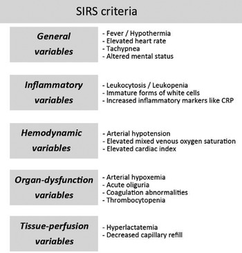
The diagnosis of sepsis is based on a confirmed infection and the presence of two or more SIRS criteria; although in some cases, the infection cannot be documented and is only suspected (Angus and van der Poll, Reference Angus and van der Poll2013; Roy et al., Reference Roy, Kwong, Lambert, Massie and Lockhart2017). The SIRS criteria (Fig. 2) can vary, depending on the author, and generally include various external symptoms such as changes in body temperature, heart rate, or breathing pattern, as well as analytical abnormalities and signs for organ or tissue perfusion dysfunction (Fecteau et al., Reference Fecteau, Smith and George2009; Taylor, Reference Taylor2015; Peach, Reference Peach2017). In human medicine, there are also score systems such as the sequential organ failure assessment score (SOFA), which evaluates breathing, circulation, liver and kidney function, coagulation, and neurologic condition. There is also a shortened version, the Quick-SOFA score, which just considers three points of the SOFA (Mccormack et al., Reference Mccormack, Kulkarni, Sperling and Keller2017; Serafim et al., Reference Serafim, Gomes, Salluh and Póvoa2018; Garbero et al., Reference Garbero, Simões, Martins, Cruz and von Zuben2019). Some scores with adaptations have also been used in the veterinary field, such as the one modified by Corley and Furr (Reference Corley and Furr2003) that showed a low negative predictive value, or the sepsis score for horses adapted from Breuer and Schusser, which includes parameters such as calcium, fibrinogen, and body inner temperature (Breuer and Schusser, Reference Breuer and Schusser2012).
Sepsis is suspected when the patient manifests two or more SIRS criteria with a possible infectious origin (Angus and van der Poll, Reference Angus and van der Poll2013).

Overall, several factors make the diagnosis of sepsis still a challenge:
• Initial clinical manifestations can be quite variable depending on individual factors such as the previous health status, the animal species, or the source of infection (Borghetti et al., Reference Borghetti, Saleri, Mocchegiani, Corradi and Martelli2009; Motzkus and Luckmann, Reference Motzkus and Luckmann2017).
• There are difficulties in many cases to detect the cause of sepsis. Although blood culture is the gold standard to detect bacteremia, it has many limitations, including a 48–72 h delay in results, a high rate of false negatives, and the possibility of false positives by sample contamination (Shozushima et al., Reference Shozushima, Takahashi, Matsumoto, Kojika, Endo and Okamura2011; Singer et al., Reference Singer, Deutschman, Seymour, Shankar-Hari, Annane, Bauer, Bellomo, Bernard, Chiche, Coopersmith, Hotchkiss, Levy, Marshall, Martin, Opal, Rubenfeld, Van, Vincent and Angus2016; Jereb et al., Reference Jereb, Mavric, Skvarc, Drobnic, Dolenc, Strunjas, Luksic and Kmet2019). In addition, there is a lack of diagnostic criteria for other causal agents such as viruses, probably resulting in underestimation of the incidence of non-bacterial sepsis (Amin and Amin, Reference Amin, Amin and Vincent2015; Gotts and Matthay, Reference Gotts and Matthay2016).
• Inflammation and SIRS without any infectious agent involved can lead to very similar clinical features (Pierrakos and Vincent, Reference Pierrakos and Vincent2010).
These challenges can limit the correct diagnosis of sepsis, and thus the initiation of a rapid and adequate treatment to ensure that the patient's life is not endangered. Sepsis treatment is complex and can require intensive care to provide vital-organ support. When the origin is bacterial, the critical element is the rapid inclusion of antibiotics, as delays in antimicrobial therapy can significantly increase the risk of dying (Martin, Reference Martin2012). Therefore, antibiotic treatment is frequently administered in a preventive approach without the confirmation of the bacterial component. This fact results in excessive and sometimes unnecessary use of antibiotics, which increase medical costs, are harmful to host microbiota, and contribute to the development of multidrug-resistant bacteria (Lin et al., Reference Lin, McGinley, Drysdale and Pollard2018; Tosi et al., Reference Tosi, Roat, De Biasi, Munari, Venturelli, Coloretti, Biagioni, Cossarizza and Girardis2018; Grondman et al., Reference Grondman, Pirvu, Riza, Ioana and Netea2020). Antibiotic resistance is increasingly leading to treatment failures and worse prognoses in both infectious and non-infectious conditions (Peralta et al., Reference Peralta, Sánchez, Garrido, De Benito, Cano, Martínez-Martínez and Roiz2007; Hocking et al., Reference Hocking, Ali, d'Angelo, Deshpande, Stevenson, Virdee and Guthrie2021). This overuse of antibiotics is widespread in farm animals, particularly serious in porcine production and remains a crucial area for improvement (Van Boeckel et al., Reference Van Boeckel, Brower, Gilbert, Grenfell, Levin, Robinson, Teillant and Laxminarayan2015; Murphy et al., Reference Murphy, Ricci, Auce, Beechinor, Bergendahl, Breathnach, Bureš, Duarte Da Silva, Hederová, Hekman, Ibrahim, Kozhuharov, Kulcsár, Lander Persson, Lenhardsson, Mačiulskis, Malemis, Markus-Cizelj, Michaelidou-Patsia, Nevalainen, Pasquali, Rouby, Schefferlie, Schlumbohm, Schmit, Spiteri, Srčič, Taban, Tiirats, Urbain, Vestergaard, Wachnik-Święcicka, Weeks, Zemann, Allende, Bolton, Chemaly, Fernandez Escamez, Girones, Herman, Koutsoumanis, Lindqvist, Nørrung, Robertson, Ru, Sanaa, Simmons, Skandamis, Snary, Speybroeck, Ter Kuile, Wahlström, Baptiste, Catry, Cocconcelli, Davies, Ducrot, Friis, Jungersen, More, Muñoz Madero, Sanders, Bos, Kunsagi, Torren Edo, Brozzi, Candiani, Guerra, Liebana, Stella, Threlfall and Jukes2017).
Roles of biomarkers in sepsis
The use of biomarkers for the early diagnosis and monitoring of sepsis can help to improve its management and treatment. Although more than 170 potential biomarkers have been described in human medicine for assessing the diagnosis and prognosis of sepsis, there is still a lack of consensus about their actual usefulness in practice (Pierrakos and Vincent, Reference Pierrakos and Vincent2010; Ivady et al., Reference Ivady, Beres and Szabo2011; Reinhart et al., Reference Reinhart, Bauer, Riedemann and Hartog2012; Liu et al., Reference Liu, Hou, Li, Chen, Wang and Wang2016).
In veterinary medicine, it is also necessary to improve both the diagnosis and prognosis of sepsis. Traditionally, the most used biomarkers to detect sepsis in animals are APPs such as CRP, as well as proinflammatory cytokines. However, these biomarkers have limitations: APPs have low specificity to detect sepsis because they increase in many non-septic conditions, while cytokines, in addition to their low specificity, have a short half-life that makes their detection difficult (Ercan et al., Reference Ercan, Tuzcu, Başbug, Tuzcu and Alim2016). New sepsis biomarkers are emerging on the veterinary side, with the intent to overcome these drawbacks (Ercan et al., Reference Ercan, Tuzcu, Başbug, Tuzcu and Alim2016; Kirbas et al., Reference Kirbas, Kandemir, Celebi, Hanedan and Timurkan2019).
Lipopolysaccharide (LPS) endotoxin concentrations have been measured with ELISA kits and reported as potential biomarkers. However, LPS molecules can decrease significantly within one hour after taking the sample, and the interpretation of the results is quite complex, as they are heterogeneous and highly variable depending on the bacteria from which they originated. In addition, LPS molecules detected in plasma could be a tiny proportion of LPS remaining after clearance of bacterial infections and would no longer represent the initial amounts of LPS capable of stimulating host cells. Therefore, LPS concentrations have limited clinical utility (Peek et al., Reference Peek, Borah, Semrad, McGuirk, Slack, Patton, Coombs, Lien and Darien2004; Senior et al., Reference Senior, Proudman, Leuwer and Carter2011; Gnauck et al., Reference Gnauck, Lentle and Kruger2016).
This review aims to update the state of the main biomarkers currently used to diagnose sepsis in large farm animals (namely pigs, horses, and cattle) and other biomarkers that could potentially be applied in veterinary medicine. For this purpose, they will be classified here into traditional biomarkers of sepsis, specific biomarkers for bacterial sepsis, and biomarkers that can provide additional information in sepsis.
Traditional biomarkers of sepsis
This category includes APPs and cytokines.
Acute phase proteins (APPs)
The acute phase response is part of the first-line host defense in response to pathogenic states such as infection, inflammation, or tissue damage. In response to these stimuli, first-line defense cells activate the release of cytokines that induce changes in the concentration of APPs. APPs are helpful to diagnose and monitor even subclinical conditions that can lead to impaired growth and reproductive performances in animals (Petersen et al., Reference Petersen, Nielsen and Heegaard2004; Quereda et al., Reference Quereda, Gómez, Seva, Ramis, Cerón, Muñoz and Pallarés2012; Schrödl et al., Reference Schrödl, Büchler, Wendler, Reinhold, Muckova, Reindl and Rhode2016).
The origin of most APPs has been traditionally associated with the liver, although there is growing evidence that it can also be extra-hepatic. According to their dynamics, APPs can be classified as positive if their concentrations increase in the acute phase reaction or as negative if their concentrations decrease. Positive APPs can also be classified as major (with very high and rapid increases upon stimulation and rapid decreases after cessation of the stimulus) and moderate (with milder and slower dynamics in general). The classification and pattern of the response differs among animal species (Eckersall and Bell, Reference Eckersall and Bell2010; Cerón et al., Reference Cerón, Martinez-Subiela, Tecles and Caldin2014).
The main advantage of APPs is that their basal levels are very low compared to other biomarkers of inflammatory responses, and, generally, marked increases in major APPs together with compatible external clinical signs can lead to the possible diagnosis of sepsis. However, rises of high magnitude can also occur in other inflammatory conditions, especially in immune-mediated pathologies, and APPs generally show a low specificity to detect bacterial sepsis (Eckersall and Bell, Reference Eckersall and Bell2010; Reczyńska et al., Reference Reczyńska, Zalewska, Czopowicz, Kaba, Zwierzchowski and Bagnicka2018). Nevertheless, because APP assays are becoming more available and affordable, they can provide valuable information in the daily farm routine, mainly in complementing the diagnosis, monitoring the development of the disease, and establishing the patient's prognosis (Cerón, Reference Cerón2019).
C-reactive protein (CRP)
This positive APP belongs to the pentraxin family and is one of the most traditionally used biomarkers in diagnosing sepsis in people and animals (Ansar and Ghosh, Reference Ansar and Ghosh2013; Enguix-Armada et al., Reference Enguix-Armada, Escobar-Conesa, La Torre and De La Torre-Prados2016). Its primary biologic functions include complement activation, opsonization, and modulation of monocytes and macrophages and cytokine production (Ballou and Lozanski, Reference Ballou and Lozanski1992; Singh et al., Reference Singh, Ngwa and Agrawal2020). In species in which it is a major APP, such as the pig, serum CRP concentration can increase greater than 100-fold; normally, it rises above physiological values as soon as 6 h after the stimulus and has a half-life of about 47 h (Jain et al., Reference Jain, Gautam and Naseem2011; Takata et al., Reference Takata, Wada, Tamura, Koide, Higaki, Mikura, Yasutake, Hirao, Nakamura, Honda, Nagatomo, Tanaka, Sohara, Watanabe, Yokoyama, Saraya, Kurai, Ishii and Goto2011). CRP measurement allows a fast and economical evaluation of the inflammatory response at an early stage, which can provide information about the severity of the pathology and the response to the treatment (Flanders et al., Reference Flanders, Stein, Shochat, Sellers, Holland, Maselli, Drew, Reingold and Gonzales2004; Schmit and Vincent, Reference Schmit and Vincent2008). However, its role in diagnosing sepsis is limited by its variable sensitivity and specificity, which, respectively, have been reported to be 30–97.2% and 75–100% among human subjects (Wyllie et al., Reference Wyllie, Bowler and Peto2005; Chan and Gu, Reference Chan and Gu2011). Therefore, in human medicine, CRP is being replaced or combined with other markers such as PCT (Müller et al., Reference Müller, Harbarth, Stolz, Bingisser, Mueller, Leuppi, Nusbaumer, Tamm and Christ-Crain2007; Qu and Summah, Reference Qu and Summah2009; Kibe et al., Reference Kibe, Adams and Barlow2011).
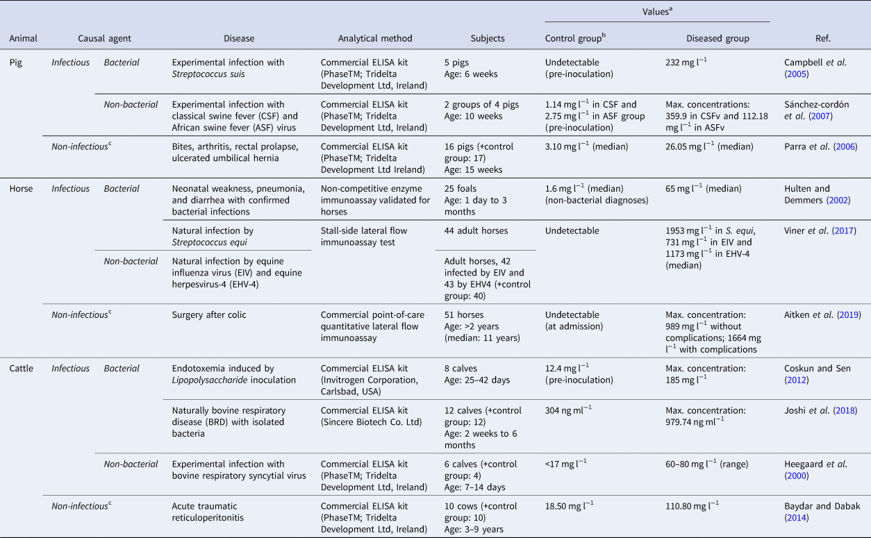
Examples of studies involving CRP in large animals have been summarized in Table 1, and are tabulated according to species, pathological condition (bacterial infections; non-bacterial infections – viral, parasitic, or fungal-; and non-septic inflammation), and analytical method used for CRP measurement. The interpretation of CRP is different for pigs, horses, and ruminants. In pigs, it is considered a major APP: CRP concentration rises in many inflammatory diseases, including those caused by bacterial infections (Lauritzen et al., Reference Lauritzen, Lykkesfeldt, Skaanild, Angen, Nielsen and Friis2003; Soni and Adebiyi, Reference Soni and Adebiyi2017; Yin et al., Reference Yin, Liu, Liu, Huang, Ci, Zhao and Yang2017), viral infections, and non-infectious etiologies (Gómez-Laguna et al., Reference Gómez-Laguna, Salguero, Pallarés, Fernández de Marco, Barranco, Cerón, Martínez-Subiela, Van Reeth and Carrasco2010; Escribano et al., Reference Escribano, Gutiérrez, Tecles and Cerón2015a). Therefore, its usefulness to discern bacterial sepsis is unclear (Heegaard et al., Reference Heegaard, Stockmarr, Pĩeiro, Carpintero, Lampreave, Campbell, Eckersall, Toussaint, Gruys and Sorensen2011). In horses, CRP is considered a moderate APP that slightly increases 3–5 days after stimuli, and does not appear useful to detect sepsis because it increases similarly to non-bacterial inflammation (Yamashita et al., Reference Yamashita, Fujinaga, Okumura, Takiguchi, Tsunoda and Mizuno1991; Do Carmo et al., Reference Do Carmo, Da Silva, Klauck, Pazinato, Moura, Duarte, Duarte, Bochi, Moresco and Stefani2015; Taylor, Reference Taylor2015; Zabrecky et al., Reference Zabrecky, Slovis, Constable and Taylor2015). In cattle, CRP is generally not considered an APP, as no acute increases have been described in certain pathologies such as respiratory disease (Nakajima et al., Reference Nakajima, Momotani, Murakami, Ishikawa, Morimatsu, Saito, Suzuki and Yasukawa1993; Prohl et al., Reference Prohl, Schroedl, Rhode and Reinhold2015), although some studies have shown mild CRP increases under conditions such as stress, intestinal obstruction, clinical mastitis, parasitic infections, or septicemia, which could make CRP useful in assessing the general status of herds (Lee et al., Reference Lee, Hsiao, Wu, Lin, Lee, Fung, Chen, Chen and Chu2003; Hussain et al., Reference Hussain, Uppal, Randhawa and Sood2015; Akgül et al., Reference Akgül, Aya, Ilmaz and Özkan2019; Appelt et al., Reference Appelt, da Silva, Cazarotto, Machado, Rodrigues, Norbury, Baldissera, Alba, Gris and Mendes2019; Dalanezi et al., Reference Dalanezi, Schmidt, Joaquim, Guimarães, Guerra, Lopes, Cerri, Chadwick and Langoni2020). Currently, because its measurement costs are similar to other routinely-measured methods, CRP could be measured on a routine basis to increase sensitivity to detect inflammation, especially in pigs (Tecles et al., Reference Tecles, Fuentes, Martínez Subiela, Parra, Muñoz and Cerón2007; Rivera-Gomis et al., Reference Rivera-Gomis, Rubio, Conesa, Salaverri, Cerón, Tortosa and Pablo2020).
Examples of research about C-reactive protein (CRP) in farm animals with different causal agents

a Values: results expressed as the arithmetic means of the values of the group unless otherwise stated.
b Control group: animals classified as healthy, unless specified that the selection procedure for the control group is different.
c Approximately (based on the graph presented in the referenced article).
d Non-infectious disease: this category includes pathologies classified as non-infectious but not necessarily confirmed to be free of secondary infectious agents.
Serum amyloid A (SAA)
This term groups a family of polymorphic apolipoproteins with a half-life of around 35 h (Çetinkaya et al., Reference Çetinkaya, Özkan, Köksal, Çelebi and Hacimustafaoǧlu2009; Takata et al., Reference Takata, Wada, Tamura, Koide, Higaki, Mikura, Yasutake, Hirao, Nakamura, Honda, Nagatomo, Tanaka, Sohara, Watanabe, Yokoyama, Saraya, Kurai, Ishii and Goto2011). SAA has several biologic functions including a chemotactic effect on monocytes, PMNs, and T cells, an inhibitory effect on IL-1, and TNF-induced fever (Shainkin-Kestenbaum et al., Reference Shainkin-Kestenbaum, Berlyne, Zimlichman, Sorin, Nyska and Danon1991; Badolato et al., Reference Badolato, Wang, Murphy, Lloyd, Michiel, Bausserman, Kelvin and Oppenheim1994). Its main use in human sepsis has been for detecting neonatal sepsis, with a sensitivity ranging from 23 to 100% and a specificity ranging from 44 to 100% (Yuan et al., Reference Yuan, Huang, Lv, Yan, Hu, Wang and Shen2013).
Studies of SAA in large animals have been summarized in Table 2, which are classified according to species, pathological condition (bacterial infections; non-bacterial infections – viral, parasitic, or fungal-; and non-septic inflammation), and analytical method used for SAA measurement. In veterinary medicine, SAA is considered the major APP in horses and one of the major APPs in cattle and pigs (Hulten and Demmers, Reference Hulten and Demmers2002; Pradeep, Reference Pradeep2014). Its concentration rises in experimental and natural infections of different natures (Campbell et al., Reference Campbell, Waterston, Andresen, Sorensen, Heegaard and Eckersall2005; Kabu et al., Reference Kabu, Elitok and Kucukkurt2016; Joshi et al., Reference Joshi, Gupta, Bhanuprakash, Mandal, Dimri and Ajith2018), including viral infections (Heegaard et al., Reference Heegaard, Godson, Toussaint, Tjørnehøj, Larsen, Viuff and Rønsholt2000; Sánchez-cordón et al., Reference Sánchez-cordón, Cerón, Núñez, Martínez-subiela, Pedrera, Romero-trevejo, Garrido and Gómez-villamandos2007) and other conditions such as local aseptic inflammation, traumatic reticuloperitonitis, non-infectious diarrhea, or surgery (Nunokawa et al., Reference Nunokawa, Fujinaga, Taira, Okumura, Yamashita, Tsunoda and Hagio1993; Jacobson et al., Reference Jacobson, Lindberg, Lindberg, Af Segerstad, Wallgren, Fellström, Hultén and Jensen-Waern2001; Parra et al., Reference Parra, Fuentes, Tecles, Martínez-Subiela, Martínez, Muñoz and Cerón2006; Pourjafar et al., Reference Pourjafar, Badiei, Nazifi and Naghib2011; Baydar and Dabak, Reference Baydar and Dabak2014). In some of these cases, SAA can remain high for some time after the trigger has disappeared (Aitken et al., Reference Aitken, Stefanovski and Southwood2019). The existence of immunoturbidimetric assays that can be adapted to automated analyzers allows the use of this APP in routine diagnostics (Franco-Martínez et al., Reference Franco-Martínez, Muñoz-Prieto, Contreras-Aguilar, Želvytė, Monkevičienė, Horvatić, Kuleš, Mrljak, Cerón and Escribano2021).
Examples of research about serum amyloid A (SAA) in farm animals with different causal agents

a Values: results expressed as the arithmetic means of the values of the group unless otherwise stated.
b Control group: animals classified as healthy, unless specified that the selection procedure for the control group is different.
c Non-infectious disease: this category includes pathologies classified as non-infectious but not necessarily confirmed to be free of secondary infectious agents.
Haptoglobin (Hp)
Hp is an α2-globulin that reduces the availability of the heme residue by binding free hemoglobin released from erythrocytes, which prevents oxidative damage to tissues and has indirect bacteriostatic and immunomodulatory effects (Petersen et al., Reference Petersen, Nielsen and Heegaard2004; Tthov et al., Reference Tthov, Nagy, Kov and Sabina2013). Hp increases moderately in experimental and natural infections, including septicemia, and has a half-life of 3–5 days (Jain et al., Reference Jain, Gautam and Naseem2011; Coskun and Sen, Reference Coskun and Sen2012; Joshi et al., Reference Joshi, Gupta, Bhanuprakash, Mandal, Dimri and Ajith2018; Kirbas et al., Reference Kirbas, Kandemir, Celebi, Hanedan and Timurkan2019). In human beings, a sensitivity of 67% and a specificity of 95% in the detection of neonatal sepsis has been reported (Emami et al., Reference Emami, Kalani and Mohaddes2016). However, this biomarker has been studied less than other biomarkers in sepsis.
Examples of studies of Hp in large animals have been summarized in Table 3, classifying them according to species, pathological condition (bacterial infections; non-bacterial infections – viral, parasitic, or fungal-; and non-septic inflammation), and analytical method used for Hp measurement. Hp is a major APP in cattle (Eckersall and Bell, Reference Eckersall and Bell2010), and in combination with SAA, Hp can potentially be used in the diagnosis and the distinction between acute and chronic inflammation (Alsemgeest et al., Reference Alsemgeest, Kalsbeek, Wensing, Koeman, van Ederen and Gruys1994; Horadagoda et al., Reference Horadagoda, Knox, Gibbs, Reid, Horadagoda, Edwards and Eckersall1999). In the pig, Hp is a moderate APP and has been included in the optimal combinations of APPs to detect infectious diseases of swine (Heegaard et al., Reference Heegaard, Stockmarr, Pĩeiro, Carpintero, Lampreave, Campbell, Eckersall, Toussaint, Gruys and Sorensen2011; Pradeep, Reference Pradeep2014). In horses, Hp also is a moderate APP; thus, its increases are usually lower and delayed, and its concentration can remain high even after the cause has been resolved (Jacobsen and Andersen, Reference Jacobsen and Andersen2007; Crisman et al., Reference Crisman, Kent Scarratt and Zimmerman2008). Among these three species, Hp concentration can also rise in non-infectious pathologies such as local aseptic inflammation, fatty liver, and after transport or surgeries (Murata and Miyamoto, Reference Murata and Miyamoto1993; Nakagawa et al., Reference Nakagawa, Yamamoto, Oikawa, Higuchi, Watanabe and Katoh1997; Parra et al., Reference Parra, Fuentes, Tecles, Martínez-Subiela, Martínez, Muñoz and Cerón2006; Guzelbektes et al., Reference Guzelbektes, Sen, Ok, Constable, Boydak and Coskun2010; Heinonen et al., Reference Heinonen, Orro, Kokkonen, Munsterhjelm, Peltoniemi and Valros2010; Zabrecky et al., Reference Zabrecky, Slovis, Constable and Taylor2015), as well as in infectious pathologies caused by viruses and parasites (Heegaard et al., Reference Heegaard, Godson, Toussaint, Tjørnehøj, Larsen, Viuff and Rønsholt2000; Pomorska-Mól et al., Reference Pomorska-Mól, Markowska-Daniel, Kwit, Czyżewska, Dors, Rachubik and Pejsak2014; El-Deeb et al., Reference El-Deeb, Iacob, Fayez, Elgioushy, Shawaf and Ibrahim2018). In addition to immunoturbidimetric methods, the existence of spectrophotometric assays that can be automated allows the use of Hp in routine diagnostics (Brady et al., Reference Brady, O'Reilly, McComb, Macrae and Eckersall2019). Hp as well as other APPs can also be measured in saliva (Cerón et al., Reference Cerón2019).
Examples of research about haptoglobin (Hp) in farm animals with different causal agents

a Values: results expressed as the arithmetic means of the values of the group unless otherwise stated.
b Control group: animals classified as healthy, unless specified that the selection procedure for the control group is different.
c Non-infectious disease: this category includes pathologies classified as non-infectious but not necessarily confirmed to be free of secondary infectious agents.
Other APPs
There are other positive APPs that could also have potential in the diagnostic and monitoring of sepsis, such as alpha 1-acid glycoprotein or porcine major acute phase protein in pigs (Moore et al., Reference Moore, Rosenfeld, Gribbon, Winlove and Tsai1997; Martín de la Fuente et al., Reference Martín de la Fuente, Carpintero, Rodríguez Ferri, Álava, Lampreave and Gutiérrez Martín2010). In addition, there are negative APPs such as albumin, apolipoprotein-1, and paraoxonase-1, which decrease in farm animals in the acute-phase response, that could be also useful in the diagnosis, prognosis, and monitoring response to treatment of sepsis (Heegaard et al., Reference Heegaard, Stockmarr, Pĩeiro, Carpintero, Lampreave, Campbell, Eckersall, Toussaint, Gruys and Sorensen2011; Schneider et al., Reference Schneider, Corrêa and Butler2013; Escribano et al., Reference Escribano, Tvarijonaviciute, Tecles and Cerón2015b; Scavone et al., Reference Scavone, Sgorbini, Borges, Oliveira-Filho, Vitale and Paltrinieri2020).
Cytokines and chemokines
Cytokines and chemokines are a large group of proteins that include those released during the innate immune response as initial mediators in the inflammatory reaction. They regulate the nature of the immune response produced after a stimulus and are essential in cell activation and recruitment, antigen presentation, cell differentiation in the bone marrow, and in general development and modulation of the inflammatory reaction. The main difference between chemokines and cytokines is that chemokines can induce chemotaxis of cells such as neutrophils. Most cytokines and chemokines are produced very early in sepsis and have a short half-life of around 4 h (Borish and Steinke, Reference Borish and Steinke2003; Orlikowsky et al., Reference Orlikowsky, Neunhoeffer, Goelz, Eichner, Henkel, Zwirner and Poets2004; Monastero and Pentyala, Reference Monastero and Pentyala2017).
Two of the most frequently studied cytokines in human beings and animals are TNF-α and IL-6. Both have been observed to increase in pigs and cattle, mainly under experimental conditions, like induced endotoxemia and bacterial infections (Basoglu et al., Reference Basoglu, Sen, Sevinc and Simsek2004; Castellheim et al., Reference Castellheim, Thorgersen, Hellerud, Pharo, Johansen, Brosstad, Gaustad, Brun, Fosse, Tønnessen, Nielsen and Mollnes2008; Ballou et al., Reference Ballou, Cobb, Hulbert and Carroll2011; Wyns et al., Reference Wyns, Croubels, Vandekerckhove, Demeyere, De Backer, Goddeeris and Meyer2015).
Cytokines could be helpful to evaluate the severity of sepsis and to help determine etiology. For example, plasma IL-6 increases more in bacterial infections, and IL-8 or IL-18 concentrations are higher when the causal agent is a Gram-negative or Gram-positive bacteria, respectively (Feezor et al., Reference Feezor, Oberholzer, Baker, Novick, Rubinstein, Moldawer, Pribble, Souza, Dinarello, Ertel and Oberholzer2003; Holub et al., Reference Holub, Lawrence, Andersen, Davidová, Beran, Marešová and Chalupa2013; Surbatovic et al., Reference Surbatovic, Popovic, Vojvodic, Milosevic, Acimovic, Stojicic, Veljovic, Jevdjic, Djordjevic and Radakovic2015). However, the short half-lives of most cytokines can lead to false-negative results, and there is often a high degree of inter-individual variability and low specificity, which makes these tests difficult to evaluate (Arnon and Litmanovitz, Reference Arnon and Litmanovitz2008; Gentile et al., Reference Gentile, Cuenca, Vanzant, Efron, Mckinley, Moore and Moldawer2014). Some authors have proposed that the development of diagnostic test panels with cytokine combinations could be beneficial in characterizing the severity of the disease (Kabir et al., Reference Kabir, Keller, Grass, Minor, Stueber, Schroeder, Putensen, Paul, Burger, Rangger, Neville and Mathiak2003; Punyadeera et al., Reference Punyadeera, Schneider, Schaffer, Hsu, Joos, Kriebel, Weiss and Verhaegh2010; Mera et al., Reference Mera, Tatulescu, Cismaru, Bondor, Slavcovici, Zanc, Carstina and Oltean2011). However, the limitations of short target half-lives, low diagnostic specificity, and a dearth of automated and affordable assays in the veterinary field restrict the use of such tests mainly to research (Scheerlinck and Yen, Reference Scheerlinck and Yen2005).
Specific biomarkers of bacterial sepsis
PCT and PSE are increasingly used in diagnosis of human sepsis, due to their higher specificity for detecting infectious disease, with strong scientific evidence of their usefulness as part of the diagnosis of sepsis (Yang et al., Reference Yang, Xiao, Zhang, Xu, Song, Liu and Sun2014; Wu et al., Reference Wu, Hu, Zhang, Wu and He2015). In addition to PCT and PSE, in this section we will describe other biomarkers that can have potential to diagnose sepsis.
Procalcitonin (PCT)
PCT is one of the most widespread biomarkers of sepsis in human medicine (Schneider and Lam, Reference Schneider and Lam2007). Normally, this glycoprotein is mainly produced in the thyroid C cells and pulmonary-endocrine cells, and is the precursor of calcitonin, a hormone with a metabolic role in calcium homeostasis (Becker et al., Reference Becker, Snider and Nylen2010). In healthy individuals, almost all PCT is converted into mature calcitonin; therefore, PCT concentrations in serum from healthy human subjects are low (Nakamura et al., Reference Nakamura, Kono, Nomura and Utsunomiya2013). In some pathologic states like sepsis, proinflammatory mediators induce massive calcitonin mRNA expression, and extra-thyroidal PCT is produced in many tissues. As there are no secretory granules in these tissues, PCT is released into the bloodstream without being converted to calcitonin (Morgenthaler et al., Reference Morgenthaler, Struck, Chancerelle, Weglöhner, Agay, Bohuon, Suarez-Domenech, Bergmann and Müller2003; Matur et al., Reference Matur, Eraslan and Çötelioğlu2017). In these cases, its concentration can rise to thousands-fold with a half-life of 25–30 h, starting to increase after 3 h and reaching maximum levels at 6 h (Nijsten et al., Reference Nijsten, Olinga, Hauw The, De Vries, Koops, Groothuis, Limburg, Ten Duis, Moshage, Hoekstra, Bijzet and Zwaveling2000; Carrol et al., Reference Carrol, Thomson and Hart2002). The degree of increase of PCT correlates with the severity and etiology of the pathology, with mild increases in local infections (Assicot et al., Reference Assicot, Gendrel, Carsin, Raymond, Guilbaud and Bohuon1993; Giamarellos-Bourboulis et al., Reference Giamarellos-Bourboulis, Grecka, Poulakou, Anargyrou, Katsilambros and Giamarellou2001).
Initial studies hypothesized that the massive production of PCT could be related to the hypocalcemia that frequently appears in patients with sepsis. However, this hypothesis was not supported because levels of mature calcitonin usually did not increase during sepsis, and it is currently hypothesized that the cause of hypocalcemia may be multifactorial (Becker et al., Reference Becker, Nylén, White, Müller and Snider2004; Holowaychuk and Martin, Reference Holowaychuk and Martin2007). Overall, the presence of hypocalcemia does not seem to be a specific biomarker of sepsis because it can be produced by other causes such as hypoparathyroidism, pseudohypoparathyroidism, hypomagnesemia, hyperphosphatemia, pancreatitis, or vitamin D deficiency, which can also be triggered by some secondary liver or intestinal problems (Kelly and Levine, Reference Kelly and Levine2013).
To date, the role of PCT in sepsis is not clear. There are studies suggesting a proinflammatory effect by increasing the expression of surface markers on neutrophils and lymphocytes or by increasing the concentration of intracellular calcium ions (Wei et al., Reference Wei, Verity, Garle, Mahajan and Wilson2008), while other studies have reported a possible anti-inflammatory role (Monneret et al., Reference Monneret, Pachot, Laroche, Picollet and Bienvenu2000; Matera et al., Reference Matera, Quirino, Giancotti, Pulicari, Rametti, Rodríguez, Liberto and Focà2012). The intraperitoneal administration of exogenous PCT to septic hamsters was toxic and increased their mortality, whereas the same administration in healthy animals did not have any effect (Becker et al., Reference Becker, Snider and Nylen2010). Therefore, deeper research in PCT functionality is necessary to better understand this biomarker.
As a biomarker of sepsis, the main advantages of PCT are the longer half-life and stability in blood than cytokines, and the higher specificity than APPs and cytokines for detecting responses to bacterial infections. Regarding specificity, in viral infections, the main mediator is Interferon-γ (IFN-γ), which inhibits transcription of PCT mRNA (Linscheid et al., Reference Linscheid, Seboek, Nylen, Langer, Schlatter, Becker, Keller and Müller2003). Increased PCT concentrations have been observed in some non-infectious inflammatory conditions such as severe burns, trauma, or major surgery (Becze, Reference Becze2016). However, these increases in PCT concentration after surgery are usually moderate and generally last less than 48 h, contrarily to infectious processes, where the increments are much higher. Overall, the diagnostic accuracy of PCT has been reported among 74.8–100% of sensitivity and 70–100% of specificity, and in some studies was able to distinguish between bacterial and non-bacterial infection better than blood culture (Aikawa et al., Reference Aikawa, Fujishima, Endo, Sekine, Kogawa, Yamamoto, Kushimoto, Yukioka, Kato, Totsuka, Kikuchi, Ikeda, Ikeda, Harada and Satomura2005; Chan and Gu, Reference Chan and Gu2011). PCT facilitates an earlier diagnosis of sepsis and correlates well with the risk of mortality (Jensen et al., Reference Jensen, Heslet, Jensen, Espersen, Steffensen and Tvede2006; Liu et al., Reference Liu, Guo, Geng and Su2015; Hamo et al., Reference Hamo, Azrad, Nitzan, Sagie, Tkhawkho, Binyamin and Peretz2017). In addition, PCT can guide clinicians to provide better antibiotic treatment to minimize the overuse of these drugs without compromising the safety of patients (Mehanic and Baljic, Reference Mehanic and Baljic2013; Sager et al., Reference Sager, Kutz, Mueller and Schuetz2017). The most commonly used PCT ranges for clinical decisions with respect to sepsis in humans are exemplified in Table 4 (Schuetz et al., Reference Schuetz, Christ-Crain and Müller2007, Reference Schuetz, Albrich and Mueller2011; Sager et al., Reference Sager, Kutz, Mueller and Schuetz2017).
Procalcitonin algorithm used in critically ill human patients with suspected sepsis (Schuetz et al., Reference Schuetz, Christ-Crain and Müller2007, Reference Schuetz, Albrich and Mueller2011; Sager et al., Reference Sager, Kutz, Mueller and Schuetz2017)

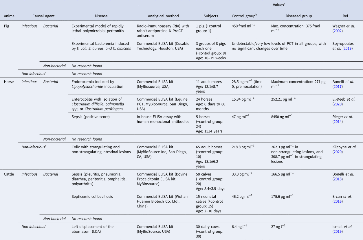
Studies about PCT in large animals have been summarized in Table 5, classifying them according to species, pathological condition (bacterial infections; non-bacterial infections – viral, parasitic, or fungal-; and non-septic inflammation), and analytical method used for PCT measurement. Compared to APPs, the number of studies on PCT in animals is relatively low. In the horse, basal levels seem to be higher than in human beings (Rieger et al., Reference Rieger, Kochleus, Teschner, Rascher, Barton, Geerlof, Kremmer, Schmid, Hartmann and Gehlen2014; Bonelli et al., Reference Bonelli, Meucci, Divers, Wagner, Intorre and Sgorbini2017). This difference could be due to interspecific differences, such as the close contact with the large amounts of LPS that are generally in the equine intestinal lumen (Dicks et al., Reference Dicks, Botha, Dicks and Botes2014). Increases in PCT concentrations have been observed in horses with natural sepsis, after induced endotoxemia and enterocolitis, and in colic (Bonelli et al., Reference Bonelli, Meucci, Divers, Radcliffe, Jose-Cunilleras, Corazza, Guidi, Tognetti, Castagnetti, Intorre and Sgorbini2015a, Reference Bonelli, Meucci, Divers, Jose-Cunilleras, Corazza, Tognetti, Guidi, Intorre and Sgorbini2015b; Teschner et al., Reference Teschner, Rieger, Koopmann and Gehlen2015; El-Deeb et al., Reference El-Deeb, Fayez, Elsohaby, Mkrtchyan and Alhaider2020). Regarding cattle, increases in PCT concentrations have been observed in systemic colibacillosis, newborn calves with sepsis, and in cows with a left abomasum displacement. In healthy calves, the concentrations were similar to those found in horses (Ercan et al., Reference Ercan, Tuzcu, Başbug, Tuzcu and Alim2016; Bonelli et al., Reference Bonelli, Meucci, Divers, Boccardo, Pravettoni, Meylan, Belloli and Sgorbini2018; Kirbas et al., Reference Kirbas, Kandemir, Celebi, Hanedan and Timurkan2019). In pigs, only a few studies with induced experimental infections found measurable concentrations of plasmatic PCT, maybe due to the lack of specificity of the methods (Wagner et al., Reference Wagner, Martinez, Vath, Snider, Nylén, Becker, Müller and White2002; Zannoni et al., Reference Zannoni, Giunti, Bernardini, Gentilini, Zaniboni, Bacci and Forni2012; Spyropoulos et al., Reference Spyropoulos, Chalkias, Georgiou, Papalois, Kouskouni, Baka and Xanthos2019; Liu et al., Reference Liu, Xu, Wang, Liang, Li, Wang, Wang, Zhu and Xiao2021). To the authors’ knowledge, very few studies have evaluated PCT in non-septic inflammation (Ismail et al., Reference Ismail, Al-Majali, Al-Rawashdeh, Daradka and Mohaffel2019; Kilcoyne et al., Reference Kilcoyne, Nieto and Dechant2020), and there are no studies about the role of PCT during viral pathologies in animals.
Examples of research about procalcitonin (PCT) in farm animals with different causal agents

a Values: results expressed as the arithmetic means of the values of the group unless otherwise stated.
b Control group: animals classified as healthy, unless specified that the selection procedure for the control group is different.
c Non-infectious disease: this category includes pathologies classified as non-infectious but not necessarily confirmed to be free of secondary infectious agents.
Presepsin (PSE)/Soluble cluster of differentiation 14 subtype (sCD14-ST)
PSE is a soluble product that comes from the cleavage of the CD14 glycoprotein. These glycoproteins are located mostly on PMN surfaces and act as receptors that can bind to bacterial structures, which can result in their proteolysis, leading to the release of sCD14 into the blood (Chenevier-Gobeaux et al., Reference Chenevier-Gobeaux, Borderie, Weiss, Mallet-Coste and Claessens2015; Memar and Baghi, Reference Memar and Baghi2019).
In human beings, PSE can be useful in the diagnosis and prognosis of local infections and in septic SIRS (Endo et al., Reference Endo, Suzuki, Takahashi, Shozushima, Ishikura, Murai, Nishida, Irie, Miura, Iguchi, Fukui, Tanaka, Nojima and Okamura2012), compared to non-infectious SIRS (Shozushima et al., Reference Shozushima, Takahashi, Matsumoto, Kojika, Endo and Okamura2011; Endo et al., Reference Endo, Suzuki, Takahashi, Shozushima, Ishikura, Murai, Nishida, Irie, Miura, Iguchi, Fukui, Tanaka, Nojima and Okamura2012; Behnes et al., Reference Behnes, Bertsch, Lepiorz, Lang, Trinkmann, Brueckmann, Borggrefe and Hoffmann2014). Some studies have concluded that PSE showed better performance in diagnosing sepsis than PCT, but other studies have not confirmed such superiority (Yaegashi et al., Reference Yaegashi, Sato, Suzuki, Kojika, Imai, Takahashi, Miyata, Endo, Shirakawa and Furusako2005; Liu et al., Reference Liu, Chen, Yin, Zhao and Li2013; Romualdo et al., Reference Romualdo, Torrella, González, Sánchez, Holgado, Freire, Acebes and Otón2014; Zhu et al., Reference Zhu, Zhong, Ji, Li, Li, Pang, Zhang and Zhao2020). Overall, the sensitivity and specificity of PSE have been reported as 76–80% and 80–85%, respectively.
The membrane CD14 complex, thus the production of PSE, can be stimulated by PAMPs other than LPS during bacterial infections, and even from other ligands derived from virus and endogenous proinflammatory mediators, but it remains unknown to what extent (Poggi et al., Reference Poggi, Vasarri, Boni, Pugni, Mosca and Dani2020). Such variabilities could explain the overlap in the concentrations reported in some studies between SIRS and sepsis (Behnes et al., Reference Behnes, Bertsch, Lepiorz, Lang, Trinkmann, Brueckmann, Borggrefe and Hoffmann2014), as well as the higher concentrations of PSE found in sepsis with a viral component or in endogenous damage such as kidney dysfunction (Chenevier-Gobeaux et al., Reference Chenevier-Gobeaux, Trabattoni, Roelens, Borderie and Claessens2014; Zaninotto et al., Reference Zaninotto, Mion, Cosma, Rinaldi and Plebani2020). In addition, the origin of PSE could also explain why increases in its concentrations occur in infections with Gram-positive bacteria, but in lower magnitude than infections with Gram-negative bacteria that have LPS as a characteristic attribute (Koizumi et al., Reference Koizumi, Sakanashi, Mohri, Watanabe, Shiota, Asai, Kato, Hagihara, Murotani, Yamagishi, Suematsu and Mikamo2020). In addition, PSE levels may be higher in some physiologic conditions like in preterm newborns and advanced age patients, in which concentrations reach 500 ng l−1, the threshold at which the diagnosis of sepsis would be highly suspected in human beings. Therefore, more studies are needed to establish more accurate ranges based on age and associated diseases (Mussap et al., Reference Mussap, Puxeddu, Burrai, Noto, Cibecchini, Testa, Puddu, Ottonello, Dessì, Irmesi, Gassa, Fanni and Fanos2012; Chenevier-Gobeaux et al., Reference Chenevier-Gobeaux, Trabattoni, Roelens, Borderie and Claessens2014).
With respect to veterinary medicine, to the authors’ knowledge, PSE has been only studied in horses. In one report, PSE concentrations did not change after the administration of LPS (Bonelli et al., Reference Bonelli, Meucci, Divers, Wagner, Intorre and Sgorbini2017). However, PSE was higher in horses classified as clinically endotoxemic patients from a veterinary teaching hospital (Fogle et al., Reference Fogle, Jacob, Blikslager, Edwards, Wagner, Dean and Fogle2017) and there was an increase of PSE in foals with septicemia and in adult horses with recurrent airway obstruction (Wagner et al., Reference Wagner, Ainsworth and Freer2013). Therefore, more research is needed to explore the potential application of this promising biomarker in farm animals.
Other biomarkers that could have potential to detect sepsis
The following biomarkers could also have potential to detect sepsis, but have been studied less than PCT and PSE:
• sTREM-1 (soluble Triggering Receptor Expressed on Myeloid Cells-1). TREM-1 is a receptor of the immunoglobulin superfamily, whose expression is regulated on neutrophils, which mediates the inflammatory response triggered by bacteria and fungi (Bouchon et al., Reference Bouchon, Facchetti, Weigand and Colonna2001). TREM-1 is released in a soluble, quantifiable form (sTREM-1) from the membrane of activated cells. Higher concentrations of this biomarker during sepsis, and low expression of the receptor in patients with non-infectious inflammatory disorders, were observed among human patients (Gibot et al., Reference Gibot, Kolopp-Sarda, Béné, Cravoisy, Levy, Faure and Bollaert2004). Regarding veterinary species, sTREM-1 has been measured in horses with inflammatory airway disease without significant changes (Bullone et al., Reference Bullone, De Lagarde, Vargas and Lavoie2015), but there are no studies in animals with sepsis to the authors’ knowledge.
• Lipopolysaccharide-binding protein (LBP). LBP is a protein that interacts with the LPS of Gram-negative bacteria and initiates a series of events that eventually lead to proinflammatory cytokine production. In several studies, LBP indicated infectious disease in human patients, but in other cases, this biomarker only distinguished infectious etiologies in patients with organ failure and its correlation with the severity of the disease was weak (Ouderkirk et al., Reference Ouderkirk, Nord, Turett and Kislak2003; Sakr et al., Reference Sakr, Burgett, Nacul, Reinhart and Brunkhorst2008). Regarding veterinary medicine, there is a study in which LBP increased significantly and faster than Hp in a group of calves experimentally infected with Mannheimia haemolytica (Schroedl et al., Reference Schroedl, Fuerll, Reinhold, Krueger and Schuett2001).
• MMP-9. Matrix-metalloproteinases (MMPs) are a family of endoproteinases involved in inflammation, healing of tissue injury, and remodeling. MMP-9 concentrations rise in people with severe sepsis, and its analysis could help determining the outcome of the illness (Hoffmann et al., Reference Hoffmann, Bertsch, Dvortsak, Liebetrau, Lang, Liebe, Huhle, Borggrefe and Brueckmann2006, Reference Hoffmann, Brueckmann and Borggrefe2009). In veterinary medicine, MMP-9 has been detected at higher activity after the administration of LPS in horses and pigs (Castellheim et al., Reference Castellheim, Thorgersen, Hellerud, Pharo, Johansen, Brosstad, Gaustad, Brun, Fosse, Tønnessen, Nielsen and Mollnes2008; Fugler et al., Reference Fugler, Eades, Moore, Koch and Keowen2013). In addition, higher concentrations of MMP-9 have been found in bronchoalveolar lavage fluid in pigs infected with porcine reproductive and respiratory syndrome virus (PRRSV) and in calves with concurrent infections of Pasteurella multocida and Mycoplasma bovirhinis (Girard et al., Reference Girard, Cléroux, Tremblay, Dea and St-Pierre2001; Simonen-Jokinen et al., Reference Simonen-Jokinen, Eskelinen, Härtel, Nikunen, Saloniemi and Maisi2005). In cattle, higher activity in milk has also been reported after endotoxin-induced mastitis and in synovial fluid after an experimentally induced septic arthritis (Raulo et al., Reference Raulo, Sorsa, Tervahartiala, Latvanen, Pirilä, Hirvonen and Maisi2002; Francoz et al., Reference Francoz, Desrochers, Simard, Saint Pierre, Fecteau, Latouche and Fortin2008). Furthermore, MMP-9 can be covalently linked to Hp and released as MMP9-Hp complexes by neutrophils when their degranulation occurs. These complexes were firstly detected in the serum of cattle with acute polymicrobial sepsis (Bannikov et al., Reference Bannikov, Mattoon, Abrahamsen, Premanandan, Green-Church, Marsh and Lakritz2007, Reference Bannikov, Hinds, Rajala-Schultz, Premanandan, Rings and Lakritz2011) and then later found at high concentrations after the infusion of LPS (Hinds et al., Reference Hinds, Niehaus, Premanandan, Rajala-Schultz, Rings and Lakritz2014).
Biomarkers that can provide additional information in sepsis
There are other biomarkers that could be useful in diagnosed cases of sepsis, for establishing the patient's prognosis by assessing endothelial damage, organ dysfunction, and alterations in the coagulation system.
• Markers of endothelial damage. The endothelial system plays an essential role in homeostasis. Endothelial damage and dysfunction can lead to complications that often accompany sepsis, such as organ dysfunction and septic shock. Examples of endothelial damage biomarkers are neopterin, angiopoietins, endocan, vascular cell adhesion molecule-1, vascular endothelial growth factor, platelet-derived growth factor, or adrenomedullin (Christ-crain et al., Reference Christ-crain, Morgenthaler, Struck, Harbarth, Bergmann and Müller2005; Pierrakos and Vincent, Reference Pierrakos and Vincent2010; Paulus et al., Reference Paulus, Jennewein and Zacharowski2011; Giannakopoulos et al., Reference Giannakopoulos, Hoffmann, Ansari, Bertsch, Borggrefe, Akin and Behnes2017). In veterinary medicine, neopterin was significantly higher in calves with septicemic colibacillosis (Ercan et al., Reference Ercan, Tuzcu, Başbug, Tuzcu and Alim2016), and adrenomedullin was found higher in critically ill neonatal foals regardless of the causal agent (Toth et al., Reference Toth, Slovis, Constable and Taylor2014).
• Markers of organic dysfunction. Some examples of these biomarkers are lactate, endothelin-1 (ET-1), and natriuretic peptides such as atrial natriuretic peptide and mid-regional pro-atrial natriuretic peptide. These markers could be valuable as early predictors of high-risk patients and survival likelihood (Brauner et al., Reference Brauner, Rohde and Clausell2000; Rivers et al., Reference Rivers, McCord, Otero, Jacobsen and Loomba2007; Mikkelsen et al., Reference Mikkelsen, Miltiades, Gaieski, Goyal, Fuchs, Shah, Bellamy and Christie2009). In animals, an increase in lactate was reported after the experimental induction of sepsis in pigs, and this marker was correlated with a higher mortality rates in horses with SIRS (Corley et al., Reference Corley, Donaldson and Furr2010; Tóth et al., Reference Tóth, Debreceni, Berhés, Hajdú, Deák, Pet, Szabó, Németh, Fülesdi and Kappelmayer2017). Regarding ET-1, higher plasma concentrations and mRNA levels in tissues were observed after the experimental induction of sepsis in pigs (Zannoni et al., Reference Zannoni, Bernardini, Gentilini, Giunti, Bacci and Forni2010).
• Markers of the coagulation system. In sepsis, it is frequent to observe changes in thrombocyte counts, antithrombin, protein C and S, activated partial thromboplastin time, D-dimer, or fibrin. These changes are not specific and can also occur in other pathologies such as cancer, trauma, and vascular or immune disorders, but their study during sepsis is helpful for prognosis and evaluation of the treatment (Pettilä et al., Reference Pettilä, Pentti, Pettilä, Takkunen and Jousela2002). In horses and cattle, the coagulation markers that seem more useful are fibrinogen and D-dimer (Giguère et al., Reference Giguère, Hernandez, Gaskin, Miller and Bowman2003; Irmak et al., Reference Irmak, Sen, Cöl, Birdane, Güzelbektes, Civelek, Yilmaz and Turgut2006; Armengou et al., Reference Armengou, Monreal, Tarancón, Navarro, Ríos and Segura2008; Hultén et al., Reference Hultén, Grönlund, Hirvonen, Tulamo, Suominen, Marhaug and Forsberg2010), but further research is needed.
Conclusions
This review has described the general concepts of sepsis and the current knowledge about three groups of potential biomarkers of sepsis in pigs, horses and cattle, namely: (1) APPs and cytokines, which have been traditionally used; (2) PCT, PSE, and other proteins that are more specific sepsis markers; and (3) other markers that can provide complementary information about this condition.
Overall, each biomarker of the three groups could have an use in sepsis and provide complementary information. APPs and cytokines, although non-specific for sepsis, are indicative of the inflammation associated with this condition and can monitor the evolution of the disease and establish the prognosis of the patient. PCT, PSE, and other molecules such as sTREM-1, LBP, and MMP-9, have huge potential because they can improve the diagnosis of bacterial sepsis. Finally, the biomarkers of the third group provide additional information for prognosis and monitoring through the assessment of endothelial damage, organic dysfunction, and alterations in the coagulation system.
Further studies should better define the ability and applications of these biomarkers, especially those with potential to differentiate sepsis from other inflammatory conditions, and establish accurate cut-off values in veterinary medicine. These are required steps to improve the diagnosis and the clinical management of sepsis in veterinary species, which is essential to increase host survival rates and achieve more rational use of antibiotics.
Acknowledgments
M.J. López-Martínez was funded by 21293/FPI/19 predoctoral grant, Fundación Séneca, Región de Murcia (Spain). This study was supported by Grant Reference PID2019-105950RB-100 funded by MCIN/AEI/10.13039/501100011033. It was also supported by a Grant Reference PCI2020-120712-2 from MCIN/AEI/10.13039/501100011033 and European Union “NextGenerationEU”/PRTR (1st ICRAD Joint Cofund Call).