




No CrossRef data available.
Article contents
Experimental infection of severe combined immunodeficient (SCID) mice with the human microsporidian Trachipleistophora hominis
Published online by Cambridge University Press: 16 April 2004
Abstract
Different courses of microsporidiosis, related to the route of infection, were observed in severe combined immunodeficient (SCID) mice inoculated with spores of the human microsporidian Trachipleistophora hominis (Phylum Microspora). After eye contamination by spores the mice became moribund within 7 to 8 weeks, showing severe infection in the conjunctiva and cornea, and lighter infections in the urinary bladder, liver and spleen. The mean survival time of intramuscularly inoculated SCID mice was 12 weeks, when heavy infection was found in muscles around the site of inoculation, and also in several viscera. Subcutaneously inoculated SCID mice developed skin lesions around the inoculation sites, and heavy urinary bladder infection, and died 6 or 7 weeks after inoculation. Intracerebrally inoculated SCID mice became moribund 5 or 6 weeks after inoculation with massive infection in the urinary bladder and liver, but none in the brain. Intraperitoneally inoculated SCID mice survived for 13 weeks and the urinary bladder and liver were the most heavily infected organs. The SCID mice, inoculated perorally and examined after 23 weeks, were uninfected. Infection was not detected in the brain of any of the inoculated SCID mice. Our results show that T. hominis has very little tissue specificity. Peroral infection seems to be ineffective in T. hominis, but eye contamination or insect bite (as mimicked by injection) are suggested as possible routes of infection under natural conditions.
Information
- Type
- Research Article
- Information
- Copyright
- © 2004 Cambridge University Press
INTRODUCTION
The phylum Microspora are intracellular protistan parasites of most animal phyla (Sprague, 1977). Although long considered to be protozoa, microsporidia are now believed to be either closely related to fungi (Keeling & McFadden, 1998; Weiss & Vossbrinck, 1998) or organisms which evolved from among the fungi (Keeling, Luker & Palmer, 2000; Keeling, 2003). Most microsporidia are parasites of arthropods and fish (Wittner, 1999). However, some microsporidia are parasites of homeothermic vertebrates, including man (Canning & Lom, 1986; Didier, Snowden & Shadduck, 1998; Wittner, 1999; Cali & Takvorian, 2003). Of special importance are microsporidia that are able to infect humans suffering immunodeficiency caused by viral infections (HIV), innate malfunctioning of the immune system or by immunosuppressive therapy (Wittner, 1999). Several species out of the 14 microsporidia belonging to 8 genera, listed as parasites of man (Curry, 1999), are evidently specific parasites of homeothermic vertebrates, including man (Enterocytozoon bieneusi, Encephalitozoon cuniculi, Encephalitozoon hellem, Encephalitozoon intestinalis). Other listed species are either incompletely known or occur sporadically. The probability that some are opportunists acquired by man from another vertebrate or invertebrate host is high. Of particular interest is the question whether microsporidia of invertebrates or poikilothermic vertebrates can adapt to life within a warm-blooded vertebrate, provided that they are able to overcome host immunity and suitable physiological conditions are met.
Trachipleistophora hominis is a candidate of a true opportunist acquired in man from an invertebrate host, as suggested by its ability to develop in mosquito tissues (Weidner et al. 1999) and by its position in the ssu rRNA phylogenetic tree (Cheney, Lafranchi-Tristem & Canning, 2000) closer to microsporidia from invertebrates than those from vertebrates. The genus and species T. hominis were originally established for a microsporidium causing myositis in an AIDS patient (Hollister et al. 1996). The parasite was found in corneal scrapings, skeletal muscles and nasopharyngeal washings (Field et al. 1996). Two cases of a generalized human infection with a similar microsporidium, but belonging to another species, T. anthropophthera have been reported (Yachnis et al. 1996; Vávra et al. 1998a). In contrast to T. hominis, T. anthropophthera is dimorphic with two kinds of sporophorous vesicles and spores (Vávra et al. 1998b). Whether the infection by this second species of the genus Trachipleistophora was acquired from a non-human source remains unknown. Due to its defective immunity, the severe combined immunodeficient mouse (SCID) is a good model for mimicking infections by opportunistic parasites in warm-blooded vertebrates. SCID mice have been used to track the course of infection of Encephalitozoon cuniculi (Koudela et al. 1993), Vittaforma corneae (Koudela et al. 1999b; šlapáková, 1999) and Brachiola algerae (Koudela et al. 2001). The purpose of the study reported here was to examine whether the SCID mouse has the potential to demonstrate how various inoculation methods influence the course of T. hominis microsporidiosis in a mammal organism with impaired immunity.
MATERIALS AND METHODS
Parasite
The human isolate of T. hominis used in this study was isolated from human muscle biopsies of an AIDS patient (Hollister et al. 1996). The organism was grown in rabbit kidney RK-13 cells in minimum essential medium (MEM) supplemented with 10% foetal calf serum.
Animals and inoculations
SCID mice were housed in groups of 2 or 4 animals per cage in flexible isolators (BEM, Znojmo, Czech Republic) with high-efficiency particulate air (HEPA) filters. All cages, food, water, and bedding were sterilized before use. A total of 30, 8 to 11-week-old SCID mice of both sexes were used.
In order to obtain infective material fully adapted to mammalian tissues the spores harvested from tissue culture (‘primary inoculum’) were inoculated into SCID mice and only spores harvested from mouse tissues (‘secondary inoculum’) were used in experiments.
‘Primary inoculum’.
Spores were obtained from the culture supernatant in culture flasks centrifuged at 2000 g at 4 °C for 10 min and were suspended in sterile phosphate-buffered saline (PBS, pH 7·2). Spores were then purified by centrifugation at 1000 g for 15 min, after layering on the top of a column of 50% Percoll in PBS. After washing and resuspension in sterile PBS, the spores were counted using a haemocytometer, and adjusted to the required concentration.
‘Secondary inoculum’.
Three SCID mice were used for preparing the secondary inoculum. One SCID mouse was injected into the gluteal muscles, a second mouse was inoculated intraperitoneally and a third was inoculated both intraperitoneally and intramuscularly. The respective mice were injected with 0·5 ml of PBS containing 116 spores of the ‘primary inoculum’. These SCID mice were euthanized and necropsied at 65 days post-infection (p.i.) and their tissues examined for the presence of microsporidia. The leg muscles from both of the intramuscularly inoculated SCID mice were excised, homogenized in sterile PBS and incubated with 0·25% trypsin for 20 min at 37 °C. The spore suspension was washed by centrifugation and resuspended in PBS and spores were then counted as described above.
The ‘inoculation mode experiment’
This experiment was designed to observe the survival times of SCID mice infected with T. hominis by different routes and to monitor the spread of the microsporidia in the host organs and tissues. Four SCID mice were used in each group and the inoculum (107 T. hominis ‘secondary inoculum’ spores suspended in 0·5 ml of PBS) was applied in 6 different ways: in drops to the eye surface, injected intramuscularly in the gluteal muscles, injected subcutaneously into the tail, injected intracerebrally, injected intraperitoneally and fed perorally by tube. Three SCID mice served as controls. All SCID mice were monitored daily for the development of clinical signs of disease and moribund SCID mice were euthanized and necropsied.
Light microscopy
At necropsy the following tissues and fluids from each mouse were examined in smears stained with Calcofluor White M2R (Vávra et al. 1993): peritoneal fluid, gluteal muscles from the site of inoculation, lungs, kidney, liver, spleen, brain, bladder and urine. Twenty-seven tissue samples (liver, pancreas, lung, cardiac muscle, spleen, brain, kidney, urinary bladder, diaphragm, stomach, duodenum, middle jejunum, ileum, caecum, colon, rectum, mesenteric lymph node, upper cheek, eyelid, nose, foot pads, tongue, jowl, scrotum or external genitalia of females, gluteal muscles, triceps brachii, inguinal mesenteric lymph nodes) from each mouse were fixed in 10% neutral buffered formalin and processed for light microscopy using standard methods. Paraffin sections were stained with haematoxylin-eosin, Masson's trichrome, Gram's method, and with the optical brightener Calcofluor White M2R as cited above.
Electron microscopy
For transmission electron microscopy (TEM), tissue samples from liver, spleen and muscles from the site where spores had been inoculated, were fixed in 2·5% glutaraldehyde in cacodylate buffer (0·1 M, pH 7·4) at 4 °C and post-fixed in 1% osmium tetroxide in the same buffer. Tissue samples were then washed 3 times in cacodylate buffer, dehydrated in a graded ethanol series and embedded in Durcupan epoxy resin. Ultrathin sections were stained with uranyl acetate and lead citrate and were examined in a JEOL 1010 transmission electron microscope.
RESULTS
‘Secondary inoculum experiment’
In the experiment aimed to multiply T. hominis spores in mice, no clinical signs of disease were seen in experimental animals before 8 weeks post-infection whatever the inoculation route. Infection did not develop in the mouse that had been inoculated only by the intraperitoneal route. Later, the SCID mice which had been inoculated intramuscularly, developed alopoecia and wasting at the site where the spores had been inoculated, and showed clinical signs of hind limb weakness and lethargy before necropsy at 65 days p.i. Infection was detectable macroscopically by the presence of alopoetic greyish areas in the leg muscles (Fig. 1A). Examination of smears, prepared from the leg muscle homogenate, revealed heavy infection and showed abundant sporophorous vesicles containing numerous spores. (Fig. 1B). Only few spores of T. hominis were found in smears prepared from peritoneal macrophages of intramuscularly inoculated SCID mice. Examination of the SCID mouse inoculated intraperitoneally as well as intramuscularly and necropsied at 65 days p.i., sporadic spores were present in peritoneal macrophages, while heavy infection was found on the serosal surface of the urinary bladder, liver and spleen and a light infection on the serosal surface of the small and large intestine. Fresh spores obtained from SCID mice measured 4·0×2·5 μm, were elongate ovoid and showed a prominent posterior vacuole (Fig. 1C). Some spores were empty. Extruded polar tubes measured about 70 μm in length.

Fig. 1. Trachipleistophora hominis in experimentally infected SCID mice. (A) Alopoetic area and necrosis (arrow) of a leg of an intramuscularly inoculated SCID mouse. (B and C) Sporophorous vesicles and spores isolated from muscles of intramuscularly inoculated SCID mouse showing the large number of spores formed in the sporophorous vesicle and the characteristic spore shape (Nomarski interference contrast). (D) Heavily infected skeletal muscle of intramuscularly inoculated SCID mice with abundant presence of fibrocytes and minimal inflammatory response (Masson's trichome stain). (E) Closely packed sporophorous vesicles within skeletal muscle fibrils (Gram stain). (F) Transversal histological section of eye showing lens (ln) and changes into corneal stroma (arrows) (H&E). (G) Toluidine blue-stained semi-thin section showing developmental stages and sporophorous vesicles of various maturity filling a skeletal muscle fibrils.
The ‘inoculation mode experiment’
Ocular inoculation.
No clinical signs of disease were seen in eye-infected SCID mice at 6 weeks after infection but, at 8 weeks, the mice developed conjunctival inflammation and intense keratoconjunctivitis (Fig. 1F) accompanied by wasting and lethargy. Moribund animals were euthanized and necropsied 8 weeks after infection. Spores of T. hominis were sporadically detected in smears prepared from peritoneal macrophages. Gram- and Calcofluor-stained histological sections revealed heavy microsporidian infection in the conjunctiva and deep into corneal stroma, and scattered microsporidian spores in the retina. Spores were also identified in the urinary bladder, liver and spleen. Microsporidia were not detected in other organs and tissues examined.
Intramuscular inoculation.
The SCID mice inoculated intramuscularly showed local alopoetic changes at the site of inoculation after 6 weeks. Later, the local alopoecia intensified, hind limb weakness occurred and the animals became lethargic. Two SCID mice died at 76 and 80 days p.i., respectively, and the remaining two SCID mice became moribund and were euthanized and necropsied at 92 days p.i. Histological sections of hind limb muscles revealed heavy infection around the site of inoculation. Pathological changes consisted of a central zone of fibrosis surrounded by degenerate muscle myofibrils containing a variable number of polygonal sporophorous vesicles. The Masson's trichrome stain demonstrated an abundant presence of fibrin fibres (Fig. 1D) and minimal inflammatory response. The central zone of fibrosis was surrounded by muscle myofibrils containing clusters of sporophorous vesicles. Gram-stained histological sections showed that these vesicles contained spore or spore precursors (Fig. 1E). Semi-thin sections demonstrated myofibrils containing various developmental stages and myofibres filled with spores between debris of sarcoplasm (Fig. 1G). A number of T. hominis spores was also found in the liver and spleen. Pancreas, lungs and urinary bladder were rarely parasitized.
Subcutaneous inoculation.
The SCID mice inoculated subcutaneously also showed local alopoetic changes at the site of inoculation and died 6 or 7 weeks after inoculation. The most heavily infected tissues were the skin and muscles around the site of inoculation and testes and urinary bladder. The kidney and rectum were only lightly infected.
Intracerebral inoculation.
Intracerebrally inoculated SCID mice were moribund 5 or 6 weeks after inoculation and the most heavily infected tissues were urinary bladder and liver (Fig. 2A and B). No microsporidia were detected in the brain of any of the intracerebrally inoculated SCID mice.

Fig. 2. Trachipleistophora hominis in experimentally infected SCID mice and T. hominis fine structure features. Histological sections (Gram stain) of intracerebrally inoculated SCID mouse showing spores within epithelial cells of urinary bladder (A) and hepatocytes (B). (C) Two meronts, 1 binucleate (m) and 3 sporophorous vesicles with mature spores (sp). (D) Mature spore showing the characteristic subapical location of the exit site of the polar filament, the very dense and compact polaroplast (po), the arrangement of polar filament coils and the large posterior vacuole (pv). (E and F) Details of the arrangement of polar filament coils. There are 8 (E) or 7 (F) coils of the same diameter in a single row, ending in 3 slightly thinner coils, displaced to the interior of the spore as a short second row. (G) Apical part of the spore showing the manubroid (mb) part of the polar filament, the anchoring disc surrounded by polar sac membranous cisterna. The polaroplast membranous stacks (cisternae) are narrow and densely packed in the apical region of the organelle and are more wide and less densely packed posteriorly. (H) Part of a sporophorous vesicle with 2 spores that have extruded their polar filaments (arrows).
Intraperitoneal inoculation.
When the moribund intraperitoneally inoculated SCID mice were examined after 13 weeks, heavy infection was found only in the urinary bladder and liver. Spores were quite common also in peritoneal macrophages, in the large intestine and in peritoneum of the lower abdomen.
Peroral inoculation.
No clinical signs of disease were seen in any of the perorally inoculated SCID mice. At necropsy, 23 weeks after inoculation, no spores or other developmental stages of T. hominis were detected in peritoneal macrophages or any organs and tissues examined.
Ultrastructure of the human isolate of T. hominis in the SCID mice
The fine structure of T. hominis, as observed in artificially infected SCID mice (Fig. 2C–H) was similar to that originally described from infection in athymic mice (Hollister et al. 1996). The microsporidia have unpaired nuclei during the whole life-cycle. Meronts usually have a single nucleus but rare cells with several (usually 2) nuclei were observed. Evidently these are forms ready for cell division. Meronts have a thick, electron-dense surface coat protruding as irregular expansions into the host cell tissue, which shows signs of digestion in the area of contact with the parasite. Sporophorous vesicles with mature spores show the characteristic dense wall (Fig. 2C). Some relatively well-preserved spores of T. hominis were found in the SCID mice, showing the subapically located site from whence the polar tube emerges during extrusion, the very dense polaroplast and slightly anisofilar polar filament arranged in a single row of 7–8 coils, terminating distally in about 3, somewhat thinner coils positioned more centripetally (Fig. 2D, E, F). The central portion of the polar filament penetrates deep into the anchoring disc, while the more external part of the filament joins just the border of the disc. At this point there is an electron-dense line representing the ‘hinge’, around which the filament inverts during extrusion (Vávra & Larsson, 1999). The anchoring disc is enveloped by the polar sac, a membranous cisterna covering apically nearly one half of the polaroplast. The well-fixed polaroplast is represented by flat, cisterna-like membrane bound stacks, each stack being 10 nm wide, separated by a narrow gap of 5 nm. Distally, the polaroplast stacks are much wider (around 23 nm), but the separating gaps are the same as in the apical portion of the polaroplast (Fig. 2G). Quite often, empty spores or intact sporophorous vesicles with spores which had extruded their polar tubes were found in areas of the host tissue considerably damaged by the microsporidian (Fig. 2H). It is believed that the infection is spread in this way within the tissues of the host.
DISCUSSION
The SCID mouse as a model for microsporidian infections in mammals
SCID mice have previously been used as models in experiments involving three microsporidian species infecting mammals. Using E. cuniculi there have been studies of the general course of infection in mice (Koudela et al. 1993), the sensitivity to albendazole (Koudela et al. 1994), the therapeutic immune reconstitution of infected mice (Heřmánek et al. 1993), the comparison of immune reactions of immunodeficient and immunocompetent hosts (Salát, Braunfuchsová & Kopecký, 2001) and the effect of storage temperature on the viability of spores (Koudela, Kučerová & Hudcovic, 1999a) SCID mice have also proved useful as laboratory models for investigation of the pathology and ultrastructure of a thermotolerant isolate of B. algerae (formerly Nosema algerae, a mosquito microsporidium) in mammals (Koudela et al. 2001). A third microsporidian introduced experimentally into SCID mice is V. cornea, in which the range of infected tissues and pathology after peroral, intraperitoneal and ocular infection was described (Koudela et al. 1999b; šlapáková, 1999). Recent studies emphasize the decisive role of cellular immune responses in defence against microsporidian infections (Khan et al. 1999; Salát et al. 2001). SCID mice are suitable models for microsporidian infections in HIV-infected humans as these mice lack functional T-lymphocytes (Bosma, 1989) and thus mimic the advanced stage of AIDS, although they also lack functional B-lymphocytes. SCID mice complement the use of athymic (nu/nu) mice which, because of being severely deficient in functional T-lymphocyte subsets, also develop acute and lethal microsporidian infections (Gannon, 1980; Hollister et al. 1996; Schmidt & Shadduck, 1983, 1984; Silveira, Canning & Shadduck, 1993; Cheney et al. 2000).
Tissues infected
The present paper supports the previous observations made on athymic (nu/nu) mice, that T. hominis has a low tissue specificity in immunocompromized hosts (Hollister et al. 1996). These authors found that the skeletal muscle in the vicinity of the injection site was readily infected after intramuscular and intraperitoneal inoculation, along with urinary bladder, spleen, lungs, peritoneum and large intestine in some of the mice. This range of susceptible tissues of athymic mice was extended by Cheney et al. (2000) to include pancreas, liver, kidneys, ovaries, heart, ascitic fluid and skin even after oral infection. Our experiments confirm these results and add cornea and retina of the eye and testes to the range of susceptible tissues. We speculate that, with the exception of cerebral tissue, all tissues of many immunocompromized mammalian hosts can be infected although the range of hosts has yet to be investigated. The absence of infection of T. hominis in cerebral tissues after oral and intraperitoneal and intramuscular inoculation (Cheney et al. 2000), and even after intracerebral inoculation (this paper), confirms the status of T. anthropophthera as a separate species since the latter causes systemic infections, including the brain, in HIV-infected humans (Vávra et al. 1998a,b). Our results differ from those of Cheney et al. (2000) in that we were not able to infect SCID mice by the oral route. Cheney et al. (2000) speculated that the AIDS patient, from whom T. hominis was first isolated, might have acquired the infection from the bite of a haematophagous insect. They based their arguments on their phylogenetic analysis of the 16S and DNA nucleotide sequence, which placed T. hominis closest to Vavraia culicis, a microsporidium infecting a range of anopheline and culicine mosquitoes and on the experiments of Weidner et al. (1999) who achieved infections in two mosquito species by feeding spores derived from culture to the larvae. Infection after simple contamination of the eye surface in the present work suggests another possible route.
Eye as portal of entry of microsporidia into mammalian organism
Our experiments demonstrate that simple contamination of the eye surface of a receptive host by T. hominis not only leads to infection of the cornea and retina but also gives rise to generalized infection of the viscera. Previously, the same phenomenon was demonstrated in V. corneae (Koudela et al. 1999b; šlapáková, 1999) and in B. Algerae (Koudela et al. 2001). In the latter case ocular contamination was surprisingly, the only method by which infection of the visual tissues of SCID mice was achieved. It was theorised that the initial infection in the eye allowed the microsporidian to adjust to the higher temperature of the mammalian body (Koudela et al. 2001). Eye-to-viscera spread of infection indicates that eye can be an important site of entry of microsporidian infections into the human body. The eye is indeed an organ targeted by several microporidia as indicated by reports of eye infections in humans and in animals indicate (see e.g. Pinnolis et al. 1981; Canning & Lom, 1986; Lowder et al. 1990; Shadduck et al. 1990; Cali et al. 1991; Didier et al. 1991; Kramer & Grossniklaus, 1991; Friedberg, Didier & Yee, 1993; Silveira & Canning, 1995; Arnesen & Nordstoga, 1997; Canning et al. 1998; Silverstein, 1998; Canny et al. 1999; Visvesvara et al. 1999; Font et al. 2000). It is of interest, that systemic infection in viscera can also lead to eye microsporidiosis as reported by Silveira et al. (1993) in intraperitoneally Vittaforma corneum-infected athymic mice.
Veronika Schacherlová is thanked for preparing tissue samples for histology and Marie Váchová for caring of experimental animals. The skilful help of Petra Masařová in processing electron microscopy samples is also gratefully acknowledged.
References
REFERENCES
ARNESEN, K. &
NORDSTOGA, K.
(1977).
Ocular encephalitozoonosis (Nosematosis) in blue foxes.
Acta Ophthalmologica
55, 641–651.CrossRefGoogle Scholar
BOSMA, M. J.
(1989).
The SCID mutation: occurrence and effect.
Current Topics in Microbiology and Immunology
152, 3–9.CrossRefGoogle Scholar
CALI, A.,
MEISLER, D. M.,
LOWDER, C. Y.,
LEMBACH, R.,
AYERS, L.,
TAKVORIAN, P. M.,
RUTHERFORD, I.,
LONGWORTH, D. L.,
McMAHON, J. &
BRYAN, R. T.
(1991).
Corneal microsporidioses: characterization and identification.
Journal of Protozoology 38,
215S–217S.Google Scholar
CALI, A. &
TAKVORIAN, P.
(2003).
Ultrastructure and development of Pleistophora ronneafiei n. sp., a microsporidium (Protista) in the skeletal muscle of an immunocompromised individual.
Journal of Eukaryotic Microbiology
50, 77–85.Google Scholar
CANNING, E. U. &
LOM, J.
(1986).
The Microsporidia of Vertebrates.
Academic Press, Inc., NY, USA.
CANNY, C. J.,
WARD, D. A.,
PATTON, S. &
OROSZ, S. E.
(1999).
Microsporidian keratoconjunctivitis in a Double Yellow-Headed Amazon Parrot (Amazona ochrocephala oratrix).
Journal of Avian Medicine and Surgery
13, 179–286.Google Scholar
CHENEY, S. A.,
LAFRANCHI-TRISTEM, N. J. &
CANNING, E. U.
(2000).
Phylogenetic relationships of Pleistophora-like microsporidia based on small subunit ribosomal DNA sequences and implications for the source of Trachipleistophora hominis infections.
Journal of Eukaryotic Microbiology
47, 280–287.CrossRefGoogle Scholar
CURRY, A.
(1999).
Human microsporidial infections and possible animal sources.
Current Opinion in Infection Diseases
12, 473–480.CrossRefGoogle Scholar
DIDIER, E. S.,
DIDIER, P. J.,
FRIEDBERG, D. N.,
STENSON, S. M.,
ORENSTEIN, J. M.,
YEE, R. W.,
TIO, F. O.,
DAVIS, R. M.,
VOSSBRINCK, C.,
MILLICHAMP, N. &
SHADDUCK, J. A.
(1991).
Isolation and characterisation of a new human microsporidian, Encephalitozoon hellem (n. sp.), from 3 AIDS patients with keratoconjuctivitis.
Journal of Infectious Diseases
163, 617–621.Google Scholar
DIDIER, E. S.,
SNOWDEN, K. F. &
SHADDUCK, J. A.
(1998).
Biology of microsporidian species infecting mammals.
Advances in Parasitology
40, 283–320.CrossRefGoogle Scholar
FIELD, A. S.,
MARRIOTT, D. J.,
MILLIKEN, S. T.,
BREW, B. J.,
CANNING, E. U.,
KENCH, J. G.,
DARVENIZA, P. &
HARKNESS, J. L.
(1996).
Myositis associated with a newly described microsporidian, Trachipleistophora hominis, in a patient with AIDS.
Journal of Clinical Microbiology
34, 2803–2811.Google Scholar
FONT, R. L.,
SAMAHA, A. N.,
KEENER, M. J.,
CHEVEZ-BARRIOS, P. &
GOOSEY, J. D.
(2000).
Corneal microsporidiosis. Report of case, including electron microscopic observations.
Ophthalmology
107, 1769–1775.CrossRefGoogle Scholar
FRIEDBERG, D. N.,
DIDIER, E. S. &
YEE, R. W.
(1993).
Microsporidial keratoconjunctivitis.
American Journal of Ophthalmology
116, 380–381.CrossRefGoogle Scholar
GANNON, J.
(1980).
The course of infection of Encephalitozoon cuniculi in immunodeficient and immunocompetent mice.
Laboratory Animals
14, 189–192.CrossRefGoogle Scholar
HEŘMÁNEK, J.,
KOUDELA, B.,
KUČEROVÁ, Z.,
DITRICH, O. &
TRÁVNÍČEK, J.
(1993).
Prophylactic and therapeutic immune reconstitution of SCID mice infected with Encephalitozoon cuniculi.
Folia Parasitologica
40, 287–291.Google Scholar
HOLLISTER, W. S.,
CANNING, E. U.,
WEIDNER, E.,
FIELD, A. S.,
KENCH, J. &
MARIOTT, D. J.
(1996).
Development and ultrastructure of Trachipleistophora hominis n. g., n. sp. after in vitro isolation from an AIDS patient and inoculation into athymic mice.
Parasitology
112, 143–154.Google Scholar
KEELING, P. J.
(2003).
Congruent evidence from alpha-tubulin and beta-tubulin gene phylogenies for a zygomycete origin of microsporidia.
Fungal Genetics and Biology
38, 298–309.CrossRefGoogle Scholar
KEELING, P. J.,
LUKER, M. A. &
PALMER, J. D.
(2000).
Evidence from beta-tubulin phylogeny that microsporidia evolved from within the fungi.
Molecular Biology and Evolution
17, 23–31.CrossRefGoogle Scholar
KEELING, P. J. &
McFADDEN, G. I.
(1998).
Origins of microsporidia.
Trends in Microbiology
6, 19–23.CrossRefGoogle Scholar
KHAN, I. A.,
SCHWARTZMAN, J. D.,
KASPER, L. H. &
MORETTO, M.
(1999).
CD8+ CTLs are essential for protective immunity against murine Encephalitozoon cuniculi infection.
Journal of Immunology
162, 6086–6091.Google Scholar
KOUDELA, B.,
VÍTOVEC, J.,
KUČEROVÁ, Z.,
DITRICH, O. &
TRÁVNÍČEK, J.
(1993).
The severe combined immunodeficient mouse as a model for Encephalitozoon cuniculi microsporidiosis.
Folia Parasitologica
40, 279–286.Google Scholar
KOUDELA, B.,
LOM, J.,
VÍTOVEC, J.,
KUČEROVÁ, Z.,
DITRICH, O. &
TRÁVNÍČEK, J.
(1994).
In vivo efficacy of albendazole against Encephalitozoon cuniculi in SCID mice.
Journal of Eukaryotic Microbiology 41,
49S–50S.Google Scholar
KOUDELA, B.,
KUČEROVÁ, š. &
HUDCOVIC, T.
(1999 a).
Effect of low and high temperatures on infectivity of Encephalitozoon cuniculi spores suspended in water.
Folia Parasitologica
46, 171–174.Google Scholar
KOUDELA, B.,
šLAPÁKOVÁ, D.,
DIDIER, P. J. &
DIDIER, E. S.
(1999 b).
Experimental infection of SCID mice as a model for Vittaforma corneae microsporidiosis. The 52nd Annual Meeting Society of Protozoology and 6th International Workshop on Opportunistic Protists. North Carolina University, Raleigh, NC, USA. Abstract No. W67.
KOUDELA, B.,
VISVESVARA, G. S.,
MOURA, H. &
VÁVRA, J.
(2001).
The human isolate of Brachiola algerae (Phylum Microspora): development in SCID mice and description of its fine structure features.
Parasitology
123, 153–162.CrossRefGoogle Scholar
KRAMER, T. &
GROSSNIKLAUS, H. E. (1991). Ocular manifestations of fungal and parasitic diseases. Current Opinion in Ophtalmology
2, 212–219.CrossRefGoogle Scholar
LOWDER, C. Y.,
MEISLER, D. M.,
McMAHON, J. T.,
LONGWORTH, D. L. &
RUTHEFORD, I.
(1990).
Microsporidia infection of the cornea in a man seropositive for human immunodeficiency virus.
American Journal of Ophthalmology
109, 242–244.CrossRefGoogle Scholar
PINNOLIS, M.,
EGBERT, P. R.,
FONT, R. L. &
WINTER, F. C.
(1981).
Nosematosis of the cornea.
Archives of Ophthalmology
99, 1044–1047.CrossRefGoogle Scholar
SALÁT, J.,
BRAUNFUCHSOVÁ, P. &
KOPECKÝ, J.
(2001).
Experimental infection of immunocompetent and immunodeficient mice with Encephalitozoon cuniculi.
Folia Parasitologica
48, 249–254.CrossRefGoogle Scholar
SCHMIDT, E. C. &
SHADDUCK, J. A.
(1983).
Murine encephalitozoonosis model for studying the host–parasite relationship of a chronic infection.
Infection and Immunity
40, 936–942.Google Scholar
SCHMIDT, E. C. &
SHADDUCK, J. A.
(1984).
Mechanisms of resistance to the intracellular protozoan Encephalitozoon cuniculi in mice.
Journal of Immunology
133, 2712–2719.Google Scholar
SHADDUCK, J. A.,
MECCOLI, R. A.,
DAVIS, R. &
FONT, R. L.
(1990).
Isolation of a microsporidian from a human patient.
Journal of Infectious Diseases
162, 773–776.CrossRefGoogle Scholar
SILVEIRA, H.,
CANNING, E. U. &
SHADDUCK, J. A.
(1993).
Experimental infection of athymic mice with the human microsporidian Nosema corneum.
Parasitology
107, 489–496.CrossRefGoogle Scholar
SILVEIRA, H. &
CANNING, E. U.
(1995).
Vittaforma corneae n. comb. for the human microsporidium Nosema corneum Shadduck, Meccoli, Davis & Font, 1990, based on its ultrastructure in the liver of experimentally infected athymic mice.
Journal of Eukaryotic Microbiology
42, 158–165.Google Scholar
SILVERSTEIN, B.
(1998).
Parasitic corneal infections.
International Ophthalmological Clinics
38, 179–182.CrossRefGoogle Scholar
šLAPÁKOVÁ, D.
(1999).
SCID mice as a model for the microsporidiosis caused by Vittaforma corneum. M.Sc. thesis, Biological Faculty of the University of South Bohemia, České Budějovice, Czech Republic [in Czech].
SPRAGUE, V.
(1977).
Systematics of the Microsporidia. In
Comparative Pathobiology
(ed. Bulla, L. A. &
Cheng, T. C.
), pp. 1–510.
Plenum Press,
New York.
VÁVRA, J.,
DAHBIOVÁ, R.,
HOLLISTER, W. S. &
CANNING, E. U.
(1993).
Staining of microsporidian spores by optical brighteners with remarks on the use of brighteners for the diagnosis of AIDS associated human microsporidioses.
Folia Parasitologica
40, 267–272.Google Scholar
VÁVRA, J. &
LARSSON, J. I. R.
(1999).
Structure of microsporidia. In
The Microsporidia and Microsporidiosis
(ed. Wittner, M. & Weiss, L. M.
), pp. 7–84.
ASM Press,
Washington DC.CrossRef
VÁVRA, J.,
YACHNIS, A. T.,
CANNING, E. U.,
CURRY, A.,
SHADDUCK, J. A. &
ORENSTEIN, J. M.
(1998 a).
A Trachipleistophora-like microsporidium of man: its dimorphic nature and relationship to Thelohania apodemi.
Folia Parasitologica
45, 157–162.Google Scholar
VÁVRA, J.,
YACHNIS, A. T.,
SHADDUCK, J. A. &
ORENSTEIN, J. M.
(1998 b).
Microsporidia of the genus Trachipleistophora – causative agents of human microsporidiosis: description of Trachipleistophora anthropophthera n. sp. (Protozoa: Microsporidia).
Journal of Eukaryotic Microbiology
45, 273–283.Google Scholar
VISVESVARA, G. S.,
BELLOSO, M.,
MOURA, H.,
DA SILVA, A. J.,
MOURA, I. N. S.,
LEITCH, G. J.,
SCHWARTZ, D. A.,
CHEVEZ-BARRIOS, P.,
WALLACE, S.,
PIENIAZEK, N. J. &
GOOSEY, J. D.
(1999).
Isolation of Nosema algerae from the cornea of an immunocompetent patient.
Journal of Eukaryotic Microbiology 46, 10S.Google Scholar
WEIDNER, E.,
CANNING, E. U.,
RUTLEDGE, R. &
MEEL, L.
(1999).
Mosquito (Diptera: Culicidae) host compatibility and vector competency for the human myositic parasite Trachipleistophora hominis (Phylum Microspora).
Journal of Medical Entomology
36, 522–525.CrossRefGoogle Scholar
WEISS, L. M. &
VOSSBRINCK, C. R.
(1998).
Microsporidiosis: molecular and diagnostic aspects.
Advances in Parasitology
40, 351–395.CrossRefGoogle Scholar
WITTNER, M.
(1999).
Historic perspectives on the microsporidia: expanding horizons. In
The Microsporidia and Microsporidiosis
(ed. Wittner, M. &
Weiss, L. M.
), pp. 1–6.
ASM Press,
Washington DC.CrossRef
YACHNIS, A. T.,
BERG, J.,
MARTINEZ-SALAZAR, A.,
BENDER, B. S.,
DIAZ, L.,
ROJIANI, A. M.,
ESKIN, T. A. &
ORENSTEIN, J. M.
(1996).
Disseminated microsporidiosis especially infecting the brain, heart and kidneys: report of a newly recognized pansporoblastic species in two symptomatic AIDS patients.
American Journal of Clinical Pathology
106, 534–543.Google Scholar

Fig. 1. Trachipleistophora hominis in experimentally infected SCID mice. (A) Alopoetic area and necrosis (arrow) of a leg of an intramuscularly inoculated SCID mouse. (B and C) Sporophorous vesicles and spores isolated from muscles of intramuscularly inoculated SCID mouse showing the large number of spores formed in the sporophorous vesicle and the characteristic spore shape (Nomarski interference contrast). (D) Heavily infected skeletal muscle of intramuscularly inoculated SCID mice with abundant presence of fibrocytes and minimal inflammatory response (Masson's trichome stain). (E) Closely packed sporophorous vesicles within skeletal muscle fibrils (Gram stain). (F) Transversal histological section of eye showing lens (ln) and changes into corneal stroma (arrows) (H&E). (G) Toluidine blue-stained semi-thin section showing developmental stages and sporophorous vesicles of various maturity filling a skeletal muscle fibrils.
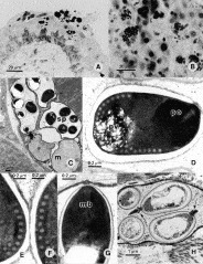
Fig. 2. Trachipleistophora hominis in experimentally infected SCID mice and T. hominis fine structure features. Histological sections (Gram stain) of intracerebrally inoculated SCID mouse showing spores within epithelial cells of urinary bladder (A) and hepatocytes (B). (C) Two meronts, 1 binucleate (m) and 3 sporophorous vesicles with mature spores (sp). (D) Mature spore showing the characteristic subapical location of the exit site of the polar filament, the very dense and compact polaroplast (po), the arrangement of polar filament coils and the large posterior vacuole (pv). (E and F) Details of the arrangement of polar filament coils. There are 8 (E) or 7 (F) coils of the same diameter in a single row, ending in 3 slightly thinner coils, displaced to the interior of the spore as a short second row. (G) Apical part of the spore showing the manubroid (mb) part of the polar filament, the anchoring disc surrounded by polar sac membranous cisterna. The polaroplast membranous stacks (cisternae) are narrow and densely packed in the apical region of the organelle and are more wide and less densely packed posteriorly. (H) Part of a sporophorous vesicle with 2 spores that have extruded their polar filaments (arrows).
 You have
Access
- 6
- Cited by