Introduction
Intracranial carotid artery pseudoaneurysms are common, but pseudoaneurysms of the extracranial carotid artery are rare. They have been reported to represent as few as one in every 800 aneurysms.Reference Dehn and Taylor1 The most common causes of this type of aneurysm include penetrating or blunt trauma and spontaneous dissection. Arteriosclerosis, Ehlers–Danlos syndrome, deep neck space infection, fibromuscular dysplasia, surgical trauma and radiotherapy are other, less frequent causes.Reference Lyons, Barker and Flood2
Perforation of the great vessels is rare. When it does happen, serious haemorrhage may occur, either at the time of perforation or later during the formation of a pseudoaneurysm. Great vessel perforation may result from oesophageal perforation, which may be iatrogenic, traumatic, spontaneous, tumour-related or due to foreign body ingestion. Foreign body ingestion may result in a combination of oesophageal perforation and traumatic injury to the common carotid artery; very rarely, this may result in pseudoaneurysm formation.Reference Kunishinge, Myojin, Ishibashi, Ishii, Kawasaki and Oka3
An aneurysm of the common carotid artery can present in several different ways. The most frequent presentations include localised symptoms due to compression, progressive enlargement with resultant rupture, and cerebral embolisation of a proximal thrombosis with subsequent resolution of all symptoms.Reference Katsantonis, Friedman and Kielmovitch4
In our patient, the offending foreign body was a thin, linear wire which was accidentally ingested, resulting in oesophageal perforation. This healed spontaneously, but a pseudoaneurysm developed due to a tear in the wall of the right common carotid artery, caused by the foreign body (which had migrated out of the oesophagus). We successfully repaired the right common carotid artery, after evacuating the infected pseudoaneurysm.
Case report
A 15-year-old boy from a remote area of Nepal presented as an emergency with a 15-day history of painful swallowing, gradually increasing in intensity. He had been treated in a local health clinic for tonsillitis. After one week, the patient had noticed a right-sided swelling of the lower neck which had progressively increased in size, associated with a fever of approximately 37.8°C without any chills or rigors (Figure 1).
The pseudoaneurysm at admission.

On direct questioning, the patient gave a history of accidental foreign body ingestion during eating, approximately 15 days ago.
On general examination, the patient's vital signs were stable. Local examination revealed a diffuse, globular, 5×5 cm mass occupying the right muscular and carotid triangles. The overlying skin was slightly erythematous. On palpation, the local skin temperature was found to be slightly raised, and the swelling was noted to be firm, smooth and tender with ill-defined margins; it was not possible to palpate the inferior border of the swelling. There were few transmitted pulsations, and the swelling was minimally fluctuant and tense.
Reducibility and trans-illumination were absent. The trachea was shifted to the left, and there were no palpable neck nodes or surgical emphysema. On auscultation, there was no audible bruit.
The results of routine blood investigations were normal.
The patient was commenced on intravenous antibiotics.
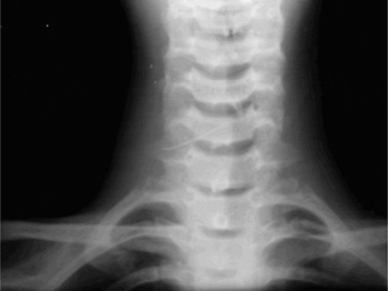
Plain X-ray of the neck showed a linear, radiopaque shadow (Figure 2). Computed tomography scanning of the neck with intravenous contrast revealed a 3 × 2.9 cm, contrast-filled space with an anterior, curvilinear, hypodense area in the right carotid space at the level of the C6 to T1 vertebrae, which also contained a linear, hyperdense foreign body.
Antero-posterior X-ray showing the thin wire.

Doppler ultrasonography of the neck was then performed, which showed a 3×3 cm pseudoaneurysm of the right common carotid artery, containing a 2.5 cm long, metallic foreign body touching the wall of the artery. The neck of the aneurysm measured 2.9 mm in diameter.
The patient was prepared for surgery under antibiotic cover. An oblique skin incision was made in the lower neck on the right. Dissection was performed with the aim of securing the right common carotid artery both below and above the aneurysm. There was little space in the neck below the aneurysm; during dissection of this region, the aneurysm was ruptured but bleeding was stopped by direct finger pressure. The dissection was thus shifted superior to the level of the aneurysm, and the common carotid artery was secured from above. The dissection was then extended below the aneurysm. There were severe adhesions between the common carotid artery and the internal jugular vein. An approximately 5 mm, linear tear was discovered in the common carotid artery wall, approximately 1 cm above the clavicle, and one end of a 3.0 cm, thin, linear, metallic wire was found piercing the artery wall. This foreign body was removed and the carotid repaired with 6-0 polypropylene sutures. The wound was sutured in layers after placing a negative pressure drain.
The patient's post-operative recovery was uneventful. There was no evidence of any neurovascular compromise.
Discussion
A pseudoaneurysm differs from a true aneurysm in that it does not contain any of the vessel wall. It is usually a result of either sharp or blunt trauma to all three layers of the artery wall, resulting in a periarterial haematoma which subsequently becomes encapsulated by the ingrowth of adjacent non-vascular and fibrotic tissue. This haematoma must continue to communicate with the artery in order to be considered a pseudoaneurysm. The central part of the haematoma can liquefy, creating a potential space for blood flow. Arterial blood pressure may lead to gradual enlargement and formation of an aneurysmal sac.Reference Beale5 Bleeding in the sub-intimal or sub-adventitial space can also result in narrowing or occlusion of the true arterial lumen.Reference Adams, Love, Jakoby, Ginsberg and Bogousslavsky6 The incidence of aneurysm formation due to such arterial wall dissection is between 10 and 30 per cent.Reference Mokri, Piepgras, Sundt and Pearson7
Traumatic pseudoaneurysms of the extracranial carotid artery are rare.Reference Doudle and Raptis8, Reference McCullum, Wheeler, Noop and DeBakey9 Most traumatic pseudoaneurysms occur in the young, in whom the risk of trauma is high.Reference Doudle and Raptis8 Pseudoaneurysms have also been reported to occur after various surgical procedures, including transsphenoidal surgery, transcavernous tumour surgery and endoscopic sinus surgery.Reference Bavinsky, Killer, Knosp, Ferraz-Leite, Gruber and Richling10, Reference Isenberg and Scott11
Repair of the pseudoaneurysm and restoration of circulation is the preferred treatment, in order to prevent emboli and compression of adjacent vessels (which may cause stroke).Reference Doudle and Raptis8, Reference McCullum, Wheeler, Noop and DeBakey9 Surgical procedures include primary excision and repair with or without a vein graft, ligation, and gradual occlusion with a clamp.Reference Purdue, Pelligrini and Arena12 Procedures using an extracranial–intracranial bypass to reduce the risk of ischaemic complications after proximal vessel occlusion have been well described.Reference Peerless and Hampf13 Immediate bleeding caused by injury to the aneurysm during localised surgical procedures should be treated with tight packing, but this may subsequently lead to carotid stenosis or complete carotid artery occlusion.Reference Chen, Concus, Halbach and Cheung14
• Extracranial carotid artery pseudoaneurysms are rare
• This paper describes an unusual case of common carotid artery pseudoaneurysm following ingestion of a thin wire
• This case was managed by neck exploration and repair of the vascular injury
• Surgical treatment procedures using an extracranial–intracranial bypass to reduce the risk of ischaemic complications after proximal vessel occlusion are well described
Our patient represents a rare case of carotid pseudoaneurysm in the neck, as a result of oesophageal perforation due to ingestion of a thin wire.
Acknowledgements
We acknowledge the help of Dr O P Pathania, Department of General Surgery, BP Koirala Institute of Health Sciences, Dharan, in the management of this case.