Over the past 25 years, non-invasive imaging, especially echocardiography, has become the primary method of evaluating the anatomy of patients with congenital cardiac disease before surgery. Although ultrasound is adequate to delineate cardiovascular anatomy for most patients, there are instances in which this modality may be inadequate, such as in patients with poor acoustic windows, or with multiple overlying or tortuous structures. In addition, standard cross-sectional echocardiography is unable to display non-fluid containing structures like the lungs or trachea and cannot display spatial relationships in three dimensions. These are all qualities that could potentially aid in planning a surgical intervention with complex anatomy. While three-dimensional echocardiography holds the potential for better delineating intracardiac anatomic relationships, it remains constrained by the physical limits of ultrasound and a relatively small field of view.
Multidetector computed tomographic angiography is an emerging non-invasive imaging modality for cardiovascular diagnosis in patients with structural cardiac disease.Reference Ou, Celermajer, Calcagni, Brunelle, Bonnet and Sidi1–Reference Spevak, Johnson and Fishman6 While computed tomographic angiography has been shown to aid the surgical planning in other specialities such as otolaryngology and neurosurgery, its utility to the surgeon in pre-operative planning for patients undergoing repair of congenital cardiac disease has not been well studied.Reference Jhang, Park, Seo, Goo and Gwak5, Reference Leong, Batra and Citardi7, Reference Pechlivanis, Schmieder, Scholz, Konig, Heuser and Harders8 Therefore, we evaluated the utility of this modality for the cardiac surgeon in the pre-operative planning of both children and adults undergoing surgery for congenital cardiac disease.
Materials and methods
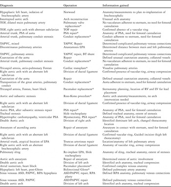
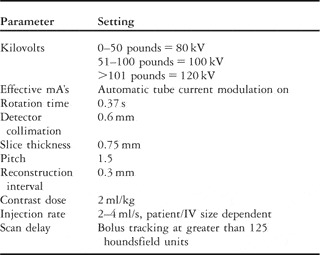
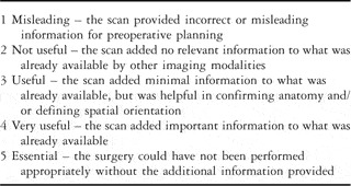
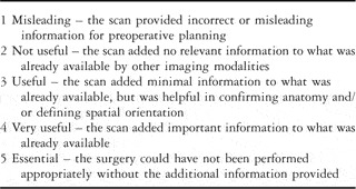
Approval was obtained from our institutional review board. Over a 12-month period from August, 2005 until August, 2006, all patients in whom computed topographic angiography of the chest was requested by the medical or surgical team for diagnostic purposes as part of their evaluation in preparation for planned congenital cardiac surgery were included in the study (33 patients; mean age = 5.6 years, range = birth to 24 years; Table 1). The surgeries were all performed by a single cardiac surgeon. In all patients, computed tomographic angiography was requested when the team felt the echocardiographic images were not adequate to completely define the cardiovascular anatomy. All patients within the cohort had their computed tomographic angiography performed by a team of paediatric cardiologists and radiologists using a standardised protocol, including radiation dose-reduction strategies and a standardised contrast dose based on weight (Table 2). The computed tomographic angiograms were all performed on a 64-slice scanner, Siemens Somatom Sensation (Siemens Medical Systems, Malvern, PA, United States of America), and the chest was scanned from the clavicles to the diaphragm. From the axial acquisitions, coronal and sagittal reconstructions were made on the scanner and subsequently, three-dimensional reconstructions were created from these data sets using an TeraRecon Aquarius workstation, San Mateo, CA, United States of America. Our dose-reduction protocol and average radiation doses have been published previously.Reference Herzog, Mulvihill and Nguyen9 As is standard of care, all patients also underwent a transthoracic echocardiogram as part of their pre-operative evaluation. After the operation, the surgeon was polled to assess the overall utility of the study in operative planning. Specifically, the surgeon was asked to grade the usefulness of the study on a 1–5 scale (Table 3), rate whether the computed tomographic angiography was useful in dictating the need for peripheral cannulation for bypass, and whether a cardiac catheterisation would have been requested if the scan were not available. For the purpose of the analyses, operations were classified according to the principal anatomic site of repair: aortic arch (sample size = 15), right ventricle to pulmonary artery conduit (sample size = 7), pulmonary arteries (sample size = 3), pulmonary veins, (sample size = 4), trachea/airway (sample size = 2), and other (sample size = 2). The “other” category included scans obtained for thoracic and vascular anatomy before pacemaker placement or pre-transplant. The usefulness score was compared among groups using a Fisher’s Exact test, SAS 9.1, SAS Institute, Cary, NC, United States of America.
List of diagnoses, interventions, and the ways in which the computed tomographic angiograms aided the surgical planning of these interventions.

ASD, atrial septal defect; BT, Blalock Taussig; LPA, left pulmonary artery; LV, left ventricle; PAPVC, partially anomalous pulmonary venous connection; PSA, pseudoaneurysm; RPA, right pulmonary artery; RV, right ventricle; TAPVC, totally anomalous pulmonary venous connection; TOF, Tetralogy of Fallot
*Repeat sternotomy
Congenital cardiac computed tomographic angiography protocol.

Rating scale for usefulness of computed tomographic angiography in preoperative planning.
Results
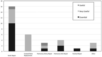
All questionnaires were completed in their entirety immediately following the surgery. Nearly all computed tomographic angiograms performed (31/33; 94%) were classified as either “very useful” or “essential” to pre-operative planning by the surgeon. The two remaining patients were scored as “useful.” The first patient had an interrupted aortic arch and, the other had tetralogy of Fallot with a right aortic arch and mirror image branching. In the surgeon’s opinion, the scans in these patients validated the echo results and only provided minimal additional information. In no cases were the anatomic findings by computed tomographic angiography found to be inaccurate by surgical inspection. The distribution of rankings for studies within diagnosis groups is shown in Figure 1. Specifically, the surgeon stated that computed tomographic angiography helped define or confirm complex vascular anatomy, describe anatomical relationships useful to surgical planning, describe severity of arterial or venous stenoses, and/or assist in planning bypass cannulation site. For each individual case, the specific utility of the CT scan is given in Table 1.
Distribution of the number of studies among anatomic groups and the frequency of the surgically determined utility of the pre-operative computed tomographic angiography within these groups.

Among surgical intervention groups, there was no difference in the utility of computed tomographic angiography for pre-operative planning (p = 0.42). However, the scans proved to be consistently useful to the surgeon for two particular intervention types: those involving the aorta (14/15, 93%) or the pulmonary veins (4/4, 100%). Computed tomographic angiography was found to be important in determining the need for peripheral cannulation in patients undergoing re-operation for conduit revision when compared with the other diagnosis groups (p = 0.02). A total of 73% of the patients for whom the scan changed cannulation site were undergoing a repeat operation and had a right ventricle to pulmonary artery conduit in place. The average age of the patient for whom computed tomographic angiography affected cannulation site was 12.9 years, compared with an average age of 2 years in the remainder of the group.
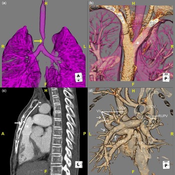
In some patients, computed tomographic angiography obviated the need for pre-operative cardiac catheterisations. In 14 out of the 33 patients (42%), the surgeon stated that he would have requested a catheterisation to further define the anatomy if the scan had not been available. Two patients in our sample underwent invasive cardiac catheterisation for haemodynamic indications before the computed topographic angiogram. In both patients, while the haemodynamic data from the catheterisation was important, the surgeon felt that the additional anatomic detail that could be provided by scan was necessary for surgical planning. Specifically, in comparison to biplane angiography, the inherent three-dimensional nature of computed tomographic angiography provided a better understanding of anatomical relationships required for pre-operative planning (Fig 2).
Three-dimensional reconstructions in four different patients to illustrate the utility of pre-operative computed tomographic angiography – (a) This volume-rendered image is a frontal view of the airway in a patient with a pulmonary sling. The vasculature has been removed through the post-processing software. Note the discrete narrowing in the distal trachea (arrow), corresponding to the location of the aberrant left pulmonary artery. In addition to confirming the presence of a pulmonary sling, this study enabled the surgeon to understand the location and degree of tracheal narrowing, allowing adequate preparation for a slide tracheoplasty. (b) Viewed from posterior–superior perspective, this volume-rendered image reveals a double aortic arch encircling the trachea. Note that the right aortic arch (asterisk) is smaller than the left, which is atypical for this anomaly and was not appreciated on echocardiography. This study convinced the surgeon to approach the repair through a right thoracotomy for division of the right arch, instead of the standard approach for a double arch repair, which is through a left thoracotomy. (c) This is a maximum-intensity projection image in the sagital plane in a patient with a common arterial trunk. The patient was scheduled for right ventricle to pulmonary artery conduit replacement through repeat sternotomy. Note that the conduit is adherent to the sternum (arrow). Given this finding, the surgeon elected to place the patient on femoral bypass before attempting the sternotomy. (d) This a volume-rendered image viewed from the posterior perspective in a patient with mixed totally anomalous pulmonary venous connections. The right and left lower pulmonary veins (RLPV and LLPV) connect to a horizontal confluence (c), which leads into a vertical vein (VV). Pulmonary venous drainage from the left upper lung empties through multiple separate veins (arrows) as the vertical vein ascends. The vertical vein dives underneath the left azygous vein (asterisk), resulting in stenosis before joining the left superior caval vein (LSCV). The right upper pulmonary vein (RUPV) connects with right superior caval vein. This study allowed the surgeon to appreciate the complexity and drainage pattern of the pulmonary veins, which was not understood completely by echocardiography, before the surgical repair.

Discussion
This descriptive cohort study highlights the utility of computed tomographic angiography in the pre-operative evaluation of children and adult patients with congenital cardiac disease. In our experience, computed tomographic angiography functions as a non-invasive complement to other imaging modalities, helping to further delineate complex anatomic relationships in an easily manipulated format. The data can be viewed in any plane and manipulated to show complex relationships of extracardiac vascular structures with other anatomic elements. Moreover, the data can be presented from a perspective that is familiar to the surgeon, allowing them to plan and visualise their procedure more readily. Furthermore, unlike echocardiography, computed tomographic angiography can illustrate adjacent thoracic structures. In this study, we found this particularly useful in the eight patients with vascular rings or slings, in which arch and tracheal structures can be simultaneously visualised in a three-dimensional fashion. In pre-operative planning conferences, the reconstructed three-dimensional computed tomography angiographic images can be shown and rotated or manipulated, demonstrating spatial relationships and true vascular dimensions in a perspective familiar to the surgeon.
Surgical planning for intervention on certain congenital cardiac lesions are more likely to benefit from computed tomographic angiography; namely, aortic arch anomalies, such as vascular rings or aneurysms, and pulmonary vein disease. We believe this is due to computed tomographic angiography’s ability to show the size, course, and contour of vessels as they course to and from the heart; something that is very difficult to re-create with two-dimensional echocardiography. Furthermore, pulmonary veins are notoriously challenging to see well by other imaging modalities.Reference Taylor10, Reference Ucar, Fitoz, Tutar, Atalay and Uysalel11
Although there was no global effect on cannulation planning by the scan, the studies were useful in older patients undergoing surgical re-intervention; particularly those involving right ventricle to pulmonary artery conduit revision or repair of a pseudoaneurysm. Specifically, the scan was helpful in delineating the proximity of vascular structures or conduit to the anterior bony chest wall. Specifically, the surgeon was particularly interested in whether vascular structures were adherent to the sternum, making it risky to re-open the chest before being placed on cardiopulmonary bypass. We feel that for many older patients needing conduit revision or pseudoaneurysm repair, obtaining computed tomographic angiography as part of the pre-operative evaluation may reduce operative and cannulation complications.
Cardiac catheterisation can provide excellent delineation of vascular structures and can provide haemodynamic information not obtainable from other modalities. However, traditional invasive angiography is limited to two simultaneous planes, usually anterior/posterior and lateral. In addition, even with digital subtraction techniques and the ability to rotate the biplane cameras, overlapping shadows on cine imaging can hamper accurate diagnosis. In contrast, the multiplanar nature of computed tomographic angiography and the ability to manipulate two- and three-dimensional images provide the ability to show individual vessels or structures as well as their anatomic relationship to one another or other structures. This is a distinct advantage for patients with complex extracardiac anatomy. In addition, computed tomographic angiography requires little to no sedation, and the patient needs only a peripheral intravenous catheter.
Another imaging modality that has gained attention in the evaluation of patients with congenital cardiac disease is magnetic resonance imaging.Reference Babu-Narayan, Gatzoulis and Kilner12–Reference Dorfman and Geva15 While magnetic resonance imaging and angiography is undoubtedly useful in this population by offering additional information about cardiac function and intra-cardiac anatomy, computed tomography offers several notable advantages over magnetic resonance imaging in certain patients.Reference Taylor10 First, as mentioned above, computed tomographic angiography allows one to simultaneously visualise vascular, airway, and adjacent bony structures in a three-dimensional fashion; something not easily obtained with magnetic resonance imaging techniques. Second, magnetic resonance imaging in younger children often requires general anaesthesia or deep sedation, while computed tomographic scans can be performed in most children with little to no sedation. Motion artefact resulting from breathing and poor cooperation are minimised by a much faster image acquisition. In our practice, given that the patient only needs to be still for a few seconds, sedation for a computed tomographic scan is only considered in patients between ages one and three, unless there is significant neurodevelopmental delay. Infants can be restrained with swaddling, and older children can usually be calmed adequately by the presence of a parent. Third, current computed tomographic scanning techniques allow for much better spatial resolution, allowing for improved visualisation and reconstruction of smaller structures, such as pulmonary veins and aortopulmonary collaterals, in infants and small children. With magnetic resonance imaging, smaller patients are faced with a lower signal to noise ratio, resulting in even poorer spatial resolution. Fourth, computed tomographic angiography is much less affected by metallic artifact.Reference Eichhorn, Jourdan, Hill, Raman, Cheatham and Long16, Reference Garg, Powell, Sena, Marshall and Geva17 In our study, this was particularly relevant in patients for whom it was important to precisely define the relationship between sternal wires and vascular structures or pseudoaneurysms in preparation for a repeat sternotomy.
Two important “savings” issues became apparent with our analysis with regards to cardiac catheterisation. First, computed tomographic angiography potentially limits the number of invasive catheterisations that are necessary when anatomical questions are the principal indication for the procedure. As shown in our results, our surgeon would have requested over 40% more catheterisations to delineate anatomic relationships had the computed tomographic angiogram not been performed. The second is an issue of cost savings. Avoiding invasive procedures or reducing the number of imaging studies not only benefits the patient, but can also bring about significant reductions in cost of medical care. The average fee for a diagnostic congenital right and left cardiac catheterisation at our institution, facility charge and supplies only; not professional fees, is $8400 whereas the same figure for a computed tomographic angiogram is only $1293, Medical University of South Carolina administration, personal communication. In addition, most computed tomographic angiographic scans can be done with little if any sedation/anaesthesia and no central vascular access, reducing both costs and potential complications. Lastly, recent work has shown that the average diagnostic cardiac catheterisation exposes the paediatric patient to significantly more ionising radiation than computed tomographic angiography.Reference Hlavacek, Mulvihill, Gonzalez, Shirali and Frey18
This study has limitations by virtue of being descriptive in nature. In addition, our study was conducted in conjunction with a single surgeon, and as such may be inherently biased. His determinations regarding the utility of the study may not be completely generalisable to the cardiac surgical community as a whole. However, the areas for which computed tomographic angiography was found to be beneficial at our programme – namely aortic and pulmonary vein pathology – are supported by literature from other institutions.Reference Taylor10, Reference DiSessa, Di Sessa, Gregory and Vranicar19 Furthermore, carrying out this study measuring whether computed tomographic angiography altered hard outcomes such as operative complications or mortality or hospital stay would likely require quite large sample sizes, but could be considered as a future research direction. Ultimately, other more rigorous studies could be conducted in the future and advance these preliminary data, further exploring the benefits of computed tomographic angiography in the congenital cardiac surgical population.
In conclusion, this study illustrates the utility of computed tomographic angiography in the pre-operative evaluation of patients with congenital cardiac disease. We feel these are preliminary data to suggest the importance of computed tomographic imaging in the clinical decision-making process for this patient group. These results may help clinicians decide which patients might benefit from a pre-operative computed tomographic angiogram, and potentially reduce the need for invasive cardiac catheterisations. In addition, data supporting the appropriateness of computed tomographic angiography in certain clinical situations may support efforts for reimbursement of this study among paediatric practitioners.
Acknowledgements
The authors are grateful to our gifted technologists for their skill and assistance with the performance of the many scans. Dr Hlavacek received an honorarium from TeraRecon for work unrelated to this study. No outside sources of funding were used for this study. The tested technology and software were purchased by the university hospital for routine patient care and not specifically for this study.