Introduction
Tremendous number of biomedical applications of various nanomaterials [Reference Gagner, Shrivastava, Qian, Dordick and Siegel1, Reference Cheng, Jiang, Wang, Chen and Liu2], including silica nanoparticles (NPs) [Reference Bitar, Ahmad, Fessi and Elaissari3, Reference Tang and Cheng4, Reference Wang, Zhao, Han, Bai, Li, Liu, Che, Hu, Zhang, Jiang and Wang5], gold NPs [Reference Tiwari, Vig, Dennis and Singh6], iron oxide NPs [Reference Ali, Zafar, Zia, Haq, Phull, Ali and Hussain7], zinc oxide nanomaterials [Reference Zhang, Nayak, Hong and Cai8], and two-dimensional nanomaterials [Reference Chimene, Alge and Gaharwar9], have been reported. In particular, silica NPs are potentially useful for nanomedicine because they are hydrophilic, have low toxicity, and are able to be labeled or modified easily [Reference Halas10]. However, some researchers are concerned about health risks induced by silica NPs [Reference Nishimori, Kondoh, Isoda, Tsunoda and Tsutsumi11, Reference Xie, Sun, Zhong, Shi and Zhang12, Reference Yu, Duan, Li, Li, Jing, Yang, Wang and Sun13]. For example, Liu et al. reported that mesoporous hollow silica NPs (MHSNs) injected in mice are captured by Kupffer cells in liver and that these MHSN-activated Kupffer cells promote the formation of silicotic nodules that induce inflammation, necrosis, and fibrosis in liver [Reference Liu, Li, Teng, Huang, Liu, Chen, Ren, He and Tang14, Reference Lin, Li, Fu, Liu, Chen and Tang15]. Electron microscopic analyses revealed that MHSNs are trapped in Kupffer cells without being degraded. If MHSNs were to be decomposed in Kupffer cells, the hepatic injury caused by activated Kupffer cells might be relieved. On the other hand, for efficient nanomedicine based on silica NPs without unwanted side effects, it is desirable that delivery systems of therapeutic proteins and drugs using silica NPs as vehicles can release the medicines under control. These two challenges can be accomplished by using stimuli-responsive degradable silica NPs [Reference Mekaru, Lu and Tamanoi16, Reference Moreira, Dias and Correia17]. Silica NPs whose degradation is responsive to external stimuli, such as light [Reference Karimi, Zangabad, Baghaee-Ravari, Ghazadeh, Mirshekari and Hamblin18] or magnetic fields [Reference Baeza, Guisasola, Ruiz-Hernández and Vallet-Regí19, Reference Saint-Cricq, Deshayes, Zink and Kasko20], can be more precisely controlled than those responsive to internal stimuli, such as low pH [Reference Lee, Cheng, Huang, Souris, Yang, Mou and Lo21, Reference Meng, Xue, Xia, Zhao, Tamanoi, Stoddart, Zink and Nel22, Reference Zhao, Li, Kabehie, Botros, Stoddart and Zink23, Reference Zhou, Wu, Yin, Jin, Zhang, Zheng, Wang and Liu24], reductive environment [Reference Giri, Trewyn, Stellmaker and Li25, Reference Cui, Dong, Cai, Wang and Li26, Reference Zhang, Liu, Nguyen, Ma, Wang, Xing and Zhao27, Reference Yang, Wan, Niu, Gu, Zhang, Yu and Yu28, Reference Prasetyanto, Bertucci, Septiadi, Corradini, Castro-Hartmann and De Cola29, Reference Hayashi, Maruhashi, Nakamura, Sakamoto and Yogo30, Reference Zhou, Du, Li, Li, Huang, Liao, Shi, Zhang and Zhang31], and enzymes [Reference Mondragón, Mas, Ferragud, de la Torre, Agostini, Martínez-Máñez, Sancenón, Amorós, Pérez-Payá and Orzáez32, Reference Liu, Zhang, Luo, Ding, Li, Dai, Zhou, Zhao, Ye and Cai33, Reference van Rijt, Bölükbas, Argyo, Datz, Lindner, Eickelberg, Königshoff, Bein and Meiners34]; however, internal stimuli–responsive degradable silica NPs have the clinical advantage that they can be controlled by biological conditions, such as intracellular environments. For instance, degradable silica NPs responsive to the reduced form of glutathione (GSH) [Reference Cui, Dong, Cai, Wang and Li26, Reference Zhang, Liu, Nguyen, Ma, Wang, Xing and Zhao27, Reference Yang, Wan, Niu, Gu, Zhang, Yu and Yu28, Reference Prasetyanto, Bertucci, Septiadi, Corradini, Castro-Hartmann and De Cola29, Reference Hayashi, Maruhashi, Nakamura, Sakamoto and Yogo30, Reference Zhou, Du, Li, Li, Huang, Liao, Shi, Zhang and Zhang31] might be used to realize a controlled drug delivery system based on silica NPs with low health risks. GSH is the most abundant antioxidant synthesized in cells, and the thiol is involved in cellular defense against oxidative stress and maintaining redox homeostasis [Reference Circu and Aw35, Reference Forman, Zhang and Rinna36]. GSH-responsive degradable silica NPs are preferentially decomposed in intracellular environments because the intracellular concentration of GSH (1–10 mM, normally 1–2 mM) is much higher than the extracellular concentration of GSH in the micromolar range [Reference Circu and Aw35, Reference Forman, Zhang and Rinna36]. In general, the chemical structures of GSH-responsive degradable silica NPs have disulfide or polysulfide bonds, which are reduced by GSH [Reference Cui, Dong, Cai, Wang and Li26, Reference Zhang, Liu, Nguyen, Ma, Wang, Xing and Zhao27, Reference Yang, Wan, Niu, Gu, Zhang, Yu and Yu28, Reference Prasetyanto, Bertucci, Septiadi, Corradini, Castro-Hartmann and De Cola29, Reference Hayashi, Maruhashi, Nakamura, Sakamoto and Yogo30, Reference Zhou, Du, Li, Li, Huang, Liao, Shi, Zhang and Zhang31].
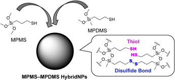
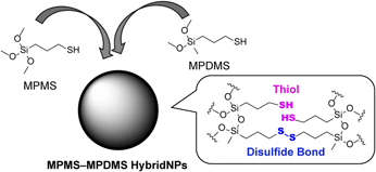
We have studied thiol-organosilica NPs as a class of silica NPs for nanomedicine. Thiol-organosilica NPs are synthesized by the sol–gel method from a thiol-organosilicate, such as 3-mercaptopropyltrimethoxysilane (MPMS) or 3-mercaptopropyl(dimethoxy)methylsilane (MPDMS) [Reference Nakamura and Ishimura37, Reference Nakamura, Ozaki, Abe, Doi, Matsumoto and Ishimura38, Reference Doura, Tamanoi and Nakamura39]. MPMS NPs are dispersive in aqueous solutions, and functionalization using thiol chemistry is easy due to the presence of abundant thiols on the surface. Hence, various biomedical applications of MPMS NPs, such as use as a fluorescence and X-ray computed tomography probe [Reference Nakamura, Awaad, Hayashi, Ochiai and Ishimura40], a polyethylene glycol (PEG)-coating nanoprobe for identification of PEG-resistant macrophages [Reference Nakamura, Hayashi, Nakano, Kanadani, Miyamoto, Kori and Horikawa41], and a multimodal imaging probe internally functionalized with iron oxide magnetic NPs and a fluorescent dye [Reference Nakamura, Hayashi, Kubo, Kanadani, Harada and Yogo42, Reference Nakamura, Hayashi, Kubo, Harada, Izumi, Tsuruo and Yogo43], have been investigated. However, the chemical structures of thiol-organosilica NPs have not been characterized in detail. Therefore, in this paper, we report the synthesis and compositional analysis of some types of thiol-organosilica NPs fabricated from MPMS and MPDMS using solid-state 13C nuclear magnetic resonance (NMR) measurements and Raman spectroscopic measurements. The results revealed that disulfide bonds in thiol-organosilica NPs were induced by existence of MPDMS constituent in thiol-organosilica NPs (Fig. 1). We also revealed that thiol-organosilica NPs, which have disulfide bonds, are GSH-responsive degradable. As an application of the GSH-responsive degradable thiol-organosilica NPs, we synthesized fluorescent thiol-organosilica NPs, including rhodamine B, and showed the GSH-responsive release of the dye. The synthesis, characterization, and application of GSH-responsive degradable thiol-organosilica NPs would lead to the further applications as a GSH-responsive drug carrier or a GSH-responsive formulation.
Fabrication of thiol-organosilica NPs, which have disulfide bonds.

Results and discussion
Synthesis of MPMS–MPDMS NPs
In previous study, we have described a one-pot synthesis of thiol-organosilica NPs using sol–gel method in a 28% ammonium hydroxide aqueous solution system [Reference Nakamura and Ishimura37, Reference Nakamura, Ozaki, Abe, Doi, Matsumoto and Ishimura38, Reference Doura, Tamanoi and Nakamura39]. Though this one-pot synthesis procedure is simple and inexpensive, the size of NPs was mainly controlled only by the concentration of the thiol-organosilicate. The size of NPs is very important for the biomedical applications, especially nanomedicine for the tumor type, because the enhanced permeability and retention effect for solid tumor targeting is exhibited by nanomaterials with the size of 10–200 nm [Reference Blanco, Shen and Ferrari44, Reference Svenson45]. Recently, by discovering that miniaturization of thiol-organosilica NPs can be induced by adding an anionic surfactant, such as sodium dodecyl sulfate (SDS) or sodium dodecylbenzenesulfonate (SDBS), we developed a method for the efficient size-controlled synthesis of small thiol-organosilica NPs with a mean diameter of less than 200 nm [Reference Doura, Tamanoi and Nakamura39]. On the other hand, in a series of publications, Khutoryanskiy and colleagues reported the controlling the size of thiol-organosilica NPs from MPMS and various biomedical application. In their article, the effect of synthetic condition, such as catalyst, solvent, temperature, and atmospheric oxygen, and the characteristic of NPs were studied. They obtained smallest particles (less than 50 nm) in the condition of NaOH as the catalyst in dimethyl sulfoxide (DMSO) solution with air bubbling [Reference Irmukhametova, Mun and Khutoryanskiy46, Reference Irmukhametova, Fraser, Keddie, Mun and Khutoryanskiy47, Reference Mahrooqi, Mun, Williams and Khutoryanskiy48]. Furthermore, they functionalized the surface of developed NPs with PEG or poly(2-alkyl-2-oxazoline) and applied to the mucus-penetrating system [Reference Mansfield, Sillence, Hole, Williams and Khutoryanskiy49, Reference Mansfield, de la Rosa, Kowalczyk, Grillo, Hoogenboom, Sillence, Hole, Williams and Khutoryanskiy50].
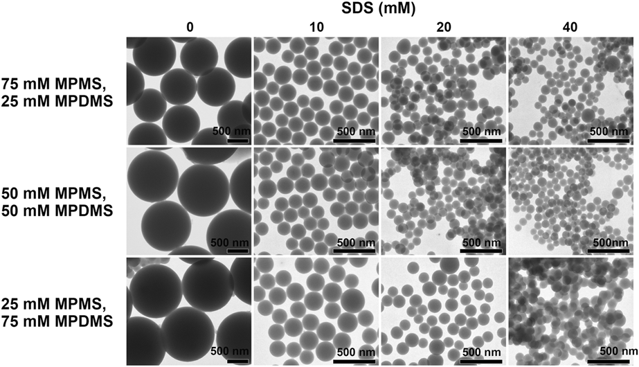
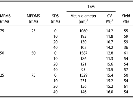
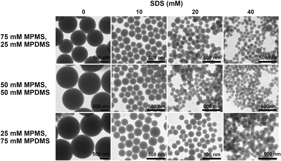
To our knowledge, though various approaches have been developed for the preparation of thiol-organosilica NPs, there are no reports about the hybrid NPs, which are composed of more than one kind of thiol-organosilicate. Attempt to synthesize and characterize hybrid thiol-organosilica NPs would open up the possibility of development of new synthetic procedure and novel function. Based on these backgrounds, we tried to synthesize the novel hybrid NPs derived from MPMS and MPDMS. Among thiol-organosilica NPs, MPDMS NPs are predicted to have disulfide bonds in the NPs, because the maximum number of siloxane bonds formed from an MPDMS is two. Therefore, we hypothesized that thiol-organosilica NPs containing MPDMS constituent have disulfide bonds, and we designed and synthesized thiol-organosilica NPs composed of MPMS constituent and MPDMS constituent, i.e., MPMS–MPDMS hybrid NPs. We examined the synthesis of MPMS–MPDMS NPs using the anionic surfactant-addition synthetic method. The size-reducing effect induced by an anionic surfactant was observed in the synthesis of these NPs. For example, while the mean diameter of MPMS–MPDMS (75:25) NPs prepared without an anionic surfactant was 1060 nm, the mean diameters of MPMS–MPDMS (75:25) NPs synthesized with 10 mM SDS, 20 mM SDS, and 40 mM SDS were 193, 130 and 102 nm, respectively (Fig. 2 and Table I); the miniaturization effect induced by 40 mM SDS was about 10.4-fold. In a similar fashion, the size-reducing effects of 40 mM SDS against MPMS–MPDMS (50:50) NPs and MPMS–MPDMS (25:75) NPs were about 16.7- and 10.5-fold, respectively. The sizes of MPMS–MPDMS (75:25) NPs and MPMS–MPDMS (50:50) NPs were smaller than those of the MPMS NPs synthesized under the same conditions [Reference Doura, Tamanoi and Nakamura39], indicating that the MPMS–MPDMS (75:25) NPs and MPMS–MPDMS (50:50) NPs are densely packed compared with MPMS NPs. The coefficients of variation (CVs) of size distribution of synthesized MPMS–MPDMS NPs were relatively small, i.e., 10.7–16.0%. MPMS–MPDMS (75:25) NPs and MPMS–MPDMS (50:50) MPs were dispersive in aqueous solution; however, MPMS–MPDMS (25:75) NPs were modestly aggregative in aqueous solution.
TEM images of MPMS–MPDMS NPs synthesized with 0, 10, 20, or 40 mM SDS.
Evaluation of MPMS–MPDMS NPs synthesized with SDS.
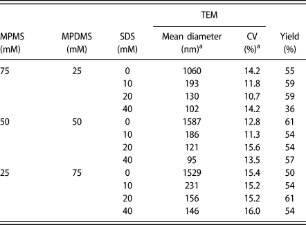
a Mean diameters and the CVs of the size distribution of thiol-organosilica NPs were calculated from more than 50 particles for each sample.
Characterization of thiol-organosilica NPs
Next, we characterized the chemical structures of MPMS NPs, MPDMS NPs, and MPMS–MPDMS hybrid NPs.
Thermal gravimetric analysis
We can measure the abundance ratio of MPMS constituent to MPDMS constituent in thiol-organosilica NPs using TGA, because the organic component of MPMS constituent is distinctively different from the organic component of MPDMS constituent. Thermal degradation of MPMS NPs, MPMS–MPDMS NPs, and MPDMS NPs was studied by TGA (Fig. S1 and Table II). At the temperatures between 150 and 800 °C, the weight loss of MPMS NPs, MPMS–MPDMS (75:25) NPs, MPMS–MPDMS (50:50) NPs, MPMS–MPDMS (25:75) NPs, and MPDMS NPs were estimated to be 44.6%, 45.2%, 47.7%, 60.4%, and 61.8%, respectively (Table II). The weight loss is derived from combustion of the organic components of MPMS NPs, MPMS–MPDMS NPs, and MPDMS NPs. Since the organic component of MPDMS constituent is larger than that of MPMS constituent, increasing the relative proportion of MPDMS constituent in the chemical structure of MPMS–MPDMS NPs is expected to increase the weight ratio of the organic component. However, the weight ratios of the organic components of MPMS–MPDMS (75:25) NPs and MPMS–MPDMS (50:50) NPs were lower than that expected from the mixing ratio of MPMS and MPDMS, indicating that MPMS constituent was preferentially incorporated into the chemical structure of these NPs. In contrast, the weight ratio of the organic part of the MPMS–MPDMS (25:75) NPs was higher than that expected from the mixing ratio of MPMS and MPDMS, indicating that MPDMS constituent was preferentially incorporated into the chemical structure of MPMS–MPDMS (25:75) NPs.
TGA of MPMS NPs, MPDMS NPs, and MPMS–MPDMS NPs.

Solid-state 13C NMR measurements
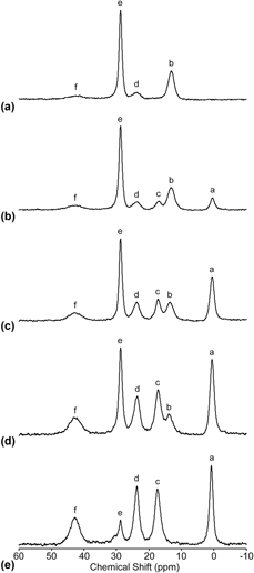
13C cross-polarization magic-angle spinning (CP MAS) NMR analyses reveal the chemical structure of hydrocarbon chains in thiol-organosilica NPs and the purity of synthesized thiol-organosilica NPs. The 13C CP MAS NMR spectra of MPMS NPs, MPMS–MPDMS NPs, and MPDMS NPs are shown in Fig. 3. The spectrum showed six characteristic peaks at 0.5 (a), 13.2 (b), 17.2 (c), 23.6 (d), 28.8 (e), and 43.2 (f) ppm. From the 13C CP MAS NMR spectra of MPMS NPs and MPDMS NPs, these peaks correspond to the aC, bC, cC, dC, eC, and fC in –Si–bCH2–eCH2–eCH2–SH and –Si–bCH2–dCH2–fCH2–S–S– derived from MPMS and aCH3–Si–cCH2–eCH2–eCH2–SH and aCH3–Si–cCH2–dCH2–fCH2–S–S– derived from MPDMS [Reference Quignard, Masse, Laurent and Coradin51]. The NMR spectrum of MPMS NPs indicates abundant thiols in the NPs, and the NMR spectrum of MPDMS NPs indicates abundant disulfide bonds in the NPs. The NMR analyses of MPMS–MPDMS NPs demonstrated that they were composed of both MPMS constituent and MPDMS constituent; additionally, peaks e and f indicated that the chemical structure contained both thiols and disulfide bonds. In the NMR spectrum of MPMS–MPDMS (75:25) NPs, strong peaks e and b indicate abundant thiols and MPMS constituent in the NPs. In the NMR spectrum of MPMS–MPDMS (25:75) NPs, enhanced peaks a, c, and f indicate abundant disulfide bonds and MPDMS constituent in the NPs. These results of the NMR analyses agree with the results of the TGA. In addition, all thiol-organosilica NPs do not include an anionic surfactant because no signals derived from an anionic surfactant in the NMR spectra.
Solid-state 13C NMR spectra of (a) MPMS NPs, MPMS–MPDMS NPs [MPDMS ratios were (b) 75:25, (c) 50:50, and (d) 25:75], and (e) MPDMS NPs.

Raman spectroscopic measurements
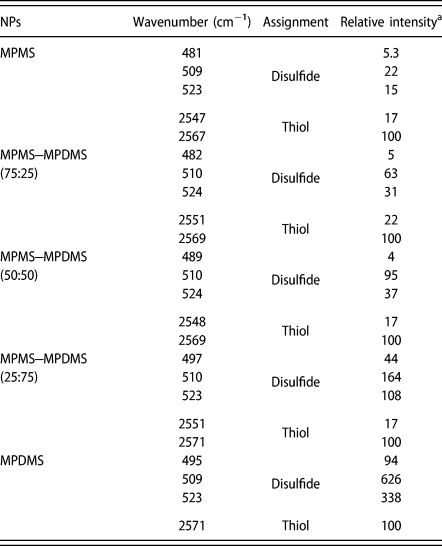
Because both disulfide bonds and terminal thiols are Raman active and display high intensity signals [Reference Bazylewski, Divigalpitiya and Fanchini52], the Raman spectroscopic measurements of thiol-organosilica NPs provide the detailed knowledge about disulfide bonds and thiols in the NPs. The Raman spectra of MPMS–MPDMS NPs are shown in Fig. S2, and the analytical data are reported in Table III. Disulfide stretching modes and thiol stretching modes of the MPMS–MPDMS NPs were located at about 480–525 and 2550–2570 cm−1, respectively [Reference Bazylewski, Divigalpitiya and Fanchini52]. The three disulfide stretching modes of each type of MPMS–MPDMS NPs are derived from three torsional conformers of a disulfide bond [Reference Van Wart and Scherag53]. The two thiol stretching modes originate from SH⋯O and SH⋯S vibrations, respectively [Reference Pawlukojć, Leciejewicz, Ramirez-Cuesta and Nowicka-Scheibe54]. The two thiol stretching modes show that thiols are close to hydroxyl groups and thiols; hence, the analyses indicate the existence of neighboring silanols and thiols in the NPs. As the proportion of MPDMS increased, the relative intensities of the peaks derived from stretching vibrations of disulfide bonds increased, indicating that the introduction of MPDMS to MPMS NPs induced the formation of disulfide bonds in MPMS–MPDMS NPs. The abundance ratio of disulfide bonds to thiols was related to the ratio of MPDMS constituent to MPMS constituent in the MPMS–MPDMS NPs. As indicated from the above NMR results, the Raman analyses confirmed that the thiol-organosilica NPs that contain MPDMS constituent have disulfide bonds in the chemical structure.
Raman modes of MPMS NPs, MPDMS NPs, and MPMS–MPDMS NPs.
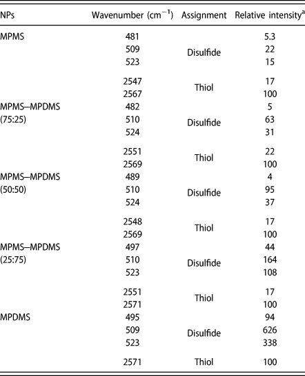
a The relative intensities of the peaks are shown based on the intensity of a thiol stretching mode (100).
The thiol content on the surface of NPs
The thiol content on the surface of NPs was determined using Ellman’s test. The content of thiol group in MPMS, MPMS-MPDMS (75:25), MPMS-MPDMS (50:50), MPMS-MPDMS (25:75), and MPDMS NPs were 4.81, 5.06, 5.58, 39.72, and 44.87 μmol/g, with a general trend of increasing content per gram with decreasing MPMS concentration. Apparently, these data disagree with the results from solid-state 13C NMR analyses and Raman spectroscopic analyses. It may be related to the difference in interior and exterior structure of the NPs synthesized under different MPMS or MPDMS concentration and availability of thiol groups for reacting with Ellman’s reagents. Khutoryanskiy and colleagues researched the physicochemical properties of thiol-organosilica NPs under various reaction conditions, and they demonstrated that the difference in the content of thiol group in NPs was caused by the effect of the catalyst, solvent, and atmospheric oxygen [Reference Irmukhametova, Mun and Khutoryanskiy46, Reference Irmukhametova, Fraser, Keddie, Mun and Khutoryanskiy47, Reference Mahrooqi, Mun, Williams and Khutoryanskiy48]. Further studies are needed in order to elucidate the interior and exterior structure of MPMS-MPDMS NPs, including specific surface area and micropore distribution.
Degradation analysis of thiol-organosilica NPs in the presence of GSH
As mentioned above, thiol-organosilica NPs containing MPDMS constituent (MPMS–MPDMS NPs and MPDMS NPs) have disulfide bonds. Because nanomaterials with disulfide bonds are potentially degradable by GSH [Reference Cui, Dong, Cai, Wang and Li26, Reference Zhang, Liu, Nguyen, Ma, Wang, Xing and Zhao27, Reference Yang, Wan, Niu, Gu, Zhang, Yu and Yu28, Reference Prasetyanto, Bertucci, Septiadi, Corradini, Castro-Hartmann and De Cola29, Reference Hayashi, Maruhashi, Nakamura, Sakamoto and Yogo30, Reference Zhou, Du, Li, Li, Huang, Liao, Shi, Zhang and Zhang31], we investigated the GSH-responsive degradability of MPMS–MPDMS NPs and MPDMS NPs compared with MPMS NPs.
Degradation analysis with 10 mM GSH
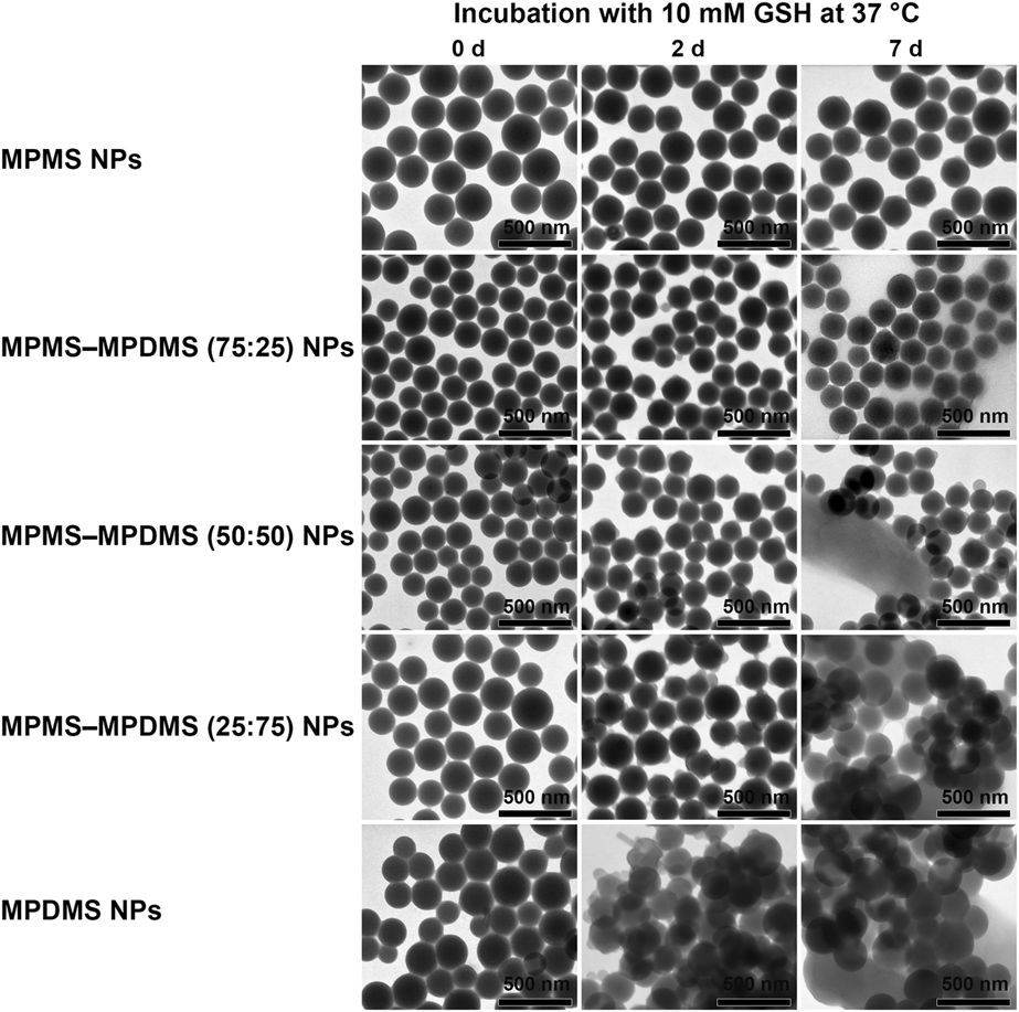
First, the GSH-responsive degradability of thiol-organosilica NPs under a concentration of GSH corresponding to the intracellular condition of cancer cells (i.e., 10 mM) was examined [Reference Yang, Wan, Niu, Gu, Zhang, Yu and Yu28]. The transmission electron microscopic (TEM) images of thiol-organosilica NPs incubated with 10 mM GSH at 37 °C for 2 or 7 days are shown in Fig. 4. The shape of MPMS NPs was hardly changed after incubation with 10 mM GSH for 2 days, but the outer edges were slightly collapsed after 7 days. The outer edges of MPMS–MPDMS (75:25) NPs were slightly damaged after 2 days, and MPMS–MPDMS (75:25) NPs were surrounded by irregular-shaped decomposed materials derived from the NPs after 7 days. The surface of MPMS–MPDMS (50:50) NPs was slightly damaged after 2 days, and not only partially collapsed MPMS–MPDMS (50:50) NPs but also aggregates of decomposed materials derived from the NPs were observed after 7 days. The surface of MPMS–MPDMS (25:75) NPs was moderately damaged after 2 days, and aggregates of the NPs and decomposed materials produced from the NPs were observed after 7 days. Aggregates of the partially collapsed MPDMS NPs were produced after 2 days, and aggregates of the NPs and destroyed materials derived from the NPs were observed after 7 days. The above finding that thiol-organosilica NPs containing disulfide bonds were partially decomposed by 10 mM GSH in vitro means that thiol-organosilica NPs, which have disulfide bonds, are able to be degraded partially in cancer cells. Our observation that the destruction of thiol-organosilica NPs containing MPDMS constituent increased as the proportion of MPDMS constituent (with high content rate of disulfide bonds) increased suggests that the content rate of disulfide bonds in the thiol-organosilica NPs predicts the GSH-responsive degradability of thiol-organosilica NPs.
TEM images of MPMS NPs, MPMS–MPDMS NPs, and MPDMS NPs incubated with 10 mM GSH at 37 °C for 0, 2, or 7 days.

Degradation analysis with 40 mM GSH
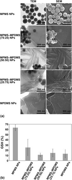
To further investigate the GSH-responsive degradability of thiol-organosilica NPs, thiol-organosilica NPs were incubated with 40 mM GSH at 80 °C for 2 months. Electron microscopic analysis of MPMS NPs revealed that although some membrane-like decomposed materials were produced, most of the MPMS NPs retained the spherical form [Fig. 5(a)]. This clarified that, as predicted from their low content rate of disulfide bonds, MPMS NPs were resistant to GSH-induced collapse. In contrast, when MPMS–MPDMS NPs were exposed to GSH, a substantial amount of irregular-shaped decomposed materials were produced. The destruction by GSH was higher for MPMS–MPDMS (50:50) NPs and MPMS–MPDMS (25:75) NPs than for MPMS–MPDMS (75:25) NPs. Most of the MPDMS NPs were also collapsed by GSH and transformed into the membrane-like irregular-shaped decomposed materials. These results indicate that both MPMS–MPDMS NPs and MPDMS NPs are susceptible to the attack by GSH.
(a) TEM images and scanning electron microscopic images of MPMS NPs, MPMS–MPDMS NPs, and MPDMS NPs incubated with 40 mM GSH at 80 °C for 2 months. (b) Percentage of GSH remaining in supernatants isolated from the samples of MPMS NPs, MPMS–MPDMS NPs, and MPDMS NPs incubated with 40 mM GSH at 80 °C for 7 days. The experiments were repeated in triplicate (n = 3).

After GSH attacks the disulfide bonds in the thiol-organosilica NPs, oxidized glutathione (GSSG) is formed through the disulfide exchange reaction. Since GSH is consumed by the series of reactions, the concentration of GSH in the supernatants isolated from the incubation samples was estimated by Ellman’s test. After 7 days of incubation with 40 mM GSH at 80 °C, we observed that 63.3% of GSH remaining in the supernatant derived from the MPMS NPs, whereas 25.3%, 11.2%, 14.9%, and 13.2% of GSH existed in the supernatant isolated from the MPMS–MPDMS (75:25) NPs, MPMS–MPDMS (50:50) NPs, MPMS–MPDMS (25:75) NPs, and MPDMS NPs, respectively [Fig. 5(b)]. Because more consumption of GSH means the higher reactivity with GSH, these results show that reactivity with GSH was higher for MPMS–MPDMS NPs and MPDMS NPs than for MPMS NPs. Additionally, the consumption of GSH by MPMS–MPDMS (50:50) NPs and the consumption of GSH by MPMS–MPDMS (25:75) NPs were almost equal to that by MPDMS NPs. These results demonstrate that MPMS–MPDMS (50:50) NPs and MPMS–MPDMS (25:75) NPs are GSH-responsive degradable nanomaterials comparable to MPDMS NPs. The surface of thiol-organosilica NPs, which have disulfide bonds, was degraded by the attack of GSH, and irregular-shaped decomposed materials were produced; but the size reduction or fragmentation of thiol-organosilica NPs containing disulfide bonds induced by the reaction with GSH was not observed. In addition, despite the increase in the abundance ratio of disulfide bonds with the introduction of MPDMS constituent, the GSH-responsive degradability of thiol-organosilica NPs containing disulfide bonds reached the maximum in the point of MPMS–MPDMS (50:50) NPs. The main reason might be the siloxane network of thiol-organosilica NPs. It is thought that the main backbone of thiol-organosilica NPs is the cross-linkage composed of siloxane bonds. Siloxane bonds are inactive against the attack of GSH, and the siloxane network are stable in solutions including GSH. The stability of siloxane network might suppress the GSH-responsive size reduction or fragmentation of thiol-organosilica NPs, which have disulfide bonds.
Rhodamine B–doped thiol-organosilica NPs containing disulfide bonds
Finally, we examined the potential of thiol-organosilica NPs, which have disulfide bonds, in biomedical applications, especially as a drug carrier for the controlled release of medical agents. As a model of drug-loaded thiol-organosilica NPs containing disulfide bonds, we synthesized rhodamine B–doped MPMS–MPDMS (25:75) NPs and checked the GSH-responsive liberation of rhodamine B from them.
Synthesis of rhodamine B–doped MPMS–MPDMS NPs
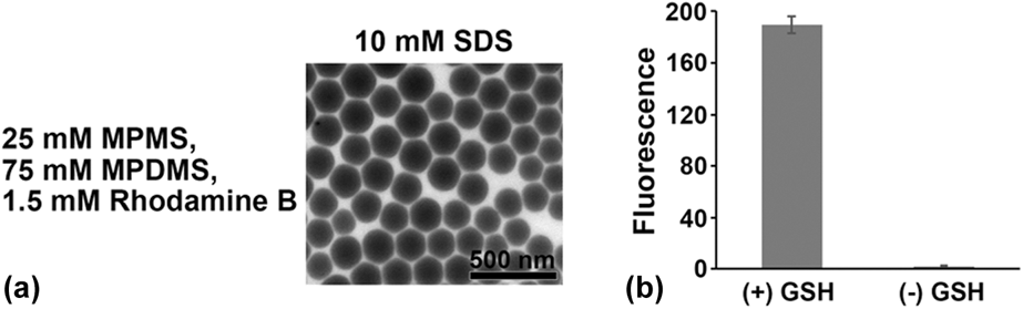
Rhodamine B–doped MPMS–MPDMS (25:75) NPs were synthesized using the anionic surfactant-addition method. The electron microscopic images of synthesized rhodamine B–doped MPMS–MPDMS (25:75) NPs are shown in Fig. 6(a). The mean diameter of the rhodamine B–doped MPMS–MPDMS (25:75) NPs was 199 nm, and the CV of the size distribution was 23.9%. The mean diameter of rhodamine B–doped MPMS–MPDMS (25:75) NPs was smaller than that of MPMS–MPDMS (25:75) NPs under the same synthetic condition (231 nm; Table I).
(a) TEM images of rhodamine B–doped MPMS–MPDMS (25:75) NPs. (b) Fluorescence intensities at 581 nm of the supernatants isolated from test samples incubated with or without 10 mM GSH for 48 h. The values represent the average and SD of three experiments (n = 3).

Release of rhodamine B from MPMS–MPDMS NPs in the presence of GSH
Next, we checked the GSH-responsive release of rhodamine B from rhodamine B–doped MPMS–MPDMS (25:75) NPs. Rhodamine B–doped MPMS–MPDMS (25:75) NPs was incubated with 10 mM GSH for 48 h, and the release of rhodamine B was estimated by measuring the fluorescence spectra of a supernatant isolated from the sample. As the control experiment, the release of rhodamine B from rhodamine B–doped MPMS–MPDMS (25:75) NPs was examined in the absence of GSH. As the result, the release of rhodamine B was 86-fold enhanced by incubation with 10 mM GSH [Fig. 6(b)]. This result demonstrates that rhodamine B–doped MPMS–MPDMS (25:75) NPs have GSH-responsive degradability and can release rhodamine B in the presence of GSH at a concentration corresponding to the intracellular condition of cancer cells [Reference Yang, Wan, Niu, Gu, Zhang, Yu and Yu28, Reference Circu and Aw35, Reference Forman, Zhang and Rinna36], suggesting that these NPs would potentially be able to release rhodamine B in cancer cells. This result is also the first report about the GSH-responsive release of functional molecules from thiol-organosilica NPs.
Conclusion
In this study, we synthesized and characterized three kinds of thiol-organosilica NPs produced from MPMS and/or MPDMS using solid-state 13C NMR analyses and Raman spectroscopic analyses and revealed that thiol-organosilica NPs containing MPDMS constituent have disulfide bonds in the chemical structure. This is the first report demonstrating the general trend that the abundance ratio of disulfide bonds to thiol was related to the ratio of MPDMS constituent to MPMS constituent in the MPMS-MPDMS NPs, experimentally. The increase in the proportion of MPDMS constituent accelerated the formation of disulfide bonds in thiol-organosilica NPs. We also revealed that thiol-organosilica NPs, which have disulfide bonds, are GSH-responsive degradable. In addition, we synthesized fluorescent dye–doped thiol-organosilica NPs, which have disulfide bonds, as a model of a GSH-responsive drug carrier and demonstrated that they can release the fluorescent dye in response to the existence of GSH gradually. This result indicates the potential ability of thiol-organosilica NPs containing disulfide bonds as a GSH-responsive drug carrier or a GSH-responsive formulation. Nowadays, we try to develop functional GSH-degradable thiol-organosilica NPs for solid tumor targeting biomedical applications because the cytosolic GSH level in cancer cells has been found to be higher than that in normal cells. We have ever mainly engaged in the basic and biomedical applicable research of MPMS NPs among thiol-organosilica NPs and have demonstrated that they are promising biomedical applicable tools [Reference Nakamura and Ishimura37, Reference Nakamura, Ozaki, Abe, Doi, Matsumoto and Ishimura38, Reference Doura, Tamanoi and Nakamura39, Reference Nakamura, Awaad, Hayashi, Ochiai and Ishimura40, Reference Nakamura, Hayashi, Nakano, Kanadani, Miyamoto, Kori and Horikawa41, Reference Nakamura, Hayashi, Kubo, Kanadani, Harada and Yogo42, Reference Nakamura, Hayashi, Kubo, Harada, Izumi, Tsuruo and Yogo43]. The development of GSH-responsive degradable thiol-organosilica NPs would open up more biomedical applications of thiol-organosilica NPs. Therefore, we believe that this report contributes the advance of GSH-responsive degradable organosilica NPs for biomedical applications, especially in the drug delivery and controlled release system for cancer therapy.
Experimental section
Materials
MPMS and MPDMS were purchased from Sigma-Aldrich Chemical Co. (St. Louis, Missouri) or Tokyo Chemical Industry Co. (Tokyo, Japan). Ammonium hydroxide (28%, w/w) and 99.5% ethanol were purchased from Kishida Chemical Co. (Osaka, Japan). Sodium dodecyl sulfate (SDS) was purchased from Wako Pure Chemical Industries (Osaka, Japan) or Tokyo Chemical Industry Co. Glutathione (reduced form, GSH) was purchased from Sigma-Aldrich Chemical Co. 5,5′-Dithiobis-(2-nitrobenzoic acid) (DTNB) was purchased from Wako Pure Chemical Industries. Disodium hydrogen phosphate dodecahydrate was purchased from Sigma-Aldrich Co., and sodium dihydrogen phosphate was purchased from Wako Pure Chemical Industries. Sodium phosphate buffer (10 mM, pH 8.0) was prepared by mixing 10 mM disodium hydrogen phosphate in Millipore water with 10 mM sodium dihydrogen phosphate in Millipore water.
Synthesis of thiol-organosilica NPs
The synthesis of MPMS NPs and MPDMS NPs was performed according to the previous report [Reference Doura, Tamanoi and Nakamura39]. The synthetic procedures for thiol-organosilica NPs composed of MPMS constituent and MPDMS constituent, i.e., MPMS–MPDMS hybrid NPs, were based on synthetic methods described in a recent paper [Reference Doura, Tamanoi and Nakamura39]. MPMS–MPDMS (75:25) NPs, MPMS–MPDMS (50:50) NPs, and MPMS–MPDMS (25:75) NPs denote thiol-organosilica NPs, which are synthesized from reaction solutions in which the molar ratio of MPMS and MPDMS is 75:25, 50:50, and 25:75, respectively. For example, in the synthesis of MPMS–MPDMS (50:50) NPs, 10 μL of 5 M MPMS in ethanol and 10 μL of 5 M MPDMS in ethanol were mixed with 10 μL of 1 M SDS in Millipore water and 970 μL of 28% ammonium hydroxide. The synthesis solution was mixed well and incubated at 100 °C for 24 h. After incubation, the cloudy reaction mixture was centrifuged at 30,000g for 10 min to remove the reagents left in the sample tube, and the synthesized MPMS–MPDMS NPs were washed with distilled water three times. After the half volumes of the suspension containing MPMS–MPDMS NPs were dried, the residues were weighed and the yields were determined. Size and morphology of the MPMS–MPDMS NPs were evaluated by electron microscopy.
Synthesis of rhodamine B–doped MPMS–MPDMS NPs
In the synthesis of rhodamine B–doped MPMS–MPDMS (25:75) NPs, 5 μL of 5 M MPMS in ethanol and 15 μL of 5 M MPDMS in ethanol were mixed with 10 μL of 1 M SDS in Millipore water, 50 μL of 30 mM rhodamine B hydrochloride in Millipore water, and 920 μL of 28% ammonium hydroxide. The synthesis solution was mixed well and incubated at 100 °C for 24 h. Washing procedures and electron microscopic analyses of the synthesized rhodamine B–doped MPMS–MPDMS NPs were similar to those used for synthetic MPMS–MPDMS NPs.
Electron microscopic analysis
Thiol-organosilica NPs dispersed in ethanol were dried and fixed on a 400-mesh copper grid (Nisshin EM Co., Tokyo, Japan) coated with polyvinyl formal. The MPMS–MPDMS NPs were coated with an extremely thin platinum layer by using an auto fine coater (JFC-1600; JEOL, Tokyo, Japan). TEM and scanning electron microscopic images of the MPMS–MPDMS NPs were obtained by using a Quanta 3D FEG electron microscope (FEI Company, Hillsboro, Oregon). The images were analyzed using Image-Pro Plus 6.1J software (Media Cybernetics, Inc., Rockville, Maryland). The CVs of the size distribution of the MPMS–MPDMS NPs were calculated using the following equation:
 $${\rm{CV}} = {{{\rm{SD}}} / M} \times 100\quad ,$$
where SD represents the standard deviation of the diameters of the NPs and M represents the mean diameter of the NPs.
Thermal gravimetric analysis
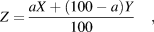
Thermal and decomposition characteristics of MPMS NPs, MPMS–MPDMS NPs, and MPDMS NPs were determined by TGA using a STA7200 thermal analysis system (Hitachi High-Tech, Tokyo, Japan) in the temperature range 30–1000 °C with a heating rate of 10 °C/min under a flow of nitrogen gas at 200 mL/min. The content rate of organic component of thiol-organosilica NPs was estimated from the difference between the weight at 150 °C and the weight at 800 °C. The abundance ratio of MPMS constituent to MPDMS constituent in MPMS–MPDMS NPs was calculated using the following equation:
 $$Z = {{aX + \left( {100 - a} \right)Y} \over {100}}\quad ,$$
where X, Y, and Z represent the content rate of organic component of MPMS NPs, MPDMS NPs, and MPMS–MPDMS NPs, respectively, and a(0 < a < 100) represents the abundance percentage of MPMS constituent in the MPMS–MPDMS NPs.
Solid-state 13C NMR measurements
13C cross-polarization magic-angle spinning (CP MAS) NMR spectra were recorded on an ECA-500 spectrometer (JEOL Ltd.) with a 6000 Hz MAS spinning rate; contact time was 2.0 ms and 600 scans with a relaxation delay of 12 s were collected. Chemical shifts are reported in parts per million (ppm). The 13C CP MAS NMR spectrum of hexamethylbenzene (typical peak, 17.17 ppm) was recorded as an external standard.
Raman spectroscopic measurements
Raman spectroscopic measurements were conducted at room temperature by using an NRS3100 laser Raman spectrophotometer (JASCO, Hachioji, Japan) equipped with a laser (532 nm). Each solid sample was filled in a holder. The obtained Raman spectra were deconvoluted by using a nonlinear least-squares curve-fitting procedure to extract single bands. A single band is assumed to be represented as a pseudo-Voigt equation:
 $${f_{\rm{v}}}\left( \nu \right) = \gamma {f_{\rm{L}}}\left( \nu \right) + \left( {1 - \gamma } \right){f_{\rm{G}}}\left( \nu \right)\quad ,$$
where f L(ν) and f G(ν) represent the Lorentzian and Gaussian components, respectively, and the parameter γ(0 < γ < 1) represents the Lorentzian component. The intensity I of a single band is evaluated according to the following equation:
 $$I = \gamma {I_{\rm{L}}} + \left( {1 - \gamma } \right){I_{\rm{G}}}\quad ,$$
where I L and I G denote the integrated intensities of the Lorentzian and Gaussian components, respectively.
Degradation analysis of thiol-organosilica NPs in the presence of 10 mM GSH
Either 5 μL of 1 M GSH in Millipore water (with 10 mM GSH) or 5 μL of Millipore water (without GSH) was added to 500 μL of 1 mg/mL MPMS NPs (mean diameter, 236 nm), MPMS–MPDMS (75:25) NPs (mean diameter, 193 nm), MPMS–MPDMS (50:50) NPs (mean diameter, 186 nm), MPMS–MPDMS (25:75) NPs (mean diameter, 231 nm), or MPDMS NPs (mean diameter, 225 nm) in 10 mM sodium phosphate buffer (pH 8.0), and the mixture was sonicated and incubated at 37 °C for 2 or 7 days. The sample was centrifuged at 30,000g for 10 min to isolate the supernatant. The residue was washed with distilled water twice, and the washed residue was analyzed using an electron microscope.
Degradation analysis of thiol-organosilica NPs in the presence of 40 mM GSH
MPMS NPs, MPDMS NPs, and MPMS–MPDMS NPs used in this experiment were the same as those used in the above degradation analyses. First, 480 μL of 10 mM sodium phosphate buffer (pH 8.0) was added to each sample of thiol-organosilica NPs. Next, 20 μL of 1 M GSH in Millipore water (with 40 mM GSH) or 20 μL of Millipore water (without GSH) was added to the suspension. The mixture was first sonicated and then incubated at 80 °C for 4–7 days and then centrifuged at 30,000g for 10 min to isolate the supernatant. Then, 500 μL of fresh 40 mM GSH in 10 mM sodium phosphate buffer (pH 8.0) or 500 μL of 10 mM sodium phosphate buffer (pH 8.0) was added to the residue, and the mixture was sonicated and incubated at 80 °C for 4 or 7 days. The procedure was repeated for 2 months. As a control, 40 mM GSH in 10 mM sodium phosphate buffer (pH 8.0) was incubated with test samples. The residue was washed with distilled water twice, and the washed residue was analyzed by electron microscopy. The concentration of residual GSH in the supernatant was evaluated using Ellman’s test.
Ellman’s test
A 1 μL aliquot of supernatant isolated from each test sample was diluted with 19 μL of 10 mM sodium phosphate buffer (pH 8.0) and then mixed with 20 μL of 2 mM DTNB in 10 mM sodium phosphate buffer (pH 8.0) and incubated at 37 °C for 30 min. The sample was then diluted with 10 mM sodium phosphate buffer (pH 8.0) to twice the original volume. The absorption spectrum of the diluted sample was recorded on an ND-1000 spectrophotometer (Thermo Fisher Scientific, Wilmington, Delaware), and the concentration of residual GSH was estimated from the absorbance at 412 nm. The thiol content on the surface of intact NPs was determined as follows: NP dispersions (10 mg/mL) were prepared in 150 μL of 50 mM phosphate buffer solution (pH 8.0) and were allowed to hydrate for 2 h. In the meantime, 1 mg of DTNB was dissolved in 3 mL of 50 mM phosphate buffer solution (pH 8.0). Then, 150 μL of DTNB solution was added to 150 μL of NPs and mixture incubated in the dark for 90 min. After that, the NP dispersions were centrifuged for 10 min at 30,000g, and the supernatant was placed in ND-1000 spectrophotometer. Absorbance was measured at 412 nm. The thiol concentration was calculated from a calibration curve of cysteine hydrochloride as a series of solution under the same conditions and with a concentration range of 0.015625–1.000 μmol/mL.
Release of rhodamine B–doped MPMS–MPDMS NPs in the presence of 10 mM GSH
Ten microliters of 1 M GSH in Millipore water or 10 μL of Millipore water was added to 1000 μL of 1.0 mg/mL rhodamine B–doped MPMS–MPDMS (25:75) NPs in Millipore water, and the mixture was sonicated and incubated at 37 °C under light shielding for 48 h. The samples were centrifuged at 30,000g for 10 min to isolate the supernatants. The fluorescence spectrum of each supernatant was recorded on an F-4500 fluorescence spectrophotometer (Hitachi High-Tech) by using 550 nm excitation light.
Notes
The authors declare no competing financial interest.
Supplementary material
To view supplementary material for this article, please visit https://doi.org/10.1557/jmr.2018.501.
Acknowledgments
This research was supported in part by JSPS KAKENHI Grant Number JP16K18909 (T.D.), YU Project for Formation of the Core Research Center (T.D.), JSPS KAKENHI Grant Number JP16K01358 (M.N.), and the JSPS Bilateral Programs (M.N.). Assistance with electron microscopic analyses was provided by Dr. Koichi Udo and the Institute for Biomedical Research and Education, Yamaguchi University Science Research Center, Japan. Assistance with solid-state 13C NMR measurements was provided by Dr. Hirotaka Fujimori, Dr. Yoshiko Murakami, Mr. Ryota Hori, Mr. Yudai Arisuda, and Mr. Yosuke Fukuzawa of Yamaguchi University. Raman spectroscopic measurements were supported by Dr. Kenta Fujii and Dr. Yanko Todorov of Yamaguchi University.