INTRODUCTION
Significant mortalities in Crassostrea gigas were first recorded along the Japanese Pacific coast in 1945 (Koganezawa, Reference Koganezawa1974) and in the 1950s in North America (Glude, Reference Glude1975). The phenomenon intensified throughout the 1960s and early 1970s in North America, particularly in Washington, and in oyster culture areas in Japan (Woelke, Reference Woelke1961; Pereyra, Reference Pereyra1964; Imai et al. Reference Imai, Numachi, Oizimu and Sato1965; Mori et al. Reference Mori, Tamate, Imai and Itikawa1965; Lipovsky and Chew, Reference Lipovsky and Chew1972; Koganezawa, Reference Koganezawa1974; Glude, Reference Glude1975; Beattie et al. Reference Beattie, Chew and Hershberger1980). More recently, significant mortalities have been recorded from a number of European countries, in particular France (Comps and Cochennec, Reference Comps and Cochennec1993; Renault et al. Reference Renault, Le Deuff, Cochennec and Maffart1994, Reference Renault, Le Deuff, Chollet, Cochennec and Gerard2000a; Goulletquer et al. Reference Goulletquer, Soletchnik, Le Moine, Razet, Geairon, Faury and Taillade1998; Soletchnik et al. Reference Soletchnik, Le Moine, Faury, Razet, Geairon and Goulletquer1999; Samain et al. Reference Samain, Boudry, Degremont, Soletchnik, Ropert, Bedier, Martin, Moal, Mathieu, Pouvreau, Lambert, Escoubas, Nicolas and Le Roux2004; Dégremont et al. Reference Dégremont, Bédier, Soletchnik, Ropert, Joly, Huvet, Moal, Samain and Boudry2005, Reference Dégremont, Ernande, Bedier and Boudry2007) and Ireland (Cotter et al., Reference Cotter, Malham, O'Keeffe, Lynch, Latchford, King, Beaumont and Culloty2010; European Food Safety Authority, 2010 (www.marine.ie/home/aboutus/newsroom/news/UpdateonOysterMortalities.htm). The main cause of ‘summer mortality syndrome’ has not been definitively determined; however, many factors including pathogens, elevated temperatures, physiological stress associated with gonadal maturation, aquacultural practices, water quality, predators, phytoplankton blooms and genetic factors are all known to contribute to C. gigas summer mortalities (Renault et al. Reference Renault, Le Deuff, Cochennec and Maffart1994; Goulletquer et al. Reference Goulletquer, Soletchnik, Le Moine, Razet, Geairon, Faury and Taillade1998; Berthelin et al. Reference Berthelin, Kellner and Mathieu2000; Lacoste et al. Reference Lacoste, Jalabert, Malham, Cueff, Gelebart, Cordevant, Lange and Poulet2001; Le Roux et al. Reference Le Roux, Gay, Lambert, Maechter, Poubalanne, Chollet, Nicolas and Berthe2002; Dégremont et al. Reference Dégremont, Boudry, Soletchnik, Bédier, Ropert, Huvet, Moal and Samain2003; Huvet et al. Reference Huvet, Herpin, Degremont, Labreuche, Samain and Cunningham2004; Malham et al. Reference Malham, Cotter, O'Keeffe, Lynch, Culloty, King, Latchford and Beaumont2009; Cotter et al., Reference Cotter, Malham, O'Keeffe, Lynch, Latchford, King, Beaumont and Culloty2010). A multi-factorial aetiology has been suggested (Dégremont et al. Reference Dégremont, Bédier, Soletchnik, Ropert, Joly, Huvet, Moal, Samain and Boudry2005; Samain et al. Reference Samain, Dégremont, Soletchnik, Haure, Bédier, Ropert, Moal, Huvet, Bacca, Van Wormhoudt, Delaporte, Costil, Pouvreau, Lambert, Boulo, Soudant, Nicolas, Le Roux, Renault, Gagnaire, Gertet, Boutet, Burgeot and Boudry2007; Soletchnik et al. Reference Soletchnik, Ropert, Mazurié, Fleury and Le Coz2007; Samain and McCombie, Reference Samain, McCombie, Samain and McCombie2008; Cotter et al. Reference Cotter, Malham, O'Keeffe, Lynch, Latchford, King, Beaumont and Culloty2010). Previous studies have investigated the role of ostreid herpesvirus 1 (OsHV-1) and variants as causative agents in C. gigas summer mortalities and have found this virus to be particularly associated with larval and juvenile mortalities (Renault et al. Reference Renault, Le Deuff, Cochennec and Maffart1994, Reference Renault, Le Deuff, Chollet, Cochennec and Gerard2000a; Le Deuff and Renault, Reference Le Deuff and Renault1999; Arzul et al. Reference Arzul, Renault, Thebault and Gérard2002; Vásquez-Yeomans et al. Reference Vásquez-Yeomans, Caceres-Martinez and Huerta2004; Friedman et al. Reference Friedman, Estes, Stokes, Burge, Hargove, Barber, Elston, Burreson and Reece2005; Burge et al. Reference Burge, Griffin and Friedman2006, Reference Burge, Judah, Conquest, Griffin, Cheney, Suhrbier, Vadopalas, Olin, Renault and Friedman2007).
Ostreid herpesvirus 1 (OsHV-1) belongs to the genus Ostreavirus, family Malacoherpesviridae which is included in the order Herpesvirales (Davison et al. Reference Davison, Eberle, Ehlers, Hayward, McGeoch, Minson, Pellet, Roizman, Studdert and Thiry2009). Viruses belonging to the order Herpesvirales are relatively large double-stranded DNA (dsDNA) viruses and can cause latent and/or lytic re-occurring infections (Sandri-Goldin, Reference Sandri-Goldin2006; Mettenleiter and Sobrino, Reference Mettenleiter, Sobrino, Thomas, Gunther and Walter2008). OsHV-1 μvar is a variant of OsHV-1 and is considered to be a more virulent strain (European Food Safety Authority, 2010). OsHV-1 μvar was originally associated with C. gigas mortalities in France in 2008 and in 2009 (Repamo, 2009; Segarra et al. Reference Segarra, Pépin, Arzul, Morga, Faury and Renault2010) and has subsequently been observed in Ireland (http://web.oie.int/wahis/public.php?page=single_report&pop=1&reportid=8503), the March region in Italy (Dundon et al. Reference Dundon, Arzul, Omnes, Robert, Magnabosco, Zambon, Gennaria, Toffan, Terregino, Capua and Arcangeli2011), the Netherlands (http://web.oie.int/wahis/public.php?page=single_report&pop=1&reportid=10749), the UK (http://web.oie.int/wahis/public.php?page=single_report&pop=1&reportid=9527) and south east Australia (http://web.oie.int/wahis/public.php?page=single_report&pop=1&reportid=10136). Other variants have been observed in a wide range of bivalves including oysters, scallops, clams and abalone (Hine and Thorn, Reference Hine and Thorn1997; Hine et al. Reference Hine, Wesney and Besant1998; Renault et al. Reference Renault, Le Deuff, Lipart and Delsert2000b; Arzul et al. Reference Arzul, Renault, Lipart and Davison2001a,b; Renault and Arzul, Reference Renault and Arzul2001; Chang et al. Reference Chang, Kuo, Lai, Yang, Ting, Hsu and Chen2005; Davison et al. Reference Davison, Trus, Cheng, Steven, Watson, Cunningham, Le Deuff and Renault2005; Hooper et al. Reference Hooper, Day, Slocombe, Handlinger and Benkendorff2007; Moss et al. Reference Moss, Burreson, Cordes, Cungan, Brown, Wang, Wu and Reece2007; Tan et al. Reference Tan, Lancaster, Hyatt, van Driel, Wong and Warner2008; Samain and McCombie, Reference Samain, McCombie, Samain and McCombie2008).
Pépin et al. (Reference Pépin, Riou and Renault2008) reported that abnormal mortalities in oyster samples were associated with a viral load range of 6 × 10³ to 5·3 × 107 copies/μl or mg. There are an estimated 1030 viruses in the worlds’ oceans and previously unknown viruses are still routinely discovered in well-studied organisms such as the panaeid shrimp (Flegel, Reference Flegel2006). In general, mortalities associated with viruses in the marine ecosystem are expected to arise sporadically when conditions become less favourable for the host, such as a physiological stress induced by significant increases in temperature, causing the host to become immunocompromised and unable to suppress an infection (Suttle, Reference Suttle2007). In the case of OsHV-1 μvar, such mortality events, which occur in an intensively cultured commercial bivalve, have become more regular and widespread.
The main objective of this study was to investigate the role of ostreid herpesvirus 1 in summer mortalities of adult and C. gigas seed and juveniles in the Irish Sea. This study was part of a larger study that looked at a range of biological and physical factors contributing to oyster mortalities along the coast of Ireland (Malham et al. Reference Malham, Cotter, O'Keeffe, Lynch, Culloty, King, Latchford and Beaumont2009; Cotter et al. Reference Cotter, Malham, O'Keeffe, Lynch, Latchford, King, Beaumont and Culloty2010).
MATERIALS AND METHODS
Study sites
Two field sampling areas were selected on the south east coast of Ireland: Dungarvan Harbour, Co. Waterford (52°03′N 007°35′W) where minimal mortalities had occurred and Bannow Bay, Co. Wexford (52°27′N 006°47′W) where more significant mortalities had previously been reported in Pacific oysters (Clarke, Reference Clarke1996). Bannow Bay, where C. gigas have been cultured since 1984, has an area of ca.1050 ha, is 8 km long, and between 1 and 3 km wide. The sea enters this shallow bay through a narrow inlet at the southern end. Little flushing occurs at low tide and water stays in the bay for 2 tidal cycles. Approximately 75% of the bay is exposed at low tide. Dungarvan Harbour is an open bay of ca. 1300 ha. On a national basis, it holds the largest standing stock of C. gigas and is the most productive area of operation in Ireland. Two sites were selected within each of the two study areas (Bannow Site A (BA), Bannow Site B (BB); Dungarvan Site A (DA) and Dungarvan Site B (DB)). These sites were located at corresponding shore heights, i.e. 4 h exposure on spring tides. BA is a muddy site where mortalities had previously occurred, BB, DA and DB are all sandy sites where minimal mortalities had been recorded. Sites BA, DA and DB were all accessible at low neap tide but spring tides were required to access BB.
Oysters
A sample of 29 market-sized C. gigas (n = 29) (24 months old, 60 + g) from Bannow Bay were provided by fishermen from stock that had been experiencing mortality in May 2003. In June 2003, a single cohort of hatchery-reared C. gigas spat (shell length ∼ 15mm) was obtained from a hatchery outside of Ireland. An initial sample of 60 of these oysters was removed for screening by polymerase chain reaction (PCR) for OsHV-1 prior to relaying oysters at both Dungarvan and Bannow Bay (Dungarvan Bay A (DA), Dungarvan Bay B (DB), Bannow Bay A (BA), and Bannow Bay B (BB)). Oysters were randomly distributed into 0·5 m2, 4 mm mesh bags on trestles at 2 densities used during the growing cycle: 1000 oysters bag−1 (n = 5 per site) and 600 oysters bag−1 (n = 3 per site). In June 2004, another single cohort of oysters was obtained from the same hatchery and an initial sample of 60 oysters was screened again prior to oysters being relayed to the same 4 sites, but at 1 density only (1000 oysters bag−1).
In both 2003 and 2004, 60 oysters were sampled from July to October at 2-week intervals from each of the sites with the exception of BB. This site was accessible only on spring tides, and was generally sampled on a monthly basis.
Mortality
A total of 200 individuals were removed randomly from each bag every 2 weeks and were counted in situ to estimate oyster mortality. The percentage mortality was determined after counting the cupped empty shell only. Empty oyster shells were discarded after each count was completed. Sixty C. gigas were removed for PCR analysis while the remaining living C. gigas were returned to the bags.
DNA extraction and PCR
In the 29 adult oysters sampled in May 2003, 100 μl of haemolymph were withdrawn from the adductor muscle sinus of each oyster and stored at −20 °C. Initially phenol/chloroform extraction of DNA was carried out. However, poor template yield led to difficulties with direct sequencing, which resulted in subsequent DNA being extracted from the frozen haemolymph using 10% chelex resin (Sigma-Aldrich) (Walsh et al. Reference Walsh, Metzger and Higuchi1991; Lynch et al. Reference Lynch, Abollo, Ramilo, Cao, Culloty and Villalba2010). Gill samples (n = 60) were collected from the initial spat samples in 2003 and 2004 and on each subsequent sampling date and frozen at −20 °C. Initially, DNA was extracted using the phenol/chloroform extraction method (Winnepenninckx et al. Reference Winnepenninckx, Backelgau and De Wachter1993); however, similar issues with the template yield led to difficulties with direct sequencing. DNA was extracted from the corresponding paraffin-embedded tissue of a subsample of individuals (Dungarvan Bay (n = 32), Bannow Bay (n = 31)) which had all amplified products in the original PCR. Deparaffinization of the tissue was carried out (Shi et al. Reference Shi, Cote, Wu, Liu, Datar, Shi, Liu, Lim and Taylor2002) and DNA extractions were undertaken using protein precipitate and cell lysis using the QIAamp DNA Mini Kit (Qiagen™).
Screening for OsHV-1 was carried out using the C2/C6 primers and PCR conditions of Renault and Arzul (2001) using a Promega dNTP mix and a Thermo Hybaid PCR express thermal cycler. This PCR identifies oyster herpes DNA (709 bp expected product size) in region C, which encodes for 2 proteins of unknown function. As this region of the genome has proven to be variable and diagnostic in previous studies of OsHV-1 type viruses (Arzul et al. Reference Arzul, Renault, Lipart and Davison2001a, Reference Arzul, Nicolas, Davison and Renaultb; Segarra et al. Reference Segarra, Pépin, Arzul, Morga, Faury and Renault2010) the present study focused on this region. Both negative and positive controls were included in each PCR, viral OsHV-1 DNA samples from heavily infected C. gigas prepared in 100 ng/l in TE buffer were used as positive controls, and deionized distilled water was used as a negative control.
Amplified PCR products from the chelex-extracted adult haemolymph and both spat subsamples were excised from the gel and products were pooled to increase the DNA concentration, 3–4 oysters approximately per pool keeping sites and adult and spat samples separate, and DNA was isolated and purified using the QiaQuick gel extraction kit (Qiagen™) prior to direct sequencing (MWG eurofins).
Construction of phylogenetic tree
Ten pooled PCR products from 33 individuals: 3 pooled samples representing 11 oysters from Dungarvan Harbour seed and juveniles, 3 pooled samples representing 10 oysters from Bannow Bay seed and juveniles and 4 pooled samples representing 12 individuals from the adult oysters, were directly sequenced. Six sequences (forward and reverse) were obtained for each of the Dungarvan Harbour and Bannow Bay seed and juveniles and 8 sequences (forward and reverse) were obtained from the adult C. gigas at Bannow Bay. Individual sequences were manually trimmed to have the same sequence length. The forward and reverse sequence reads were aligned using the ClustalW algorithm (Geneious v.5.1.3, www.geneious.com, using the default options). The consensus sequence, based only on sequence for which both strands were present, of each alignment was matched against the National Centre for Biotechnology Information (NCBI) nucleotide database with BLASTn (Basic Local Alignment Search Tool) to confirm that the sequences were similar from the OsHV-1 genome. Alignments were edited to count each putative indel as one mutational tranversion (transition/transversion ratio of 0·1288) and the phylogenetic relationship among sequence variants was visualised using a Maximum Likelihood tree, with 1000 bootstraps, based on the Kimura 2 parameter model (Kimura, Reference Kimura1980) distances using the software package MEGA v.5.05 (Tamura et al. Reference Tamura, Peterson, Peterson, Stecher, Nei and Kumar2011).
RESULTS
Mortality
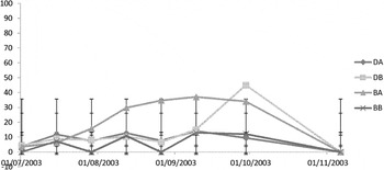
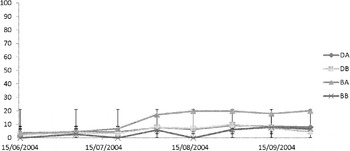
The percentage mortality was not quantified in the adult market-sized oysters but the oyster farmers had reported approximately 10% mortalities in the stocks at the time of sampling. During summer 2003, percentage mortality in the seed and juveniles varied from 14·2% in DA to 45% in DB. There was greater mortality in the higher density bags (1000 oysters bag-1) early in the season; however, there was no significant effect of density on mortality at the end of the sampling period (ANOVA P = 0·06) (Fig. 1). Mortality levels were much lower in 2004 varying from 8% at BB to 19·8% in BA (Fig. 2).
Mean mortality (%) in Crassostrea gigas at Irish sites in 2003.

Mean mortality (%) in Crassostrea gigas at Irish sites in 2004.

PCR analysis
In the adult C. gigas haemolymph samples, products were originally amplified in 7% (2/29) of the oysters screened. In the PCR re-screening of the Chelex extracted adult C. gigas, DNA products of the expected size (709 bp) were amplified in 90% (26/29) of the sample.
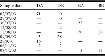
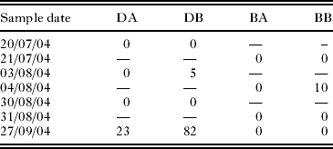
The initial samples of C. gigas spat taken prior to relaying to both Irish locations in 2003 and 2004 were negative for OsHV-1 when screened by PCR. 900 and 960 individual seed and juvenile C. gigas were screened by PCR in 2003 and 2004 respectively (Tables 1 and 2). In 2003, products were amplified in 13 of the 15 sample periods (87%) screened. Products were initially observed in DA at 2 weeks (71% prevalence) and at all sites by 8 weeks post-relaying. Peaks in prevalence varied between sites at different times from a peak of 28% at DB in September to a peak of 71% at DA in July (Table 1). In 2004, products were amplified in 4 of the 16 samples (25%) screened. In 2004, it was 8 weeks post-relaying prior to positive results being obtained by PCR (DB and BB) and positive results were initially obtained in DA at 16 weeks post-relaying. No positive results were obtained from BA. The highest prevalence of OsHV-1 was observed in September 2004 at 16 weeks post-relaying at DB (82%) and at DA (23%). At BB, a single sample amplified products (10%) in August, 8 weeks post-relaying (Table 2).
Results (% positive) from cPCR screening of Crassostrea gigas samples (n = 60) from Bannow Bay and Dungrvan Bay in 2003
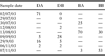
— Sample not screened.
Results (% positive) from cPCR screening of Crassostrea gigas samples (n = 60) from Bannow Bay and Dungrvan Bay in 2004
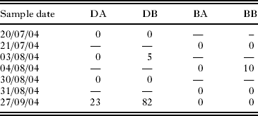
— Sample not screened.
In the PCR re-screening of the subsample of Dungarvan Harbour and Bannow Bay seed and juveniles in which DNA was extracted from paraffin-embedded tissue, products were visualized in 34% (11/32) of the Dungarvan Harbour sample and 32% (10/31) of the Bannow Bay sample.
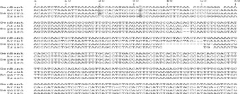
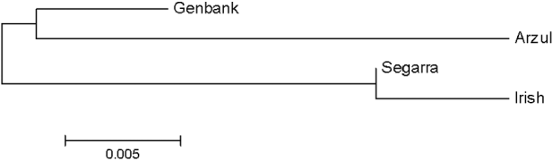
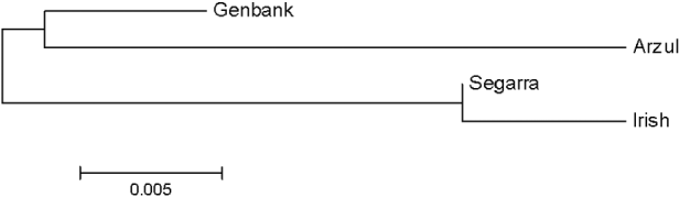
Sequencing confirmed that the DNA sequences obtained from the adult oysters and the seed and juveniles at the two sites in Ireland were identical. In the BLASTn analysis, the Irish sequences (GenBank Accession number JQ963169) were similar to OsHV-1 (GenBank Accession number AY509253). Further, sequences were aligned and genetic variation was detected in the C region in the Irish OsHV-1 μvar sequences compared to all previously published sequences (i.e. GenBank Accession number AY509253, Arzul et al. Reference Arzul, Renault, Lipart and Davison2001a,b; Segarra et al. Reference Segarra, Pépin, Arzul, Morga, Faury and Renault2010, Fig. 3). The Irish sequence was almost identical to the OsHV-1 μvar strain recently identified in France (Segarra et al. Reference Segarra, Pépin, Arzul, Morga, Faury and Renault2010) and now epizootic in Europe. The differences consisted of 1 nucleotide deletion (adenine/thymine) and 1 nucleotide insertion (guanine/cytosine) over the entire 360 bp (Fig. 3) indicating a new variant genotype at the Irish sites. This sequence has a deletion at 20 bp from the 5′ end which is shared with the French strain (Arzul et al. Reference Arzul, Renault, Lipart and Davison2001a, Reference Arzul, Nicolas, Davison and Renaultb) but was absent in the AY509253 and OsHV-1 μvar variants. In addition, the Irish OsHV-1 μvar variant genotype shows an insertion (guanine/cytosine) at 32 bp from the 5′end of the fragment, which is not present in any of the three French strains. In the Maximum Likelihood tree (Fig. 4), OsHV-1 μvar and the Irish OsHV-1 μvar variant genotype are closely related. The phylogenetic relationship between OsHV-1 μvar and the Irish OsHV-1 μvar variant genotype showed good bootstrap support (85%. cf. Fig. 4).
Aligned DNA sequences of the C region amplified by the C2/C6 primer pair. Sequences were edited to show identical sequence length. Note the indels at 20 and 32 bp when comparing the French and Irish Sequences. Sequences are coded as follows; GenBank Accession number AY509253, Davison et al. Reference Davison, Trus, Cheng, Steven, Watson, Cunningham, Le Deuff and Renault2005); OsHV-1 var (Arzul et al. Reference Arzul, Renault, Lipart and Davison2001a, Reference Arzul, Nicolas, Davison and Renaultb); OsHV-1 μvar (Segarra et al. Reference Segarra, Pépin, Arzul, Morga, Faury and Renault2010); and Irish variant genotype (GenBank Accession number JQ963169).

Maximum likelihood tree showing the phylogentic (Kimura 2 parameter model distances) relationship among four OsHV-1 sequence variants, using all available variability. Sequences are coded as follows; GenBank, (GenBank Accession number Y509253, Davison et al. Reference Davison, Trus, Cheng, Steven, Watson, Cunningham, Le Deuff and Renault2005); Arzul, (Arzul et al. Reference Arzul, Renault, Lipart and Davison2000 a, Reference Arzul, Nicolas, Davison and Renaultb); Segarra, (Segarra et al. Reference Segarra, Pépin, Arzul, Morga, Faury and Renault2010); and Irish variant genotype (GenBank Accession number JQ963169).
DISCUSSION
In this study, an undescribed Irish OsHV-1 μvar variant genotype was detected at 2 Irish C. gigas cultivation sites and was associated with on-going mortalities. The phylogenetic relationship among the 4 variants of OsHV-1 (Figs 3 and 4) shows that OsHV-1 μvar and the Irish variant genotype are most similar to each other. OsHV-1 μvar was described as a new OsHV-1 variant genotype based on 5 deletions (including a deletion of 4 microsatellite motifs; TAC) in the C region compared to OsHV-1. These deletions are also present in the Irish OsHV-1 μvar variant genotype with an additional 2 indels. These additional indels observed in the Irish variant genotype were not present in OsHV-1 μvar. Although, the 2 indels this study observed do not permit for strong conclusions about which virus variant is derived from which further sequence information is needed before a strong analysis of the relationship between OsHV-1 μvar and the Irish variant genotype can be established. However, the presence of 2 additional indels in the Irish OsHV-1 μvar could suggest that it may be derived from OsHV-1 μvar and represent a new variant of the virus. Alternatively, OsHV-1 μvar is derived from the Irish OsHV-1 μvar variant genotype, although that would demand that the 2 indels observed in the Irish variant genotype are ancestral for the clade containing OsHV-1 μvar and the Irish variant genotype, and have consequently been lost in OsHV-1 μvar. It is not clear if the Irish OsHV-1 μvar variant genotype differs from OsHV-1 μvar in virulence, tissue tropism etc. and it is obvious that more research is needed to confirm any biologically significant differences between the variant genotypes. Nevertheless, the close resemblance to OsHV-1 μvar in oysters sampled prior to 2008 in France (Segarra et al. Reference Segarra, Pépin, Arzul, Morga, Faury and Renault2010) may indicate that OsHV-1 μvar has been present for longer than thought, but had escaped detection until 2008. While the Irish OsHV-1 μvar variant genotype represents a previously undocumented strain, there is no information of its biology being different than that of OsHV-1 μvar and future studies should examine additional areas of its genome and study the pathology to elucidate potential biological differences among the strains/genotypes. Herpes-like particles were detected by chance in a marine fungoid protist (thraustochytrid-like organism) (Arzul et al. 2001) and it has been suggested by Hurst et al. (Reference Hurst2011) that marine protists may possibly act as vectors and/hosts for OsHV-1. A higher number of viruses are found in shallower coastal waters as more host organisms are available (Suttle, Reference Suttle2007).
In this study, an Irish OsHV-1 μvar variant genotype was detected in both adult C. gigas and C. gigas seed and juveniles. A lower percentage of seed and juveniles was observed to be infected compared to the adult oysters; however, the seed and juveniles had only been exposed for several weeks and months while the adult C. gigas had been at the Irish site for several years and screening indicated that no OsHV-1 or variant sequences were detected in initial samples of spat screened prior to relaying. It has been reported that C. gigas older than 18 months can be a source of the OsHV-1 and variants (European Food Safety Authority, 2010). Previous studies have shown that OsHV-1 can persist in surviving adult C. gigas following primary infection (Arzul et al. Reference Arzul, Renault, Thebault and Gérard2002; Lipart and Renault, Reference Lipart and Renault2002; Renault, Reference Renault, Mahy and Van Regenmortel2008; Hurst et al. Reference Hurst2011) and it is recommended that movements and mixing of different age groups should be avoided (European Food Safety Authority, 2010).
The mortalities at both sites were associated with a complex aetiology including elevated temperature, nutrient and plankton levels (Malham et al. Reference Malham, Cotter, O'Keeffe, Lynch, Culloty, King, Latchford and Beaumont2009; Cotter et al. Reference Cotter, Malham, O'Keeffe, Lynch, Latchford, King, Beaumont and Culloty2010). Malham et al. (Reference Malham, Cotter, O'Keeffe, Lynch, Culloty, King, Latchford and Beaumont2009) demonstrated that oysters exposed to the environmental conditions recorded in this study were immunocompromised. A combination of these unfavourable environmental factors may have made the oysters more susceptible to the Irish OsHV-1 μvar variant genotype and eventually succumb to infection.
ACKNOWLEDGEMENTS
The authors thank Cliff Cunningham for help with constructing the phylogenetic tree. Many thanks to the shellfish growers who provided the shellfish material for this study.
Financial Support
The project was part funded by the European Regional Development Fund (ERDF) through the Ireland Wales programme (INTERREG 4A) and the Beaufort Marine Research Award in Fish Population Genetics funded by the Irish Government under the Sea Change Programme.