Case description
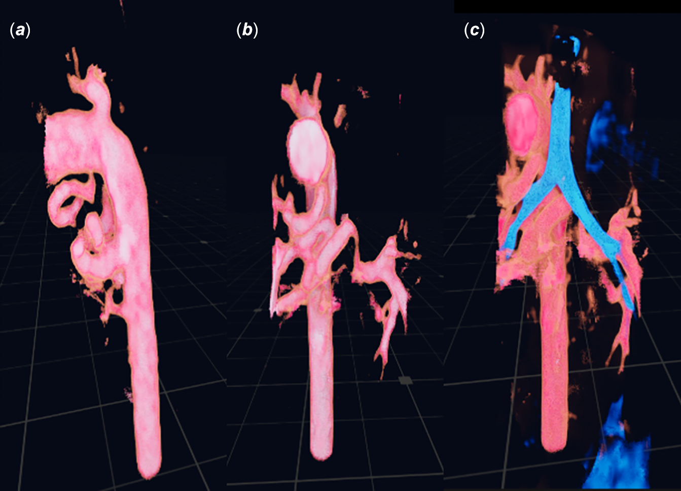
A 3.7 kg term male infant was admitted with a known diagnosis of pulmonary atresia with ventricular septal defect and major aortopulmonary collateral arteries due to baseline desaturation. Despite non-invasive respiratory support, he experienced extremely labile oxygen saturations. A CT scan was performed (Figure 1), outlining the pulmonary arterial circulation.
Virtual reality 3D reconstructions from the pre-procedural CT scan (Vea Simulations, Warsaw, Poland). Panel a is a lateral projection demonstrating the origins of the 3 MAPCAs— one in the ductal position, a major vessel more caudally, and one smaller vessel further down. Panel b is an anterior projection of the pulmonary supply, with the most cranial vessel feeding the right upper lobe, the middle vessel offering supply to the majority of both lungs with a severe stenosis to the left lung vessel, but the course of the most caudal vessel is poorly demonstrated. Panel c is the same projection as b with additional windowing for the airways.

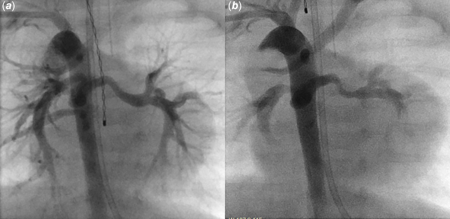
Cardiac catheterisation was then undertaken with a view to potential stenting . Initial angiography (Figure 2 a ; video 1, supplementary materials) was performed demonstrating a major aortopulmonary collateral arteries off the underside of the arch feeding the right upper lobe, a larger vessel more caudally bifurcating to supply the entire left lung and the majority of the right, and a lower vessel supplying the right lower lobe. Notably, the branch to the left lung looked larger than on the CT scan.
Angiographic procedural still frames in the PA projection. Panel a the first angiogram, taken during a period of clinical stability. During an unprecipitated period of marked desaturation, the angiogram in panel b demonstrates clear distortion of the MAPCA supplying the left lung, and no flow into the most caudal MAPCA.

We tried unsuccessfully to enter the most cranial major aortopulmonary collateral arteries with a catheter, but during a catheter exchange, the patient suffered an acute, severe desaturation (to unrecordable levels) alongside an increase in the diastolic blood pressure from 18 mmHg to the low 30s. A repeat angiogram this point (Figure 2 b ; video 2, supplementary materials) performed from the same site with the same volume injection and at the same frame rate demonstrated complete occlusion of the lowest vessel, with no flow to the right lower lobe, and a marked confirmational change to the portion of the middle major aortopulmonary collateral arteries supplying the left lung. There had been no catheter manipulation in these territories prior to vessel spasm.
There were ongoing episodes of severe desaturation throughout the case, despite introduction of a glyceryl trinitrate infusion (chosen for immediate availability and from prior clinician experience).
We feel these images offer an insight into the potential behaviour of major aortopulmonary collateral arteries, with spontaneous obstruction of several vessels simultaneously, explaining the clinical picture of unproked transient desaturation on the ward.
Supplementary material
The supplementary material for this article can be found at https://doi.org/10.1017/S1047951126124329
Financial support
None.
Competing interests
None declared.