Cross-sectional echocardiography is currently the cornerstone for the assessment of congenital cardiac malformations.1 In congenital valvar aortic stenosis, it has been used to define the anatomy of the valve, the size of the aortic root, the morphology of the left ventricle, as well as haemodynamic characteristics of the obstruction. Valvar morphology may predict the likelihood of successful treatment using catheter-based as opposed to surgical intervention.2, 3 Complete visualization of the aortic valve, nonetheless, is often sub-optimal with standard cross-sectional echocardiography, thus limiting this kind of assessment. Recent advances in real time three-dimensional echocardiography have significantly improved the appreciation of the complex three-dimensional functional anatomy of cardiac structures.4 It is now feasible not only to define detailed functional anatomy, but it is also possible accurately and directly to quantitate the volumes of chambers and vessels without making geometric assumptions about these structures.5 In this prospective comparative observational study, we evaluated the ability of the new technique to delineate the detailed anatomy and morphometry of the severely stenotic aortic valve, comparing our findings with observations made during surgical evaluation of the valvar anatomy.
Methods
Patients: We examined all patients with severe congenital aortic valvar stenosis undergoing surgical valvotomy from 1st December 2003 through 31st July 2004. It is our practice to perform real time three-dimensional echocardiogram as a routine to assess patients with aortic valvar disease, and surgical valvotomy is our treatment of choice for those shown to have severe stenosis. In the patients studied, the initial diagnosis was confirmed by conventional trans-thoracic cross-sectional and Doppler echocardiography. Patients with associated coarctation of the aorta or hypoplastic left ventricles were excluded. The data was acquired prospectively, with the comparative analysis made retrospectively.
Acquisition of images: All images were obtained with the Phillips Sonos 7500 echocardiographic system, using a 4 megaherz, 4X Matrix transducer. Additional sedation or anaesthesia was not required. Older cooperative children were asked to hold their breath during acquisition of images to optimise their quality. Three dimensional full volume loops were acquired in planes that included the aortic valve along with a segment of the ascending aorta and left ventricular outflow tract. A minimum of 3 images was acquired for each patient. These images were acquired from modified parasternal and apical 5 chamber views. A second investigator, blinded to the standard cross-sectional echocardiographic and operative findings, analysed the stored trans-thoracic real time three-dimensional echocardiographic image loops. Three-dimensional echocardiographic findings were then compared with anatomy as described by the surgeon. The surgeon himself was unaware of the three-dimensional echocardiographic findings. No photographic recordings were made during the operation, but rather the operative notes were used to obtain the findings.
Analysis: The stored full volume loops were analysed using “Q lab 4.1” software, Philips Ultrasound 1997–2005, United States of America. Images were analysed in any plane providing good visualisation of the aortic valve. The three-dimensional volume was sliced, and/ or cropped, in a variety of three-dimensional planes in order selectively to display the relevant cardiac structures. The dimensions of the left ventricular outflow, the aortic valve, and the aortic root were measured in end-systole, as an average of three measurements. The valvar apparatus was described using the various three dimensional planes, see Figure 1. Assessment also included measurements of the diameter of the aortic valve at the level of the basal attachment of the leaflets, assessment of the number of leaflets, identification of sites of fusion between leaflets, and presence of nodules or excrescences on the surface of the leaflets.

Figure 1. An illustration of aortic valvar stenosis as displayed by real time three dimensional echocardiography. Figure 1a depicts the three-dimensional full volume before slicing and cropping. The three different planes are depicted as blue (b), green (c) and red (d). The blue plane (b) cuts the outflow tract, passing through the aortic valve in the long axis across the non and left coronary arterial leaflets. The green plane (c) passes through the right and non-coronary leaflets at right angle to plane (b). The red plane (d) runs perpendicular to the blue plane, showing the aortic valve in short axis. The aortic valvar leaflets are marked as R (right), L (left) and N (non) respectively.
Surgical evaluation: At the time of surgery, the aortic root, aortic valve, and sub-valvar region were inspected through a standard aortotomy. The number of leaflets, sites of abnormal fusion, and presence of additional abnormalities were noted. The dimensions of the aortic valvar orifice were determined by passage of progressively large Hegar dilators through the aortic valve, once the valvotomy had been performed.
Statistical methods: Median and ranges defined the continuous variables. Simple non-parametric correlations were performed utilizing the Spearman-Rho correlation coefficient. All analyses were done using SPSS version 12.0.1.
Results
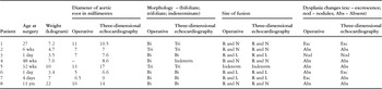
We evaluated 8 consecutive patients, with a median age of 16 weeks, and a range from 1day to 11 years, with a median weight of 5.96 kilogram, this ranging from 2.78 to 22 kilograms. The demographics, and characteristics of the abnormal valves, are displayed in Table 1.
Table 1. Demographic details, along with operative and three-dimensional echocardiographic findings.

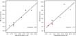
Size of the aortic valve: We were able to make direct comparisons between echocardiographic measurements and surgical findings in all patients bar one, where the operative sizing of the orificial diameter was missing. The median diameter as judged from three-dimensional echocardiography was 8.5 millimetres, with a range from 6.5 to 14 millimetres. The median diameter as judged by cross-sectional echocardiography was 8.8 millimetres, with a range from 6.6 to 17 millimetres. The median diameter as judged from surgical assessment was 7 millimetres, with a range from 5 to 14 millimetres. The diameter as derived from three-dimensional reconstructions strongly correlated with the values obtained during operative assessment (rs equals 0.92), being better than measurements made at cross-sectional echocardiography (rs equals 0.78), see Figure 2.

Figure 2. Scatter plot comparison and correlation between operative measurement of the size of the aortic root with three-dimensional (blue circles) and cross-sectional (red circles) echocardiographic measurements.
Aortic valvar morphology: When compared with the surgical findings, three-dimensional echocardiography correctly identified a valve having 3 leaflets in 4 patients, bifoliate morphology in three patients, and an indeterminate arrangement in the final patient. This strongly correlated with operative findings (rs equals 0.69) in comparison with cross-sectional echocardiography (rs equals 0.13), the difference being statistically significant (p less than 0.05 – see Table 2).
Table 2. Spearman's correlation (rs) of three-dimensional and cross-sectional echocardiographic findings with operative findings.

Fusion between the right coronary arterial and non-coronary leaflets was identified from the reconstructions in four patients, between the right and left coronary arterial leaflets in three patients, and could not be determined in the final patient. The predicted sites of fusion were all confirmed by the operative findings, whereas standard cross-sectional echocardiography proved much worse (rs equals 0.49), this difference again being statistically significant (p less than 0.05), see Table 2.
Dysplastic changes as identified on three-dimensional echocardiography: In 5 patients, we identified dysplastic changes on the right coronary arterial leaflet. In an additional 3 patients, we found thickening and/or nodules. Of these, 1 patient required surgical debridement of the nodule. In the other two patients, the valve thickening was diffuse, and this was trimmed at surgery. The nodule that required surgical excision measured 2 by 3 millimetres. We separated the dysplastic changes observed on three-dimensional echocardiography into excrescences and nodules, defining excrescences as diffuse thickening of the leaflets, but measuring less than 2 by 3 millimetres, and nodules as focal thickenings measuring greater than 2 by 3 millimetres. Nodularity as defined above on three-dimensional echo (rs equals 0.61) correlated well with operative findings, in comparison with cross-sectional echocardiography, which performed less well (rs equals 0.35), albeit that the difference was not statistically significant (see Table 2).
Discussion
Real time three-dimensional echocardiography has the potential to provide valuable additional information to conventional cross-sectional echocardiography for the assessment of patients with congenitally malformed hearts. Despite the small numbers in our series, we were able to demonstrate that three-dimensional echocardiographic assessment of the congenitally stenotic aortic valve gives accurate information about valvar dimensions and morphology, and provides valuable additional information about morphological aberrations of the valve that may not be appreciable by standard cross-sectional-echocardiography. Three-dimensional echocardiography has previously been shown to provide good anatomical and pathological correlation in the identification of bifoliate aortic valves in post-mortem specimens.6 We provide evidence here for similar accuracy in the clinical setting. There was excellent concordance between our reconstruction and the surgical assessment. Half of our patients had fusion between the right coronary and the non-coronary arterial leaflets. Other investigators have reported the commonest site of fusion to be between the right and left coronary arterial leaflets. As all of our patients had surgical valvotomy, they had by definition severe aortic stenosis, presumably sub-selecting the group with fusion between the right and non-coronary leaflets. Other investigators have assessed the accuracy of multi-plane transoesophageal cross-sectional echocardiography in predicting aortic valvar morphology. This demonstrated sensitivity and specificity of 87% and 91% respectively.7 Our series, though small, showed a strong correlation for measurements of the diameter of the valvar orifice, the valvar morphology, and identification of dysplastic leaflets. Our reconstructions proved completely sensitive and specific in predicting the site of fusion between the leaflets. The technique, therefore, may have the potential for assessing abnormal valvar morphology more accurately than conventional echocardiographic modalities.
In 3 patients, our reconstruction demonstrated diffuse thickening of the leaflets, and/or the presence of nodules on their surface. Such information has the potential to inform the assessing cardiologist about the most appropriate route for therapeutic intervention, surgical as opposed to trans-catheter balloon dilation. In all three cases, the surgeon was able to address these morphological aberrations at operation. Such patients are perhaps more appropriately managed surgically, although conclusive data in this regard is lacking.
We recognize that our study has its limitations. Although the dataset was collected prospectively, our numbers were small, and hence our study remains largely descriptive. Larger studies are required to verify our findings. The planes used to acquire the images are non-standard, and vary significantly in each study, depending on the quality of the images and the physical properties of the ultrasonic probe. Analysis is also subjective, and depends on the expertise of the observer. There was no second observer, hence we were unable to assess inter observer variability. Despite these limitations, we conclude that real time three-dimensional echocardiography accurately defines the morphometric characteristics of the congenitally stenosed aortic valve. It also delineates additional valvar pathology that may be missed by conventional echocardiographic assessment, such as the exact site of fusion of between leaflets, and the presence of dysplastic changes in the leaflets. It has the potential to inform the clinician about the most appropriate therapeutic modality. We believe the technique should be used routinely in the assessment of stenotic aortic valves in children.