An atrial septal defect is one of the frequent congenital cardiac malformations, accounting for up to two-fifths of congenital cardiac disease in adults, and one-tenth of the malformations seen in children. The lesions, if needing closure, have traditionally been treated surgically. The surgical approach, however, is traumatic and leads to relatively long periods of convalesence. Such defects were first closed by transcatheter insertion of a device as long ago as 1976, and transcatheter closure of defects within the oval fossa is now a recommended option for treatment, with ongoing evolution of techniques and devices.Reference Kardon, Sokoloski, Levi, Perry, Schneider, Allada and Moore1–Reference Rosas and Attie4 In this investigation, we have taken advantage of the availability of patients undergoing transcatheter closure to study echocardiographically the changing conditions of the right heart before and after closure.
Patients and methods
General data
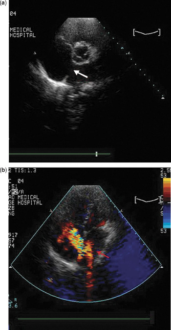
Between March of 2005 and October of 2007, 28 male and 45 female patients aged between 5 and 61 years were selected for transcatheter closure from 233 patients diagnosed as having defects within the oval fossa at Qingdao University, China. We performed ultrasonic investigations 1 day before, and 3 days and 3 months after the interventional closure. The criterions for selection for interventional closure included a maximum relaxed diameter for the defect between 8 and 33 millimetres, with distances of at least 4 millimetres between the margins of the defect and the superior and inferior caval veins, the right superior pulmonary veins, the coronary sinus, and the hinges of the atrioventricular valves, a distance of at least 2 millimetres between the margin of the defect and the non-coronary sinus of the aorta, shunting from left to right, and no other complicating abnormalities (Fig. 1). We also required that the electrocardiogram shows no evidence of frequent atrial premature contraction or atrial fibrillation, that pulmonary plethora was evident radiologically, and that the echocardiographic recordings were of suitable quality. All the ultrasonic interrogations and measurements were performed by the same investigator.
Cross-sectional and colour Doppler images of a patient with an atrial septal defect (arrow).

We used a Philips Sonos7500 coloured Doppler ultrasonic system, coupled with S3 and S4 electronic phased array transducers having frequencies from 1.0 to 4.0 megaherz.
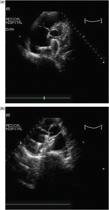
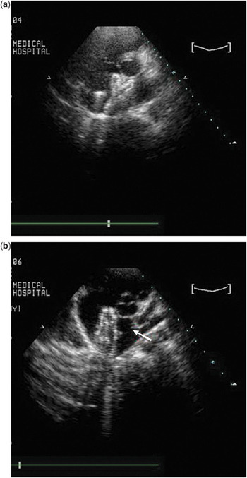
The size of the device required for occlusion was determined after estimating the size, appearance, and relationships of the defect to adjacent structures using multiple views. The device was inserted using general anaesthesia for children aged under 8 years, and local anaesthesia for older patients. The device was inserted following right femoral arterial puncture, using echocardiographic and fluoroscopic monitoring (Figs 2 and 3).Reference Santoro, Pascotto and Caputo5
The procedure of transcatheter closure, showing (a) the occluder in the left atrium and (b) the occluder positioned across the atrial septum.

Panel a shows the device embracing the aorta, while panel b shows the device alongside the aorta (arrow) in the patient in whom it was necessary to close the defect eventually by surgery.

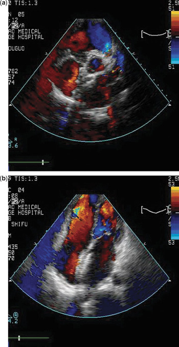
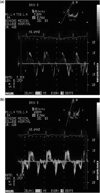
We measured the antero-posterior diameter of the right ventricle, the transverse diameter and length of the right atrium, the inner diameter of the pulmonary trunk, and observed the geometric appearance of the right atrium, focusing on both its parietal wall and the septum, and interrogating flows across the mitral, tricuspid, and aortic valves. We also excluded any obstruction in the systemic and pulmonary venous channels (Fig. 4). In addition to cross-sectional imaging, we used tissue Doppler to calculate the systolic and early and late diastolic velocities of the basal parietal walls of the right ventricle (Fig. 5), also using Doppler interrogation to measure the highest rate of tricuspid regurgitation.Reference Jiang, Levine and Weyman6, Reference Veldtman, Razack and Siu7 Comparison between measurements was done using Student’s t-test, taking values for P of less than 0.05 as being statistically significant.
Panel a shows the obliteration of atrial shunting after closure of the defect, with panel b showing reduced dimensions of the right-sided chambers.

Tissue Doppler measurements are shown (a) one day before closure and (b) 3 days after closure.

Results
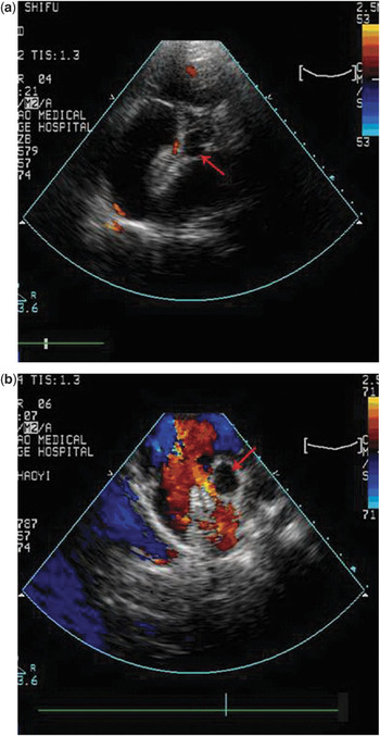
The diastolic diameter of the defects measured from 8 to 33 millimetres, and the defects were closed using devices measuring from 10 to 40 millimetres. Echocardiographic interrogation after insertion showed normal morphology of all devices, which had been well incorporated after 3 months into the surrounding atrial tissues. The defects had been completely occluded in 56 patients, with 13 patients shown to have a trace of residual shunting (17.8%), and 4 to have small shunts (5.4%). We deemed small shunts to be those measured echocardiographically at no more than 2 millimetres in width (Fig. 6).
The images show (a) a small residual shunt (arrow) and (b) moderate residual shunting (arrow). The patient shown in panel b subsequently required surgical removal of the device and closure of the defect (See Figure 3b).

Within 3 days of closure, shortening in the diameter and length of the right atrium was seen in 66 patients, and shortening in the antero-posterior diameter of the right ventricle in 37 patients. After 3 months, the antero-posterior diameter of the right ventricle, and the dimensions of the right atrium, had returned to normal values in 70 patients. Another 3 patients still had moderate pulmonary hypertension with mild dilation of the right ventricle. After follow-up of 1 year, their pulmonary arterial pressures gradually declined and the dimensions of the right heart returned to normal. By this time, evidence of minimal residual shunting had disappeared in 13 patients, while the mild shunting in the 4 patients had disappeared in one, but increased to moderate shunting in another. After a further 6 months, we removed the occluder in this patient and closed the defect surgically (Figs 3b and 6b).
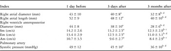
The measurements made using tissue Doppler showed that all measured velocities, along with right ventricular pressures, declined subsequent to interventional closure, the differences already reaching statistical significance after 3 days (p less than 0.05), and becoming more significant after 3 months (p less than 0.01 – Table 1.)
Functional and morphological changes subsequent to interventional closure of atrial septal defects.

Note: Difference between 3 days after and 1 day before the surgerya p < 0.05; that between 3 month and 1 day after the surgeryb p < 0.01; 3 months and 3 days after the surgeryc p < 0.05; months and 3 days after the surgeryd p < 0.01.
Key to abbreviations: Right ventricular lateral wall velocity in systole (Sm), early diastole (Em), late diastole (Am).
Discussion
In patients with atrial septal defects, one of the commonest congenital cardiac diseases, shunts of significant volume can, over the long term, lead to overload of the right ventricle with decreased ventricular function, increased pulmonary arterial pressures, backward cardiac failure and atrial arrhythmias, and even progress to produce Eisenmenger’s syndrome. Our echocardiographic study has confirmed that transcatheter closure of such defects rapidly obliterates the left to right shunt, reverses the abnormal haemodynamics, and reduces the ventricular preload. Already by 3 days, our measurements show significant reductions in the volume and dimensions of the chambers of the right heart, with the improvements more marked in the right atrium than the right ventricle. We also observed a decline in pulmonary arterial pressures, mainly related to the lowered right ventricular preload and the mitigation of the hyperkinetic state subsequent to closure.Reference Diab, Cao, Bacha and Hijazi8–Reference Kort, Balzer and Johnson10
Our study confirms, therefore, that the geometric structure of the right heart begins to normalize within 3 days of transcatheter closure of atrial septal defects.Reference Carlson, Justino and O’Brien11, Reference Fischer, Stieh, Uebing, Hoffmann, Morf and Kramer12 In the subsequent 3 months, the right atrium and right ventricle show further reduction in size, along with significant ongoing decline in pulmonary arterial pressure, ameliorating still further the hyperkinetic state of the right ventricle.Reference Pascotto, Santoro and Cerrato13
During the subsequent 18 months, the right ventricular structure showed still further improvement concomitant with myocardial remodelling.
The complex morphology of the right ventricle produces difficulties in assessing accurately its function, albeit that such assessment is important when seeking to evaluate the condition of the patients, and when making decisions concerning optimal treatment and judging prognosis.Reference Santoro, Pascotto and Sarubbi14 We are still assessing such changes during ongoing follow up. Our study shows, nonetheless, that echocardiographic monitoring not only serves to guide the interventional closure of atrial septal defects, but is also invaluable in the evaluation of right ventricular structure and function subsequent to closure, as well as contributing to assessment of the long-term efficacy of the closure.