Introduction
Bilateral vocal fold immobility results in airway obstruction. It has multiple causes, including bilateral recurrent laryngeal nerve paralysis, bilateral cricoarytenoid joint ankylosis and posterior glottic stenosis.Reference Hillel, Benninger, Blitzer, Crumley, Flint and Kashima1 Airway obstruction can also be caused by laryngeal synkinesis – an aberrant reinnervation of one vocal fold – that occurs following recurrent laryngeal nerve paralysis and which results in excessive adduction.Reference Crumley2 In all of these cases, phonation is usually not severely impaired and may even be normal; however, respiration is often impaired by the narrow airway. The inability to recover the normal function of at least one of the vocal folds, or the lack of an adequate glottic aperture, with persistent airway symptoms, are indications for surgical treatment.
There are multiple surgical options to treat vocal fold immobility. Surgical treatment was originally described by Chevalier Jackson, who used cordectomy through a laryngofissure to open the airway. Others have described: arytenoidectomy, through the thyroid alar window;Reference Jackson3 arytenoid abduction with lateralisation sutures;Reference Kelly4 and the use of a carbon dioxide (CO2) laser technique for a transoral cordectomy, withReference Woodman5 or withoutReference Ossoff, Sisson, Duncavage, Moselle, Andrews and McMillan6 transoral arytenoidectomy. However, all of these surgical procedures reduce airway obstruction at the expense of phonation.
This study aimed to demonstrate the efficacy of a novel technique – endoscopic partial arytenoidectomy with true vocal fold lateralisation – in the treatment of bilateral vocal fold immobility.
Materials and methods
Patient selection and evaluation
The charts of 18 patients (10 women and 8 men), with a mean age of 57.1 years (range, 23–73 years), were retrospectively reviewed at tertiary referral centres. Post-operative follow-up duration ranged from one to five years. All patients had been treated by a tracheostomy because of bilateral vocal fold immobility, and decannulation had failed prior to being referred to our centres. Aetiologies of the bilateral vocal fold immobility included post-thyroidectomy complications, neck radiotherapy and cerebrovascular accident (Table 1).
Bilateral vocal fold immobility aetiologies and Voice Handicap Index scores

Pt. no. = patient number; pre-op = pre-operative; VHI = Voice Handicap Index; post-op = post-operative; F = female; M = male; CVA = cerebrovascular accident
Pre-operatively, all patients underwent transnasal and trans-stomal endoscopy, with retrograde examination of the vocal folds and the subglottis using flexible laryngo-tracheoscopy, under local anaesthesia, to confirm the diagnosis of bilateral true vocal fold immobility, and to identify any associated tracheal or subglottic obstructive lesion(s).
Each patient's voice was evaluated using the Voice Handicap Index before and after the surgery. The pre-operative Voice Handicap Index average score was 30.8 (range, 26–36) (Table 1). Patients completed the Voice Handicap Index based on their initial obstructive symptoms (before tracheostomy insertion) and weeks after the lateralisation surgery (after tracheostomy removal).
Surgical technique
Here, we describe the endoscopic arytenoidectomy technique. The reconstructive transoral laser microsurgery procedure was performed under general anaesthesia. Ventilation was maintained through the trans-stomal placement of an appropriate cuffed endotracheal tube.
A Lindholm laryngoscope (model 8587A; Karl Storz, Tuttlingen, Germany) was used for the suspension microlaryngoscopy (using a dental guard for protection), providing a wide aperture for the bimanual manipulation of instruments and tissues. The larynx was then examined with a 30° Hopkins rod telescope (model 49046 BA; Karl Storz). The mobility of the cricoarytenoid joint was tested using a laryngeal spatula. Associated unilateral or bilateral arytenoid fibrosis and/or cricoarytenoid ankylosis was also assessed for planned additional treatment. The same surgical approach was used for all cases in this series, with the addition of mobilisation of cricoarytenoid joints that were found ankylosed and/or fibrosed.
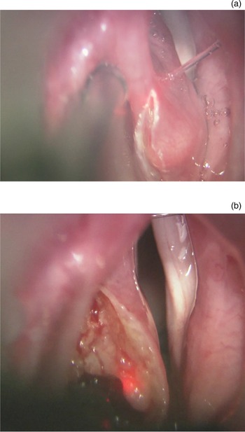
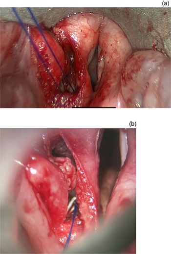
An operating microscope, with a microspot CO2 laser (1.2 mm diameter) and a digital AccuBlade® robotic joystick controller (with a 0.1 second pattern delay), was used to incise the epithelium overlying the arytenoid tower cartilage. A sigmoid incision was made over these structures and took the medial incision through to the subglottis. The incision effectively severed the medial face of the arytenoid body in preparation for the creation of an advancement flap, leaving it attached to the medial mucosa, the vocal process of the arytenoid and the thyroarytenoid muscle. We flanked this incision into the interarytenoid mucosa, posteriorly, and into the deep tissues of the piriform sinus, laterally, circumferentially isolating the central arytenoid, in preparation for its removal. We then tipped the central arytenoid medially and incised its deep attachments that are frequently fibrosed to the cricoid cartilage, consistent with the clinical impression as described above. This is similar to a type IV central arytenoidectomy surgical technique (previously described by Atallah et al.Reference Atallah, Mk, Al Omari, Righini and Castellanos7) used to make room for a wider airway.
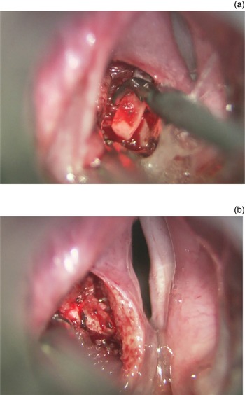
After we removed the central arytenoid, we used topical adrenaline on cottonoids or low-wattage suction cautery for haemostasis, as needed (Figures 1 and 2).
(a & b) Sigmoid incision of epithelium overlying the arytenoid tower cartilage. The medial incision is extended to the subglottis for creation of an advancement flap.

(a & b) The central arytenoid is circumferentially isolated and removed.

We then mobilised the medial face of the residual arytenoid body and vocal process by making anterior incisions through these soft tissues and drawing this structure posterolaterally. Once it was sufficiently loose, we considered the flap prepared for lateralisation and reattachment to the lateral residual arytenoid (its muscular process).
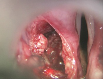
We irrigated the wound with copious amounts of sterile saline and then turned our attention to attaching this advancement flap posterolaterally (Figure 3).
After removal of the central arytenoid, the wound is irrigated and the flap is made ready for posterolateral attachment.

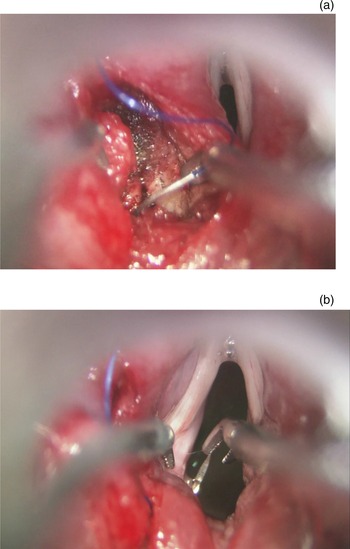
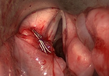
Using size 4-0 Prolene sutures on a P3 cutting needle, we passed first through the residual muscular process, and then out into the airway lumen underneath the vocal process. The needle was then passed back to the wound above the vocal process, through the cartilaginous residual medial face of the arytenoid, and then back through the deep soft tissues again to broadly draw the arytenoid posterolaterally. This suturing method enabled us to lateralise the patient's glottis and prevent drifting of the fold medially (Figure 4). The suture ends were brought to the outside of the laryngoscope and clipped with titanium clips (Figure 5). The excess suture was cut by a 1.5 mm laser line with a 0.1 second delay.
(a & b) A size 4-0 Prolene suture is passed through the residual muscular process into the airway lumen, underneath the vocal process, back to the wound above the vocal process and then back through the deep soft tissues.

(a & b) The suture ends are brought to the outside of the laryngoscope then clipped with titanium clips.

We closed the superficial wound, incorporating the interarytenoid mucosa with the superior edge of the residual cartilage face in a figure-of-eight configuration. We clipped all sutures in the manner of Atallah et al.,Reference Atallah, Manjunath, Omari, Righini and Castellanos8 laser lysing the excess suture in order to fully close the wound (Figure 6).
The wound is superficially closed with a figure-of-eight configuration and all sutures are clipped.

Post-operative care
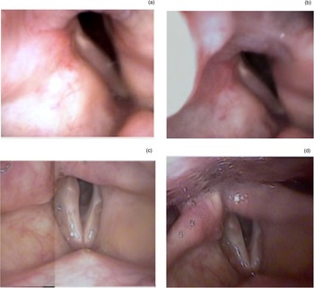
Post-operatively, we admitted the patients for 2–4 days for airway observation. They were given a prophylactic broad-spectrum intravenous antibiotic. They also received respiratory care, including nebulised 3 per cent sodium chloride to help clear airway secretions. After discharge, the patients were followed up in the out-patient clinic at two and six weeks post-operatively, where we repeated the fibrotic examination to confirm that the lateralised vocal fold was in an optimal position with a reasonable glottis gap, as shown in Figure 7. Patients were readmitted once they were ready for decannulation, and placed on close observation during tracheostomy removal.
(a–d) A laryngeal examination conducted six weeks after surgery showed the position of the lateralised vocal fold with a reasonable glottis gap.

Results
Eighteen patients were included in the study, including 10 women and 8 men. Their mean age was 57.1 years, ranging from 23 to 73 years. All patients were tracheostomy dependent – decannulation had failed prior to them being referred to our care. The most common aetiology of bilateral true vocal fold immobility was thyroidectomy (61.11 per cent).
All patients were treated with the reconstructive transoral laser microsurgical procedure described above, via a suspension laryngoscopy approach, under general anaesthesia. Endoscopic resection of obstructive tracheal granulation tissue with a laser was also performed in six patients, with or without local corticosteroid injection. Tracheal balloon dilatation was performed in two patients. Two patients needed a second-stage surgery for a minor revision. There were no post-operative local complications related to surgery.
All patients with a tracheostomy were successfully decannulated. This was accomplished either after the initial or the revision procedures (range, 2–8 weeks). In all patients, the transoral partial arytenoidectomy with true vocal fold lateralisation showed complete healing by 8–12 weeks, with no residual clips (on the transoral sutures) on out-patient laryngeal examination.
The voice outcome after the surgery was evaluated using the Voice Handicap Index. The average score was 15 (range, 12–20), indicating a significant improvement in patients’ voice outcome (p < 0.001).
Discussion
Bilateral vocal fold immobility is a rare condition that can be due to vocal fold paralysis or cricoarytenoid joint fixation. It is frequently associated with significant morbidity and disability, most significantly tracheostomy dependence. Varying degrees of dyspnoea, dysphonia and dysphagia occur, depending on the underlying aetiology, vocal fold position and compensatory behaviour(s).Reference Olthoff, Zeiss, Laskawi, Kruse and Steiner9–Reference Segas, Stavroulakis, Manolopoulos, Yiotakis and Adamopoulos11 Thyroid surgery remains the most common cause of bilateral vocal fold immobility.Reference Rosenthal, Benninger and Deeb12
Oral or nasal endotracheal tubes, or tracheostomy tubes, are most commonly used to deliver mechanical ventilatory support in respiratory failure.Reference Stauffer, Olson and Petty13 Tracheostomy is known to have deleterious effects on voice and swallowing, and vastly diminishes quality of life.Reference Bennett14–Reference Citta-Pietrolungo, Alexander, Cook and Padman16 There is also a risk of airway stenosis, as tracheostomy can cause laryngeal stenosis and/or tracheal stenosis.Reference Grillo, Donahue, Mathisen, Wain and Wright17 However, tracheostomy may be required if uncompensated bilateral vocal fold immobility is not adequately addressed.
• Bilateral vocal fold immobility is a rare condition caused by vocal fold paralysis or cricoarytenoid joint fixation
• Bilateral vocal fold immobility is frequently associated with significant morbidity and disability
• Reconstructive transoral laser microsurgery is a minimally invasive, safe and effective treatment option
• The use of an endoscopic (rather than a laryngofissure) approach in bilateral vocal fold immobility reduces iatrogenic harm to the larynx and trachea
There are a number of surgical techniques described in the literature designed to address bilateral vocal fold immobility. The endoscopic reconstructive transoral laser microsurgery based partial arytenoidectomy with true vocal fold lateralisation described in this paper is a minimally invasive, safe, effective and reproducible technique.
A decannulation rate of 100 per cent was obtained in tracheostomy dependent patients, with a small percentage of patients requiring one or two additional minor procedures.
Although endoscopic suturing can be challenging, it is facilitated by the use of vascular clips to secure the stitches in a way that is much simpler, more reliable and timesaving than endoscopic knot tying.Reference Atallah, Manjunath, Omari, Righini and Castellanos8 It is important to stress that the titanium ligation clips present no clinical danger whatsoever. In fact, the use of haemostatic clips in transoral laryngeal surgery has already been reported in the literature in transoral laser cancer resection techniques, to control bleeding from medium to larger size vessels.Reference Steiner18,Reference Canis, Ihler, Martin, Wolff, Matthias and Steiner19 The titanium clips used are very small, light and biocompatible. Most of the clips are swallowed or coughed out during the early post-operative period.
Conclusion
Reconstructive transoral laser microsurgery based partial arytenoidectomy with true vocal fold lateralisation is a safe, effective and well-tolerated choice for airway reconstruction in those with airway obstruction due to bilateral true vocal fold immobility. It requires the combined techniques of careful laser-based partial arytenoidectomy and lateralisation, facilitated by the use of vascular clips to secure the stitches in a simple and reliable way. The use of an endoscopic approach rather than a laryngofissure approach reduces iatrogenic harm to the larynx and trachea. Additionally, it can be performed by laryngologists familiar with laryngeal microsurgery using commonly available laryngeal instruments and a scanning CO2 laser.
Competing interests
None declared