Introduction
Within the past decade greater attention has been given to arthropod-borne zoonotic diseases in many countries of Europe, due to their increasing prevalence. This is presumably caused by changed climatic conditions, in particular global warming, extensive rainfall and floods. Also the spread of a new mosquito species has been recorded in several European countries during recent years (Roiz et al., Reference Roiz, Eritja, Escosa, Lucientes, Marques, Melero-Alcibar, Ruiz and Molina2007, Reference Roiz, Neteler, Castellani, Arnoldi and Rizzoli2011), which could be the potential vector of Dirofilaria species. Currently, subcutaneous dirofilariosis caused by the nematode D. repens is regarded as the arthropod-borne disease with the fastest spread (Genchi et al., Reference Genchi, Rinaldi, Cascone, Mortarino and Cringoli2005) and, regarding its zoonotic character, infection represents a high infection risk for both animals and humans. Dogs, and to a lesser extent cats and wild carnivores, are the main natural hosts for D. repens and the related species D. immitis (Magi et al., Reference Magi, Calderini, Gabrielli, Dell'Omodarme, Macchioni, Prati and Cancrini2008). Humans are only accidental and dead-end hosts, which harbour the L3 stage of larvae after being bitten by infected insect vectors.
In the past, dirofilariosis was strictly regarded as a parasitic disease of warm climatic zones in the world. In Europe, infection was endemic in the Mediterranean region, with the highest incidence of both human infection (Pampiglione et al., Reference Pampiglione, Canestri-Trotti and Rivasi1995) and canine infection detected in Italy (Pampiglione et al., Reference Pampiglione, Rivasi, Angeli, Boldorini, Incensati, Pastormerlo, Pavesi and Ramponi2001). The incidence of autochthonous infections was also reported in other countries (reviewed by Genchi et al., Reference Genchi, Kramer and Rivasi2011). But within the past 10 years, increasing numbers of canine and human cases have also been reported from central European countries, and Slovakia was one of the first central-European countries with confirmed canine subcutaneous dirofilariosis. In 2005 the causative agent of infection was diagnosed among dogs in the south-western region (Svobodová et al., Reference Svobodová, Svobodová, Beladičová and Valentová2005) and infection caused by D. repens was diagnosed in almost 35.0% of dogs originating from southern Slovakia; all these cases were confirmed as autochthonous (Miterpáková et al., Reference Miterpáková, Antolová, Hurníková and Dubinský2008, Reference Miterpáková, Hurníková, Antolová and Dubinský2009). Canine dirofilariosis was also confirmed in neighbouring countries. In the Czech Republic and Hungary reports of autochthonous cases of infection were published in 2006 and 2007, respectively (Svobodová et al., Reference Svobodová, Svobodová, Genchi and Forejtek2006; Fok et al., Reference Fok, Kiss, Majoros, Jacsó, Farkas and Gyurkovszky2007). Shortly after detecting D. repens in dogs in Slovakia, the first autochthonous human case of subcutaneous infection was recorded by Babál et al. (Reference Babál, Kobzova, Novák, Dubinský and Jalili2008) in the same territory. Unlike dogs, where diagnosis is based on the presence of microfilaria in the blood or subcutaneously, in humans adult D. repens infection is nearly exclusively present. The reviews of human dirofilariosis (Pampiglione et al., Reference Pampiglione, Canestri-Trotti and Rivasi1995; Genchi et al., Reference Genchi, Kramer and Rivasi2011) indicate that in almost 47% of infections, the parasite was found in subcutaneous nodules in the head region, particularly in ocular or periocular sites. In Slovakia, three human cases of D. repens have been recorded so far, and in the present paper we report a fourth case. In the second case (described by Ondriska et al., Reference Ondriska, Lengyel, Miterpáková, Lengyelová, Strehárová and Dubinský2010) an intact worm of D. repens was isolated from a nodule on the forearm, whereas diagnosis of the first (Babál et al., Reference Babál, Kobzova, Novák, Dubinský and Jalili2008) and the third (Nováková et al., Reference Nováková, Kinčeková, Adamicová, Kompaníková, Švihrová, Šimeková, Krause, Pavlínová and Dvorožňáková2011) human cases was based on histological examination of excised tissues containing a worm.
Whereas ocular localization of the adult worm causes painful symptoms, subcutaneous dirofilariosis is usually manifest by a painless and/or swelling nodule or other unspecific symptoms. Moreover, it can be detected many years after the initial infection and differential diagnosis often includes dermal cysts, fibroma, rheumatic diseases, tumours and other pathologies (Jelínek et al., Reference Jelínek, Schulte-Hillen and Löscher1996). It is highly possible that, in the new endemic regions, many cases have been misdiagnosed due to problems with correct identification of D. repens on histological sections by non-experienced pathologists.
This present human case is the first one in the new endemic region of Slovakia, where D. repens infection in a patient was confirmed by computed tomography (CT) scans, histological examination and polymerase chain reaction (PCR)-based detection of a D. repens-specific DNA fragment.
Materials and methods
Clinical and histological examination
A 58-year-old female patient visited the local hospital seeking medical examination of a painful skin thickening situated on the lower eyelid close to the nose. Clinical examination revealed a pea-sized palpable nodule located in the nervus trigeminus. There was a visible swelling around the nodule and the overlying skin did not show evidence of inflammation. Otolaryngological examination did not show a problem in the nasal cavities. The patient received antibiotic therapy in combination with a steroid drug, which did not result in improvement; therefore CT examination of the face was performed. The nodule was surgically removed and subjected to histological analysis. Tissue was fixed in 10% non-buffered formalin for 7 days, embedded in paraffin and tissue sections were stained with haematoxylin–eosin (HE) in a local histological laboratory, using a routine procedure. Microscopic examination revealed a worm-like structure embedded in the striated muscles, therefore tissue specimens were sent to the Institute of Parasitology in Košice for morphological and molecular identification of the parasite. Seven-micron-thick sections from the nodule were prepared and used for both histological examination following staining with HE and molecular analysis of parasitic DNA.
Molecular analysis
Histological examination of the tissue sections revealed the presence of a dirofilaria-like nematode and a PCR-based approach was used for species identification. From the several unstained sections of two paraffin blocks prepared from the nodule, containing only a small amount of surrounding human tissue, worm tissue was excised under the microscope. Excised tissue was processed by the modified protocol described by Džamič et al. (Reference Džamič, Čolovič, Arsič-Arsenijevič, Stepanovič, Boričić, Džamić, Mitrović, Rašić, Stefanović, Latković and Kranjić-Zec2009) and then subjected to a re-hydratation step in a graded ethanol series prior to DNA extraction with NucleoSpin Tissue DNA-extraction KIT (Macherrey-Nagel, Germany). Isolated DNA was eluted into 50 μl of elution buffer (Tris-EDTA, pH 8.0) and the total amount of DNA (52 μg) and protein impurities were determined spectrophotometrically. In the PCR reactions the primer set specific for a conserved repetitive sequence in the cytochrome c oxidase subunit 1 (CO1)-encoding gene of D. repens (DIR3/DIR4) was used (Vakalis et al., Reference Vakalis, Spanakos, Patsoula and Vamvakopoulos1999). PCR amplifications were performed in 50 μl reactions containing 5 μl 10 × PCR buffer, 3 μl of MgCl2 (50 mm concentration), 0.25 μl (1U) GoTaq Hot Start polymerase (all from Promega, Madison, Wisconsin, USA), 1 μl of deoxynucleoside triphosphates (dNTPs) mix (10 mm concentration) and 3 μl (10 mm concentration) of each primer. In a set of PCR reactions, increasing DNA concentrations from 0.5 to 4.5 μg/reaction were used as the template, and amplification conditions were the same as described by Vakalis et al. (Reference Vakalis, Spanakos, Patsoula and Vamvakopoulos1999). Stained gels were photographed on a Lumibis gel imaging system (DNR, Germany). The positive control in the PCR reaction represented DNA isolated from a live D. repens adult worm obtained from the nodule of another patient (Ondriska et al., Reference Ondriska, Lengyel, Miterpáková, Lengyelová, Strehárová and Dubinský2010).
Results
Clinical and histopathological observations
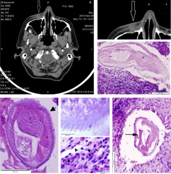
The primary diagnosis indicated fibroma faciei–susp. neurofibroma and CT examination revealed a discrete fibrous nodule approximately 0.8 cm average diameter (fig. 1A,a). The patient had no fever and differential diagnosis did not show malignancy. The nodule was surgically removed and, as the first histological examination performed in the hospital did not confirm the presence of D. repens, DNA-based molecular analysis was proposed. Following the second histological examination performed at the Institute of Parasitology, infection with D. repens adult worm was confirmed, indicated by the external cuticular ridges and the robust muscle cells (fig. 1B and D). In cross-sections of the worm, only one tube beside the intestine was visible in the body cavity, representing a single male sex organ (testes) (fig. 1C). It seems that the D. repens worm was alive at the time of examination, indicated by the thick muscle layer un-detached from the cuticle, cells visible in the muscle layers and the presence of spermatozoa in the sex tube. The thickness of the cuticle in the intact worm was 9.1 ± 1.9 μm and the average measurements of the oval-shaped intact worm were 235.8 ± 8.8 × 157.8 ± 3.2 μm. The worm was deeply embedded in the muscles and surrounded by a granulomatous inflammatory lesion where eosinophils and neutrophils were the most frequent inflammatory cells. In the outside layers of the parasitic granuloma, large cells with two or more nuclei and pink cytoplasm, probably of macrophage origin, were often present (fig. 1E). However, only a small portion of the worm retained well-preserved morphology and the rest of the worm body seemed to be damaged during histological processing (fig. 1F). One year after surgery, the patient has had no further problems.
(A–F) CT and microscopic analysis of the subcutaneous nodule in the patient. (A, a) CT scan of patient's head, showing an intense swelling on the left side with subcutaneous nodule (arrow). (B) Transverse section of D. repens adult worm localized in the granulomatous inflammatory lesion forming the nodule; M, muscles. (C) Cross-section of intact portion of the worm with undetached muscles (M), one tube representing male sex organ (testes, T) beside the intestine (In); note external cuticular ridges (arrow). (D) Higher magnification of the surface of D. repens with well-discernible ridges. (E) In the outside layers of the granuloma, large cells with two or more nuclei and pink cytoplasm were often seen (arrowheads). (F) An example of the worm cross-section with damaged morphology lacking the external ridges, note detached muscle cells (arrow), (haematoxylin-eosin staining, HE). Scale bars: (B, C and F) 100 μm; (D and E) 20 μm. (A colour version of this figure can be found online at http://www.journals.cambridge.org/jhl).

Molecular diagnosis
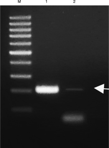
Identification of the D. repens specimen in the nodule was confirmed following amplification of the 246 bp D. repens-specific PCR product. In PCR reactions using DNA isolated previously from the intact D. repens worm at a final concentration of 0.2 μg, the intense 246 bp specific DNA fragment was amplified (fig. 2). As the deleterious effect of formalin fixation on DNA integrity is well known, we performed several PCR reactions, in which different DNA concentrations were used. No specific product was amplified in PCR reactions on DNA obtained from the tissue sections at concentrations below 4.5 μg/reaction. In the PCR reaction where approximately 4.5 μg of DNA served as the template, the faint D. repens-specific 246 bp band and the intense unspecific band corresponding to the excess of primers and fragmented DNA were obtained (fig. 2). No other PCR products were seen on the gel after visualization.
Molecular diagnostic assay based on polymerase chain reaction (PCR) with set of primers, DIR3/DIR4, specific for D. repens. Lanes: M, 100 bp DNA ladder (Promega); 1, (positive control) amplification of the specific 246 bp product on the DNA template isolated from live D. repens obtained previously from another human patient; 2, result from polymerase chain reaction on DNA extracted from formalin-fixed and paraffin embedded tissue of the nodule from our patient. The weak D. repens 246 bp specific product (arrow) was detected on 1.5% agarose gel stained with ethidium bromide.

Discussion
Epidemiological studies carried out within the past 10 years in Europe indicate that the occurrence of human subcutaneous dirofilariosis caused by D. repens is increasing (Pampiglione et al., Reference Pampiglione, Rivasi, Angeli, Boldorini, Incensati, Pastormerlo, Pavesi and Ramponi2001; Genchi et al., Reference Genchi, Kramer and Rivasi2011). In view of the recent rise of human D. repens infection not only in Europe, but also in Asia (Dissanaike et al., Reference Dissanaike, Abeyewickreme, Wijesundera, Weerasooriya and Ismail1997; Nadgir et al., Reference Nadgir, Tallur, Mangoli, Halesh and Krishna2001), it is considered an emerging zoonosis in these continents.
Since 2005, when for the first time D. repens in dogs was reported in the territory of Slovakia by Svobodová et al. (Reference Svobodová, Svobodová, Beladičová and Valentová2005), four human cases of the infection have been diagnosed. Three of these patients, including the case described in our paper, live in the south of country (Bratislava, Komárno) and south-western part of Slovakia (Trnava). The autochthonous character of the case reported in the present paper can be suggested by the patient's epidemiological history. She often visited a garden located near an irrigation canal colonized with a high number of mosquitos, which were probably the source of infection. Moreover, southern parts of Slovakia are characterized as the warmest regions of the country, with periodic floods and frequent mosquito infestation. Neighbouring Hungary is also considered to be a ‘new’ endemic region in Europe, where 16 human cases were described between 2001 and 2006 (Szénási et al., Reference Szénási, Kovács, Pampiglione, Fioravanti, Kucsera, Tánczos and Tiszlavicz2008).
Due to the relatively rare incidence of human dirofilariosis in new endemic countries, the primary diagnosis of subcutaneous nodules performed by general practitioners depends on microscopic examination of tissue cross-sections, but helminth infection is not usually considered. Most cases have been diagnosed retrospectively, when the histopathological sections of biopsy or the excision material were examined in the Central Parasitological laboratories. In the case of our patient, initial therapy was with antibiotic treatment combined with steroid anti-inflammatory drugs, which had no effect on the size of the nodule. CT examination of the patient's head did not indicate the presence of the worm in the lower eyelid close to the nose, where intense swelling was present. Histological examination by the pathologists showed the presence of an unspecified worm. After examination of many tissue sections we were able to identify a male D. repens nematode by means of characteristic multilayered cuticle, external cuticular ridges and the robust muscle cells. However, in the majority of cross-sections the morphology of the specimen was highly damaged; this probably hindered species identification in the hospital. The characteristic worm morphology is usually lost in dead worms in the case of old infections or it can be a result of the host inflammatory reaction in relatively recent infections (Pampiglione et al., Reference Pampiglione, Rivasi and Canestri-Trotti1999). Considering the fact that the period between transmission of larvae to humans and an appearance of clinical signs can take from 2 to up to 12 months (Pampiglione & Rivasi, Reference Pampiglione and Rivasi2000), and our patient asked for the medical examination soon after discovery of the nodule on her face, we suppose that D. repens was alive at the time of surgery. Therefore it seems that worm morphology was markedly altered during the tissue processing. After surgery, the routine examination of excised tissues comprises formalin fixation followed by embedding in paraffin blocks. However, the quality of formalin (buffered or non-buffered) and length of fixation can affect the quality of final histological sections considerably (personal observations) and, even more, the quality of DNA (Hamazaki et al., Reference Hamazaki, Koshiba, Habuchi, Takahashi and Sugiyama1993; Koshiba et al., Reference Koshiba, Ogawa, Hamazaki, Sugiyama, Ogawa and Kitayima1993).
Up to now, the majority of published human cases of subcutaneous dirofilariosis have insisted on the histological examination of excised tissue and identification of morphological features of this nematode (Pampiglione et al., Reference Pampiglione, Canestri-Trotti and Rivasi1995). However, morphological deformations of the worms, mainly loss of the surface cuticular ridges due to the factors mentioned above, can render diagnosis almost impossible. Molecular diagnostic procedures utilize PCR-based amplification of the worm-specific DNA fragments. This is a quite straightforward approach when the DNA template is isolated from the intact worm (Ondriska et al., Reference Ondriska, Lengyel, Miterpáková, Lengyelová, Strehárová and Dubinský2010). On the contrary, the PCR method often fails on DNA samples extracted from formalin-fixed and paraffin-embedded tissue sections (Favia et al., Reference Favia, Lanfrancotti, Della Torre, Cancrini and Coluzzi1996), which seems to be the result of DNA fragmentation. Koshiba et al. (Reference Koshiba, Ogawa, Hamazaki, Sugiyama, Ogawa and Kitayima1993) found that formalin fixative resulted in DNA degradation when tissue was stored at room temperature and non-buffered formalin with acidic pH was used. In our molecular analysis on DNA extracted from the tissue sections we used the set of primers DIR3/DIR4, which is highly specific for D. repens CO1 gene and amplifies a 246 bp product (Vakalis et al., Reference Vakalis, Spanakos, Patsoula and Vamvakopoulos1999). In PCR reactions amplification of this specific product was successful only when a much higher amount of DNA template was used in the comparison with DNA from intact D. repens, indicating massive DNA damage caused by formalin fixation. In successful diagnostic PCR procedures, the period of formalin fixation may vary from up to 20 days (Vakalis et al., Reference Vakalis, Vougioukas, Patsoula, Spanakos, Sioutopoulous and Vamvakopoulos2002) to several months (Džamič et al., Reference Džamič, Čolovič, Arsič-Arsenijevič, Stepanovič, Boričić, Džamić, Mitrović, Rašić, Stefanović, Latković and Kranjić-Zec2009), but the fixation procedure itself seems to be crucial for retaining the quality of DNA. It was shown that fixation with buffered formalin with neutral pH, performed at 4°C, permitted extraction of DNA of sufficient quality even for Southern blot analysis (Koshiba et al., Reference Koshiba, Ogawa, Hamazaki, Sugiyama, Ogawa and Kitayima1993). In our study, identification of D. repens in the nodule was confirmed by both microscopic examination and PCR-based amplification of a specific DNA product by the modified procedure. Such an approach would help to avoid misdiagnosis of subcutaneous nodules with unclear aetiology when morphology of the nematode in the tissue is damaged.
Dirofilariosis threatens public health via common domestic animal hosts and insect vectors, the population of which has increased in the southern part of Slovakia (Jalili, Reference Jalili2010). It is expected that spread of the disease will continue due to climatic changes in central Europe, and this warrants a higher awareness among clinicians, who will be the initial healthcare practitioners contacted by patients.
We conclude that the spread of subcutaneous dirofilariosis to new European regions requires standardization of diagnostic methods and close co-operation between physicians, surgeons and parasitologists. Taking into account that histological identification of the parasite has to be unequivocal in all cases, diagnostic methods based on the effective isolation of DNA from paraffin sections and PCR-based amplification of the worm-specific products should be standardized.
Acknowledgements
The main support for this work was provided by the Scientific Grant Agency VEGA of the Ministry of Education and the Slovak Academy of Sciences (Project no. 2/0188/10 and Project no. 2/0011/12). The technical support for the realization of this work was provided within the frame of the project ‘Centrum of Excellence for Parasitology’ No. 26220120022 supported by the operating programme ‘Research and Development’ funded by the European Fund for Regional Development (0.4).