Introduction
Chagas disease (CD) is a Neglected Tropical Disease that represents a public health problem, showing high morbidity and mortality, in affected populations (Rassi and Marin-Neto, Reference Rassi and Marin-Neto2010; Lidani et al. Reference Lidani, Bavia, Ambrosio, De Messias-reason and Barbosa2017). Endemic in Latin America, CD in last years has received increasing attention as an emerging problem in North America and Europe due to international migrations (Martins-Melo et al. Reference Martins-Melo, Ramos, Alencar and Heukelbach2014). The World Health Organization (WHO) estimates that 6–7 million people are infected with Trypanosoma cruzi worldwide (WHO, 2015). Benznidazole (BZN), the standard drug available for treatment, has limited effectiveness and can cause severe adverse effects, due to its toxicity (Vendrametto et al. Reference Vendrametto, Santos, Nakamura, Dias Filho, Cortez and Ueda-Nakamura2010; Sousa et al. Reference Sousa, Oliveira, Tessarolo, Menezes, Sampaio, Canuto and Martins2017). This drug is more effective in the acute phase of the disease, rarely showing benefits in the chronic phase (Adade et al. Reference Adade, Chagas and Souto-Padrón2012; Hasslocher-moreno et al. Reference Hasslocher-moreno, Brasil, Sousa, Xavier, Chambela and Sperandio2012).
Several advantages have been shown by nanoparticle drug delivery systems compared with the traditional therapy. Recently, the production of CaCO3 nanoparticles for targeted and selective release of toxic drugs, used as chemotherapy agents for the treatment of cancer, allows high doses to be delivered to the desired target tissue, minimizing the exposure of healthy tissues to drug side-effects (Zhao et al. Reference Zhao, Lu, Hu, Li, Dong and Lin2010; Yang et al. Reference Yang, Li, Li, Zhang, Liu, Liang and Sun2017).
Nanosystems can help to overcome toxicity problems and increase drug efficacy (Zazo et al. Reference Zazo, Colino and Lanao2016), because they easily perform membrane transposition and controlled release of the therapeutic molecule, reducing plasma drug concentrations (Romero and Morilla, Reference Romero and Morilla2010).
Therefore, the present work aimed to evaluate if BZN nanoencapsulation in calcium carbonate improves its effect in all evolutive forms of T. cruzi.
Materials and methods
Compounds
BZN was kindly donated by Pharmaceutical Laboratory of Pernambuco-Brazil (LAFEPE®, Brazil). Stock solutions were prepared in DMSO. Serial 2-fold dilutions were performed do obtain working solutions, with DMSO at a maximum concentration of 0.5%, which has no toxicity towards T. cruzi or mammalian host cells. Benznidazole-loaded calcium carbonate nanoparticles (BZN@CaCO3) were dispersed in sterile phosphate buffer solution (PBS) and incubated in a sonicator for 40 min at 37° C to disaggregate the agglomerated particles. Serial dispersions in working concentrations were prepared for in vitro biological assays. Calcium chloride, sodium dodecyl sulphate (SDS), Pluronic F-68, sodium carbonate and sodium citrate were used in the production of calcium carbonate nanoparticles.
Production of drug-free calcium carbonate nanoparticles (@CaCO3)
Calcium chloride (50 mL)(0.05 M), SDS (10 mL)(1%) and Pluronic F-68 (10 mL)(1%) were mixed under continuous stirring (300 rpm) for 24 h forming solution A. Separately, 50 mL of sodium carbonate (0.025 M), 50 mL of sodium citrate (0.025 M), 10 mL of 1% SDS and 10 mL of 1% Pluronic F-68 were mixed under continuous stirring (300 rpm) for 24 h forming solution B. Solution B was slowly added to solution A at a rate of 10 mL.h−1, under constant stirring of 350 rpm at 30° C for a period of 72 h. Then the mixture was centrifuged at 15 000 rpm for 15 min. Nanoparticles were washed several times with 75% ethanol, redispensed in 15 mL of bidistilled water and submitted to ultrasound (frequency 22 kHz, power take 120 W) for 30 min. After that, nanoparticles were dispersed in the solution frozen at low temperatures and then lyophilized for a period of 48 h. Drug-free calcium carbonate nanoparticles (@CaCO3) were produced in Laboratório de Ciências e Tecnologia dos Materias of Universidade Federal do Ceará, Brazil.
Characterization of @CaCO3 by atomic force microscopy (AFM)
To obtain AFM height data, 1.0 mg of lyophilized @CaCO3 was previously dissolved in 1.0 mL of Milli-Q water and immediately pipetted on a coverslip. The coverslip was air-dried for 5 min, and placed on steel sample disks covered with double-sided adhesive tape. Sample surfaces were scanned in air with an AFM Shimadzu SPM-9700 using the tapping mode at a scan of about 1 Hz, resonance frequencies of about 200–380 kHz, with crystal silicon cantilevers at a spring constant of ~51 N m−1 and tip radius of 15 nm. The scan size performed was 1.00 × 1.00 µm2. AFM scan controls were adequately adjusted (sufficient contact force and high gains) to prevent tip artefacts during sample scanning. The height data were processed with Shimadzu SPM-9700 software aimed at the visualization of two-dimensional (2D) images. We used the particle analysis software SPM9700 by the Shimadzu Corporation to study the size distribution of the AFM height data. The Shapiro–Wilks test was performed to determine whether or not a random sample of values followed a normal distribution (with a significance level of 0.01). The mean particle size and confidence interval (CI) were determined.
Production of BZN@CaCO3
The procedure for BZN encapsulation in CaCO3 nanoparticles is very similar to the procedure for the creation of drug-free calcium carbonate nanoparticles (@CaCO3). The only difference lies in the preparation phase of solution A. Approximately 1 mg of BZN, previously dissolved in 20 mL of water with a minimal quantity of ethanol, is immediately added to solution A prior to its blending with solution B. Subsequently, solution B is slowly added to solution A as described above. BZN -loaded calcium carbonate nanoparticles (BZN@CaCO3) were produced in Laboratório de Ciências e Tecnologia dos Materias of Universidade Federal do Ceará, Brazil.
Quantification of BZN in the BZN@CaCO3 conjugate
For the analysis of the presence of BZN in the BZN@CaCO3 conjugate, samples of BZN, @CaCO3, and BZN@CaCO3 were separately mixed and pressed with KBr powder at the concentration of about 5% (w/w) for the preparation of pellets for infrared absorption measurements. Infrared absorption spectroscopy was performed using an absorption spectrometer (model FTLA 2000) in the region of 400–4000 cm−1. The measurement of BZN concentration in the BZN@CaCO3 conjugate was performed by UV–VIS spectrophotometry (Cary 5000 UV–vis–NIR), in the 200–450 nm range (Bezerra et al. Manuja et al. Reference Manuja, Kumar, Dilbaghi, Kumar, Manuja and Singh2013). To obtain a calibration curve, different dilutions of BZN were prepared within the range of concentrations of 5.07–38 mg L−1. BZN@CaCO3 was diluted in distilled water at 260, 130 and 86.7 mg L−1, and then mixed for 4 h at 37 °C to allow complete drug release. Finally, the solution was analysed in a UV spectrophotometer and the BZN concentration was calculated by interpolation on BZN standard curve (Unciti-Broceta et al. Reference Unciti-Broceta, Arias, Maceira, Soriano, Ortiz-González, Hernández-Quero, Munoz-Torres, Koning, Magez and Garcia-Salcedo2015).
Parasites and cell culture
Trypanosoama cruzi Y-strain epimastigotes and trypomastigotes were donated by Laboratório de Bioquímica of Chemical Institute of Universidade de São Paulo. Epimastigotes were maintained at 28° C in Liver Infusion Tryptose (LIT) medium supplemented with 10% fetal bovine serum (FBS). The cells were cultured in the Dulbecco's modified Eagle's medium (DMEM) medium containing 10% FBS and maintained at 37° C in a 5% CO2 atmosphere. The trypomastigote forms of T. cruzi obtained by infecting LLCMK2 cells, were incubated at 37 °C in an atmosphere with 5% CO2 in the DMEM medium (Vitrocell, São Paulo, Brazil) supplemented with antibiotics and 2% of FBS (Meira et al. Reference Meira, Guimarães, Santos, Moreira, Nogueira, Tomassini, Ribeiro, Souza, Santos and Soares2015). Intracellular amastigotes were obtained and cultured as described below.
Effect of BZN@CaCO3 on T. cruzi epimastigote forms
Epimastigote forms were seeded in 96-well plates at a density of 106 cells mL−1, untreated or treated with BZN, BZN@CaCO3 and @CaCO3 (6.25–50 µg mL−1) and incubated for 24, 48 and 72 h. After these periods, parasite death rates were determined by light microscopy through direct quantification of the number of viable parasites using a Neubauer chamber, and IC50 (compound concentrations that inhibit 50% of epimastigote growth) was calculated by non-linear regression (Nogueira et al. Reference Nogueira, Saraiva, Sultano, Cunha, Laranja, Justo, Sabino, Coelho, Rossini, Atella and Paes2015).
Effect of BZN@CaCO3 on T. cruzi trypomastigote forms
Trypomastigotes were seeded in 96-well plates at 106 cells mL−1, untreated and treated with BZN, BZN@CaCO3 and @CaCO3, and incubated for 24 h. Cell viability evaluation was assessed by counting and the LC50 (compound concentrations that kill 50% of trypomastigotes) was determined (Borges et al. Reference Borges, Aires, Higino, Medeiros, Citó, Lopes and Figueiredo2012; Meira et al. Reference Meira, Guimarães, Santos, Moreira, Nogueira, Tomassini, Ribeiro, Souza, Santos and Soares2015).
Cytotoxicity to mammalian cells (MTT assay)
LLC-MK2 cells were seeded in 96-well plates at 105 cells mL−1, maintained in the DMEM and incubated for 24 h in the presence or absence of serial dilutions of BZN, BZN@CaCO3 and @CaCO3 (6.25–50 µg mL−1). After incubation, the cytotoxic effect was assessed using MTT (Amresco, Ohio, USA; 5 mg mL−1) assay. The absorbance was measured in a microplate reader spectrophotometer (Biochrom® Asys Expert Plus) at 570 nm. Controls without cells and with cells treated only with the diluent were run in parallel. The 50% cytotoxic concentration (CC50) was determined by non-linear regression. In addition, the selectivity index (SeI) was determined as the CC50/LC50 ratio (Marín et al. Reference Marín, Ramírez-Macías, Rosales, Muro, Reviriego, Navarro, Aran and Sánchez-Moreno2015).
Effect of BZN@CaCO3 on T. cruzi amastigote forms
To investigate the effect of BZN@CaCO3 on amastigotes, LLC-MK2 cells were seeded in plates containing glass coverslips, cultivated in 10% FBS DMEM and maintained at 37° C in a 5% CO2 atmosphere for 24 h. Thereafter, the cultures were washed, infected with trypomastigotes (parasite: host cell ratio of 20:1) and incubated for 24 h on 2% FBS DMEM. After the infection period, non-internalized parasites were removed by washing with PBS, and the cells were cultivated in fresh 2% FBS DMEM and treated with Bz-NP (8.7 and 17.4 µg mL−1) and Bz (56.7 and 113.4 µg mL−1) for 24 h (Hernández-Chinea et al. Reference Hernández-Chinea, Carbajo, Sojo, Arvelo, Kouznetsov, Romero-Bohórquez and Romero2015). Untreated infected cells were used as negative control. The experimental groups were washed with PBS, fixed in Bouin's solution, stained with Giemsa and mounted on glass slides. The number of infected cells and amastigotes per infected cell was evaluated by random counting of 300 cells, in three different experiments. Survival Index (SuI) was determined (SuI = % of infected cells × number of amastigotes/number of infected cells) (Vendrametto et al. Reference Vendrametto, Santos, Nakamura, Dias Filho, Cortez and Ueda-Nakamura2010).
Flow cytometry analysis
Aiming to evaluate cell-death pathway, the epimastigote forms were seeded in plates and incubated for 24 h in the presence or absence of Bz-NP and Bz at their IC50 (8.72 µg mL−1 and 56.7 µg mL−1, respectively). To investigate membrane damage and phosphatidylserine externalization, the parasites were washed and labelled with 7-aminoactinomycin (7AAD) and Annexin V-PE according to the manufacturer's instructions (BD Biosciences, CA, USA). To evaluate mitochondrial transmembrane potential alterations, the parasites were washed and incubated with Rho 123 (Sigma–Aldrich™, St. Louis, USA) (10 µg mL−1) for 30 min in the dark. Finally, the cells were washed and analysed to evaluate the red fluorescence staining. The level of mitochondrial labelling, in comparison with that for untreated parasites, was established by determining the fold-change (treated/non-treated cell ratio) of the geometric mean ([(X 1), (X 2), …, (X N)]1/N, where X is the fluorescence intensity of each event and N is the total number of events). Cell populations were evaluated by flow cytometry. Acquisition and analyses were performed using FACSCalibur flow cytometer (BD Bioscences, CA, USA) and CellQuest software.
Scanning electron microscopy (S.E.M.)
Epimastigote forms were treated with IC50 (8.7 µg mL−1) and 2 × IC50 (17.4 µg mL−1) of BZN@CaCO3 for 24 h. After incubation, the parasites were fixed for 2 h with 2.5% glutaraldehyde (Electron Microscopy Sciences, Hatfield, Pennsylvania) washed, dehydrated, dried with CO2, coated in gold and analysed in a FEG Quanta 450 scanning electron microscope (FEI, Oregon, USA). Digital images were acquired and stored in a computer.
Statistical analysis
All assays were run at least three times in triplicates. Values were expressed as mean ± s.e.m. in three independent experiments. Statistical comparisons were analysed using GraphPad Prism 5.0 program by ANOVA with Bonferroni's post-test. Significance criteria were set as P < 0.05.
Results
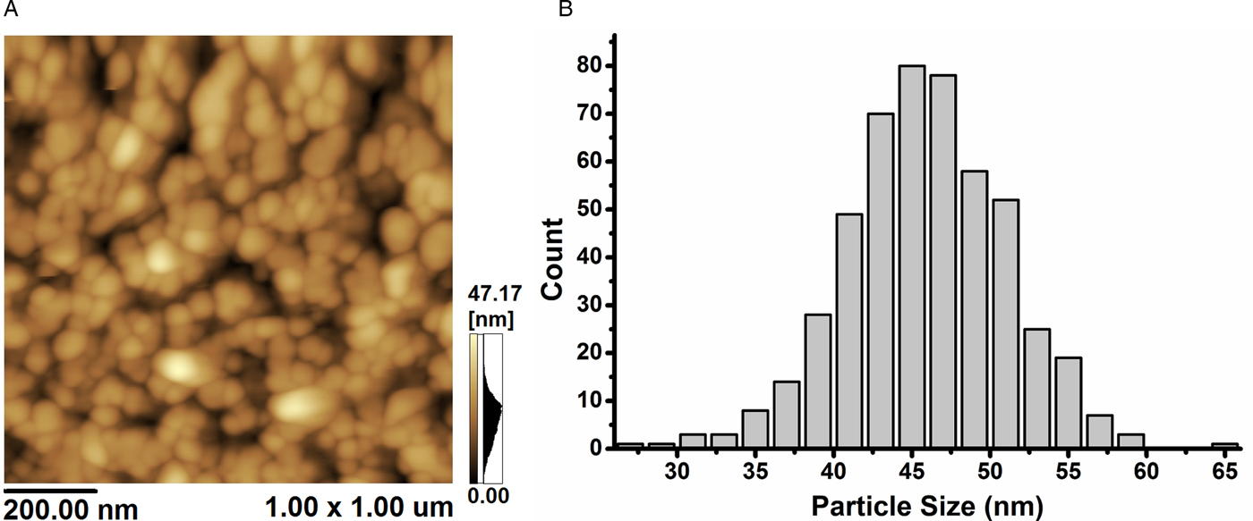
Fig. 1 shows the AFM image of @CaCO3 along with the particle size distribution. The distribution was Gaussian, with a mean size of 41.81 nm and standard deviation of 5.18 nm. The @CaCO3 showed a minimum particle size of 27.83 nm and a maximum of 64.01 nm.
AFM analyses of @CaCO3: (A) Tape mode AFM image of CaCO3 nanoparticles in 2.50 × 2.50 nm2 scale; (B) Particle size distribution graph.

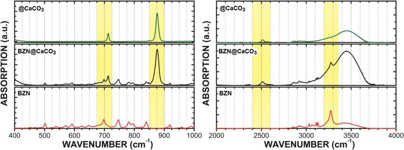
The Infrared spectroscopy analysis showed characteristic absorption bands of BZN and @CaCO3 in the BZN@CaCO3 indicating that the conjugation occurred (Fig. 2).
Infrared analyses showing the main absorption bands (yellow region) present in the BZN, @CaCO3 and BZN@CaCO3

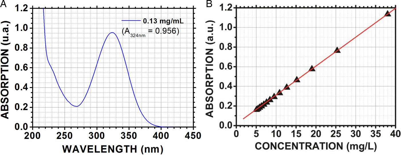
The result of the spectrophotometric assays (Fig. 3) showed that the BZN@CaCO3 had an absorbance peak at 324 nm, the same observed for BZN (Bezerra et al. Reference Bezerra, Bezerra-Neto, Sales, Santos, Martins, De Lima-Neto, Caetano, Albuquerque and Freire2014). Through the linear interpolation of the BZN absorbance (at 324 nm) in different dilutions (calibration curve), the BZN concentration was determined in the conjugated form as being ~25%.
Spectrophotometric analyses: (A) Absorption curve to BZN@CaCO3; (B) Curve of absorption vs. concentration of BZN (in solution) used to determination of amount of BZN present in the BZN@CaCO3 conjugated.

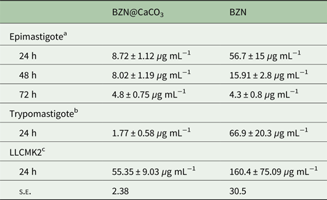
BZN@CaCO3 showed trypanocidal activity against epimastigote forms, inhibiting parasite growth at the three treatment times. The treatment of T. cruzi epimastigotes with BZN@CaCO3 and BZN resulted in dose-dependent growth inhibition, with an IC50/24 h treatment of 8.72 µg mL−1, IC50/48 h of 8.02 µg mL−1 and IC50/72 h of 4.8 µg mL−1 while BZN showed IC50/24 h treatment of 56.7 µg mL−1, IC50/48 h of 15.91 µg mL−1 and IC50/72 h of 4.3 µg mL−1 (Table 1).
Effect of benznidazole-loaded calcium carbonate nanoparticles (BZN@CaCO3) and benznidazole (BZN) on LLCKK2 mammalian cells, epimastigote and trypomastigote forms of Trypanosoma cruzi Y strain

(CC50, IC50 and LD50 values are expressed in μg mL. Selectivity index (SeI) was determined by the ratio between CC50/LC50 values. Data are expressed as means ± s.e.m. in three independent experiments.)
BZN@CaCO3, benznidazole-loaded calcium carbonate nanoparticles; BZN, benznidazole.
a IC50 compound concentrations that inhibits 50% of epimastigote growth.
b LC50 compound concentrations that kills 50% of trypomastigotes.
c CC50 compound concentrations that lyses 50% of LLCMK2 cells.
BZN@CaCO3 killed trypomastigotes in all tested concentrations (50–0.39 µg mL−1) after 24 h of treatment. BZN reduced trypomastigote viability only at highest tested concentrations (50, 25 and 12.5 µg mL−1). When compared with BZN, BZN@CaCO3 showed a significantly higher effect over trypomastigotes, even at lower concentrations. BZN LC50 was 66.9 ± 20.3 µg mL−1, while BZN@CaCO3 LC50 was 1.77 ± 0.58 µg mL−1 (Table 1).
When treated with BZN and BZN@CaCO3, LLC-MK2 cells were evaluated by the MTT reduction method. The data showed that both formulations decreased viability levels, with a CC50 of 55.35 ± 9.03 µg mL−1 for BZN@CaCO3 and 160.4 ± 75.09 µg mL−1 for BZN (Table 1). Despite this, the SeI obtained was 30.5 for BZN@CaCO3 and 2.38 for BZN, showing that BZN@CaCO3 is more selective towards T. cruzi than BZN (Table 1).
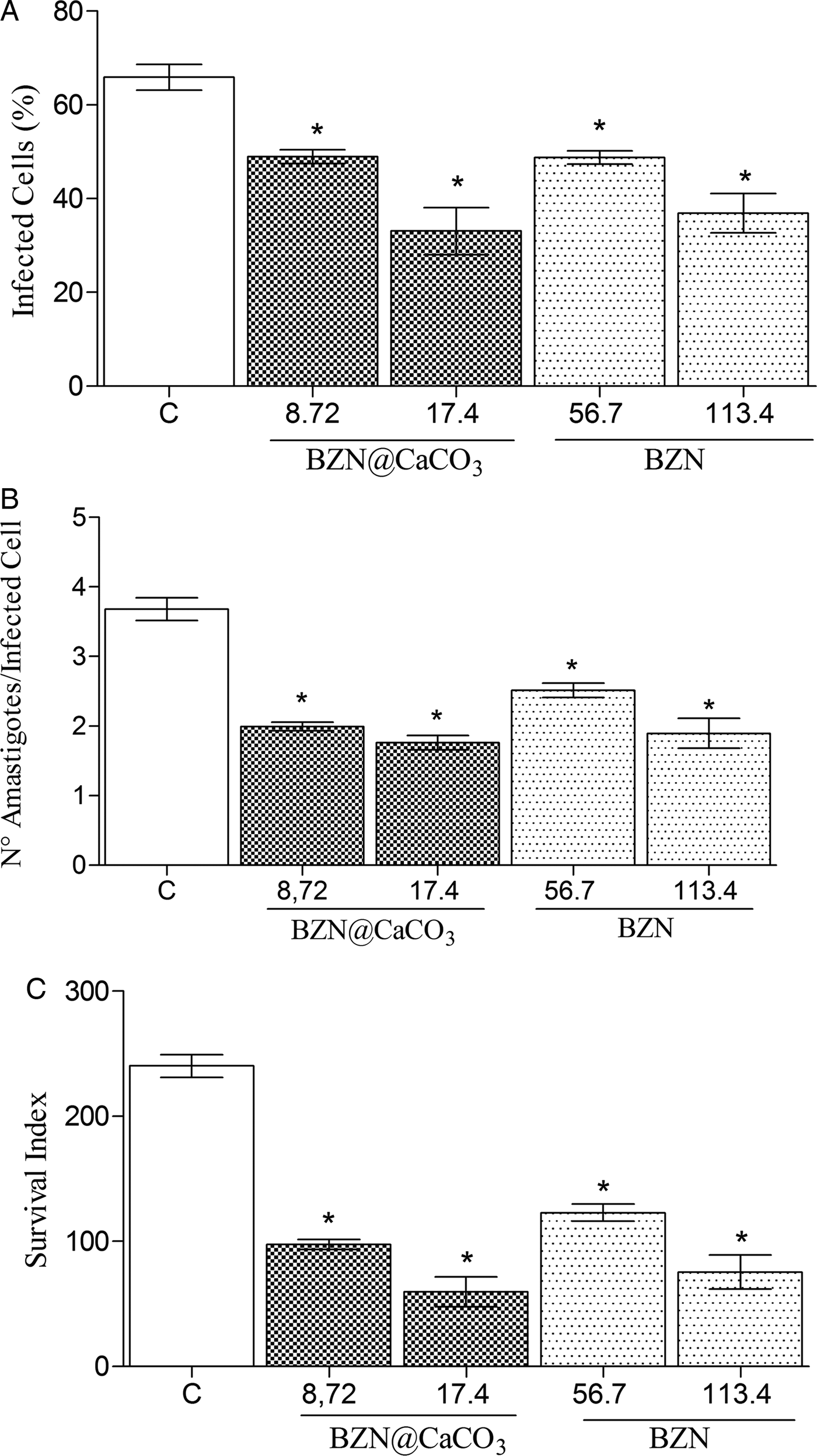
In intracellular amastigote forms, results showed that BZN@CaCO3 IC50 (8.72 µg mL−1) reduced by 25% the mean percentage of infected cells, and by 46% the number of amastigotes per cell. BZN IC50 (56.7 µg mL−1) reduced by 25% the mean percentage of infected cells and by approximately 32% the number of amastigotes per cell (Fig. 4A and B). Incubation with BZN@CaCO3 2 × IC50 (17.4 µg mL−1) reduced the mean percentage of infected cells and the number of amastigotes per cell by approximately 50% (Fig. 4A and B) while incubation with BZN 2 × IC50 (113.4 µg mL−1) reduced by 44% the mean percentage of infected cells and 50% the number of amastigotes per cell (Fig. 4A and B). The decrease in SuI reached 75% of reduction for BZN@CaCO3 2 × IC50 and 68.59% for BZN 2 × IC50 (Fig. 4C).
Effect of benznidazole-loaded calcium carbonate nanoparticles (BZN@CaCO3) and benznidazole (BZN) after 24 h of treatment on intracellular amastigotes in a percentage of infected cells (A), in number of amastigote per cells (B) and in survive index (C).

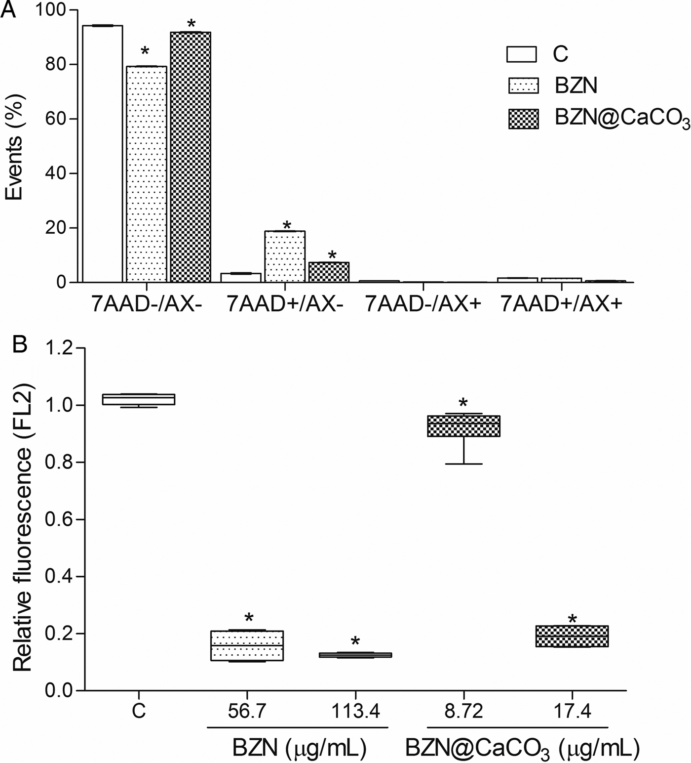
In the flow cytometry analysis, epimastigote forms treated with BZN@CaCO3 IC50 and BZN IC50 for 24 h showed an increase of 7-AAD+ cells (Fig. 5A). In the Rho 123 assay, epimastigote forms treated with BZN@CaCO3 IC50 and 2 × IC50 decreased Rho 123 fluorescence. This Rho 123 fluorescence reduction was observed after treatment with BZN IC50, as well as with 2 × IC50 (Fig. 5B).
(A) Epimastigote forms death was measured by 7-aminoactinomycin (7AAD) and Annexin V-PE by flow cytometry analysis after 24 h treatment with benznidazoleloaded calcium carbonate nanoparticles (BZN@CaCO3) and benznidazole (BZN) IC50. (B) Mitochondrial transmembrane potential alterations in epimastigote forms was evaluated, after 24 h treatment with BZN@CaCO3 and BZN (IC50 and 2×IC50), by flow cytometry by Rho 123.

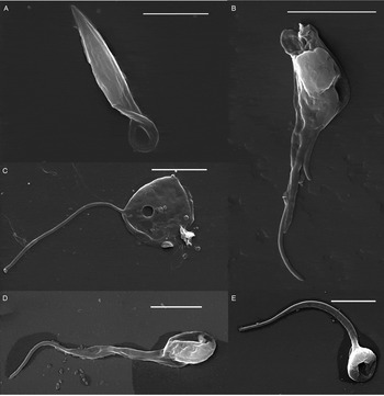
Epimastigote forms treated with BZN@CaCO3 and BZN showed morphological alterations that were observed by s.e.m. The parasites showed loss of membrane integrity with the presence of pores after treatment with BZN@CaCO3 IC50 and 2 × IC50 (Fig. 6B and C), as well BZN IC50 and 2 × IC50 (Fig. 6D and E). Total loss of intracellular volume was seen after BZN@CaCO3 2 × IC50 treatment (Fig. 6C).
Scanning electron microscope of control epimastigotes (A), epimastigotes after 24 h treatment with Bz-NP IC50 (B) and double IC50 (C), epimastigotes after 24 h treatment with Bz IC50 (D) and double IC50 (E).

Discussion
Calcium carbonate (@CaCO3) nanoparticles are biodegradable, biocompatible and have low toxicity (Fujiwara et al. Reference Fujiwara, Shiokawa, Morigaki, Zhu and Nakahara2008; Cheang et al. Reference Cheang, Wang, Hu, Xing, Chang, Yao, Liu and Xu2010; Qian et al. Reference Qian, Shi, Tang, Zhang, Liu and Cao2011). It has been widely used as a drug carrier (Wei et al. Reference Wei, Ma, Hu, Yu, Mcleish, Su and Shen2010; Sharma et al. Reference Sharma, Verma, Teja, Pandey, Mittapelly, Trivedi and Mishra2015). The zeta potential of @CaCO3 positive in distilled water (+16.37 mV), the solubility of biological matrices (Lee et al. Reference Lee, Kim, Kim, Lee, Jeong, Kim, Oh and Choi2015) and the low cost in its production became it a promising nanoparticle. Benzidazole, a standard drug for CD treatment, have poor permeation across the plasma membrane, being required a huge concentration gradient of BZN to gain access the cytoplasmic amastigotes. The toxicity and low permeability of BZN are the factors which contribute to failing in treatment. Nanosystems easily perform membrane transposition and they could help the poor permeation of BZN (Morilla and Romero, Reference Morilla and Romero2015). In this study, we investigated the effect of BZN -loaded calcium carbonate nanoparticles (BZN@CaCO3) against the epimastigote, trypomastigote and amastigote forms of T. cruzi Y strain, and compared it with the BZN effect.
In epimastigote forms, BZN@CaCO3 showed a better trypanocidal effect than BZN. The nanoparticles inhibited the growth of epimastigotes at the three incubation times. The effect at 72 h was similar, but it was better at 24 and 48 h, suggesting a quicker action of the nanoparticle in comparison with BZN.
Chitosan nanoparticles loaded with the trypanocidal drug pentamidine and coated with a single-domain nanobody that specifically targets the surface of African trypanosomes increases drug efficacy. The pentamidine nanoparticle concentration that kills 50% of trypomastigotes (LC50) was approximately 14 times lower than that of free pentamidine (Unciti-Broceta et al. Reference Unciti-Broceta, Arias, Maceira, Soriano, Ortiz-González, Hernández-Quero, Munoz-Torres, Koning, Magez and Garcia-Salcedo2015). This study shows that BZN@CaCO3 LC50 was 37 times lower than the BZN LC50 when tested in trypomastigote forms.
One of the criteria for the production of infective cell-guided therapeutics is the level of selectivity. The leishmanicidal effect of the saponin b-escin and its poly(lactic-co-glycolic acid) (PLGA) nanoparticle showed that the nanoparticle is four times more selective for promastigote forms of Leishmania than the free saponin (Van de Ven et al. Reference Van de Ven, Vermeersch, Matheeussen, Vandervoort, Weyenberg, Apers, Cos, Maes and Ludwig2011). Leishmania amazonensis and Leishmania chagasi showed that the SeI of amphotericin B-chitosan-chondroitin nanoparticles is 10 times higher than that of free amphotericin (Ribeiro et al. Reference Ribeiro, Chávez-Fumagall, Valadares, França, Rodrigues, Duarte, Lage, Andrade, Lage, Arruda, Abanades, Costa, Martins, Tavares, Castilho, Coelho and Faraco2014). In this work, we demonstrated that BZN@CaCO3 had a 12-fold higher SeI than BZN for T. cruzi.
Previously, it was demonstrated that nifurtimox nanoparticles have antiamastigote effects similar to that of free nifurtimox at lower concentrations (Sánchez et al. Reference Sánchez, Cuellar, Zulantay, Gajardo and González-Martin2002). In vitro tests with andrographolide nanoparticles (diterpenoid lactones extracted from the leaves of Andrographis paniculata) found that this formulation has a higher trypanocidal effect over amastigotes in resistant strains of Leishmania donovani than the free substance (Mondal et al. Reference Mondal, Roy, Das, Halder, Mukherjee and Bera2013). In this study, regarding the intracellular form of T. cruzi, the BZN@CaCO3 was capable of reducing the percentage of infected cells and the number of amastigotes/cell, consequently reducing the survival index. The reduction of the survival index in the groups treated with BZN@CaCO3 at concentrations of 8.7 and 17.4 µg mL−1 was similar to the one observed in the group treated with BZN (56.7 and 113.4 µg mL−1, respectively), showing that BZN@CaCO3 also is effective against amastigote forms.
Cell death by necrosis is commonly characterized by a fast decrease in cytoplasmic volume, the collapse of organelles and membrane rupture (Krysko et al. Reference Krysko, Vanden Berghe, Herde and Vandenabeele2008). The loss of the intracellular oxidoreduction control is an important event associated with the mechanism of cytotoxic drugs (Waypa et al. Reference Waypa, Smith and Schumacker2016), including BZN. It has been described that the BZN action is strictly associated with oxidative stress induction in parasites (Maya et al. Reference Maya, Cassels, Iturriaga-vásquez, Ferreira, Faúndez, Galanti, Ferreira and Morello2007), causing reduction of nitro compounds and binding to T. cruzi macromolecules. In the flow cytometry assays, epimastigote cultures treated with BZN and BZN@CaCO3 showed an increase in 7-AAD labelling, suggesting cell death by necrosis.
The mitochondria are essential organelles for cell survival, as they have a central role in the energy production mechanism. Mitochondrial signalling mechanisms are sensitive to ATP reduction and, consequently, activate the oxidative phosphorylation pathways (Wen et al. Reference Wen, Banik, Pathak, Kumar, Kolishetti and Dhar2016). Therefore, drugs capable of interfering with this mechanism may cause cell lesion due to ATP synthesis collapse. To continue the action mechanism studies, parasites treated with BZN@CaCO3 and BZN were submitted to flow cytometry analysis to assess the transmembrane mitochondrial potential. A reduction in the accumulation of mitochondrial fluorescence was observed in both groups, indicating that cell death induced by free BZN and BZN@CaCO3 are related to the interference in the mitochondrial metabolism.
BZN@CaCO3 showed a similar effect on mitochondria, suggesting that the effects found for this substance are induced by the BZN contained in this nanoparticle. Using scanning electron microscopy, we also observed morphological alterations, characteristic of necrotic cell death in BZN and BZN@CaCO3-treated parasites.
It is important to highlight that the real concentration of BZN in the nanoparticle is approximately one-quarter of its weight. The BZN -loaded calcium carbonate nanoparticle showed a trypanocidal effect on the amastigote forms, as well as free BZN, but at lower concentrations. It could be explained by an increase of permeability of BZN promoted by the nanoencapsulation (Higaki et al. Reference Higaki, Kameyama, Udagawa, Ueno, Yamaguchi, Igarashi, Ishihara and Mizushima2006). All these experiments were performed with T. cruzi Y strain, known as nitro-derivative-moderately resistant strain (Branquinho et al. Reference Branquinho, Mosqueira, de Oliveira-Silva, Simões-Silva, Saúde-Guimarães and de Lana2014). BZN@CaCO3 showed a SeI 12-fold higher than BZN.
In this work, we performed all the assays with a BZN -resistant T. cruzi strain and BZN@CaCO3 showed an effect in lower concentrations than free BZN. We also observed an increase of the selective index, decreasing the cytotoxicity against mammalian cells and increasing the toxicity to all developmental forms of the parasite. BZN have poor permeation and a high gradient of concentration is needed to gain the amastigote forms, the responsible forms of maintain the infection. We suggest based on the properties of nanosystems, the increase of permeability of BZN, becoming this drug less toxic and more effective.
In conclusion, CaCO3 nanoencapsulation of BZN promoted lower toxicity and higher selectivity to T. cruzi, with lower concentrations of BZN@CaCO3 being required to result in a trypanocidal effect against all developmental forms of T. cruzi.
Acknowledgement
We also thank Analytic Center – UFC/CT – INFRA / Pro-Equipment CAPES.
Financial support
This research was partially supported by Brazilian Research Agency CNPq-INCT-Nano(Bio)-Simes, Project 573925/2008-9.