Coarctation of aorta is one of the uncommon complications of transcatheter occlusion of patent ductus arteriosus in infants. Reference Zahn, Peck and Phillips1,Reference Sathanandam, Gutfinger and Morray2 Several previous studies have considered this condition as iatrogenic consequence, most of which is trivial and could be self-improved. Reference Zahn, Peck and Phillips1–Reference Duke and Chan6 However, potential genetic predisposition towards coarctation of aorta post-patent ductus arteriosus occlusion cannot be excluded in infants with patent ductus arteriosus complicated by complex malformations, especially bicuspid aortic valve. As evidenced, Reference Siu and Silversides7 bicuspid aortic valve significantly increases risk of infants developing into aortic valve disease or aortopathy including coarctation of aorta by adolescence. For severe cases, surgical treatment needs to be considered.
Under this context, we would like to report a special case of infant with patent ductus arteriosus complicated by bicuspid aortic valve, who presented coarctation of aorta together with significant aortic valve stenosis and regurgitation three years after percutaneous patent ductus arteriosus occlusion. A genetic rather than iatrogenic predisposition has been discussed to indicate genetic predetermination potentiating aortopathy in such cases and highlight the importance of carefully pre-procedural decision-making and close follow-up.
Case presentation
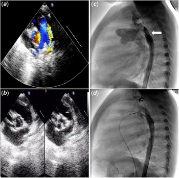
A 14-month-old preterm male infant (Ht 72 cm, Wt 8 kg) was referred for patent ductus arteriosus (Fig. 1A). Bicuspid aortic valve (Fig. 1B) with mild aortic valve stenosis and aortic valve regurgitation was also observed by outpatient echocardiography. Physical examination detected continuous murmur in left second intercostal space and enhanced pulmonic second sound.
Perioperative images. Outpatient echocardiography showed patent ductus arteriosus ( A ) and bicuspid aortic valve ( B ). Pre- ( C ) and post-procedural ( D ) aortography. White arrow indicated aortic dysplasia.

Right cardiac catheterisation showed pulmonary arterial pressure was 59/15 mmHg, and the mean pulmonary arterial pressure was 29.67 mmHg. Left cardiac catheterisation recorded 35 mmHg pressure gradient across aortic valve. Aortography delineated a tubular patent ductus arteriosus (10 mm in length, 3.5 and 6 mm in diameter of the pulmonary and aortic end) and slight aortic dysplasia as indicated by the white arrow in Figure 1C. An Amplatzer Duct Occluder II (St Jude Medical, Plymouth, MN) sized 4/6 was well positioned at the lesion. No residual shunt or occluder protrusion into the left pulmonary artery or aortic lumen was detected by post-procedural aortography (Fig. 1D). Pulmonary arterial pressure post-occlusion was 40/16 mmHg and mean pulmonary arterial pressure dropped to 24 mmHg. No pressure gradient across the occluder was recorded by continuous pressure monitoring. During regular follow-up, echocardiography revealed no residual shunt or significant progression of aortic valve stenosis or aortic valve regurgitation. Importantly, only mild flow acceleration (<1.5 m/s) in the descending aorta was observed.
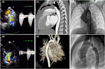
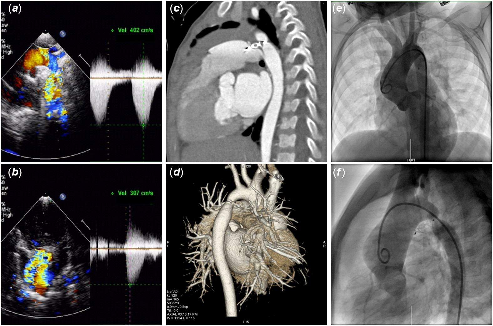
However, at the third-year visit, outpatient echocardiography detected 4 m/s flow acceleration in descending aorta (Fig. 2A). Moderate aortic valve stenosis and severe aortic valve regurgitation were also identified (Fig. 2B). Further CT angiography explicitly demonstrated coarctation of aorta (Fig. 2C and D). Then, the patient was admitted for re-intervention. Aortography showed coarctation of aorta close to Amplatzer Duct Occluder II aortic retention disc, severe aortic valve regurgitation and significant enlargement of ascending aorta (Fig. 2E and F). Right cardiac catheterisation showed pulmonary arterial pressure was 48/16 mmHg, and mean pulmonary arterial pressure was 26.67 mmHg. Left catheterisation recorded 45 and 24 mmHg pressure gradient across aortic valve and coarctation of aorta, respectively. Eventually, the patient received Ross procedure and artificial vascular connection.
Images in follow-up. Echocardiography showed 4 m/s flow acceleration in descending aorta ( A ) and aortic valve regurgitation ( B ). Sagittal view ( C ) and three-dimensional reconstruction ( D ) of CT angiography. Antero- ( E ) and lateral-posterior ( F ) aortography.
Discussion
Transcatheter occlusion of patent ductus arteriosus has been widely acknowledged as a preferred safe and effective therapeutic option for most patent ductus arteriosus patients. With the development of new occlusive devices, increasing underweight infants with patent ductus arteriosus have benefited from this minimally invasive therapy. Reference Zahn, Peck and Phillips1,Reference Tomasulo, Gillespie and Munson3,Reference Baspinar, Irdem, Sivasli, Sahin and Kilinc5 As reported by recent studies, Reference Zahn, Peck and Phillips1–Reference Baspinar, Irdem, Sivasli, Sahin and Kilinc5 coarctation of aorta is an uncommon iatrogenic complication of transcatheter patent ductus arteriosus occlusion in infants. Coarctation of aorta post-patent ductus arteriosus occlusion is mostly trivial and does not need further treatment. Catherine et al. Reference Tomasulo, Gillespie and Munson3 retrospectively observed 3 mild coarctation of aorta (flow acceleration >2 m/s) and 4 flow acceleration (1.5–2 m/s) in descending aorta among 44 small infants undergoing transcatheter patent ductus arteriosus occlusion with different devices. All the seven cases were self-improved in short- to mid-term follow-up without further re-intervention. Another study by Osman et al. Reference Baspinar, Irdem, Sivasli, Sahin and Kilinc5 reported 4 mild coarctation of aorta post-transcatheter patent ductus arteriosus occlusion in 77 patients weighing <10 kg. At half year visit, coarctation of aorta no longer existed in one patient and did not require further intervention in the other 3. Samer and colleagues Reference Masri, El Rassi, Arabi, Tabbakh and Bitar4 found 16% aortic protrusion rate in a retrospective cohort study involving 50 consecutive subjects receiving patent ductus arteriosus occlusion with Amplatzer Duct Occluder II devices, among whom no coarctation of aorta due to aortic protrusion was detected. Besides, an increasing trend of Amplatzer Duct Occluder II aortic retention disc protrusion occurrence attributable to its flexible structure was also indicated, Reference Baspinar, Irdem, Sivasli, Sahin and Kilinc5,Reference Forsey, Kenny and Morgan8 especially in non-conical patent ductus arteriosus infants. Reference Masri, El Rassi, Arabi, Tabbakh and Bitar4 However, these studies are mainly focused on infants with single lesion of patent ductus arteriosus, sometimes complicated by simple intracardiac defects. Relevant data still lack for infants with patent ductus arteriosus complicated by bicuspid aortic valve and aortic dysplasia.
It has been documented that coarctation of aorta is the most common form of congenital vascular defects coexisting with bicuspid aortic valve. Reference Siu and Silversides7,Reference Roos-Hesselink9 And approximately 50%–70% of coarctation of aorta patients have bicuspid aortic valve. Reference Roos-Hesselink9 Besides growth retardation, bicuspid aortic valve has even more profound effects on the development of aortic valve and aorta. In the Toronto cohort study, Reference Tzemos10 22% of patients with bicuspid aortic valve required intervention within 9 years of follow-up. Hereby, many cardiologists hold the notion that most patients with bicuspid aortic valve would require some form of treatment for aortic valve disease or aortopathy in certain stage of life. Reference Siu and Silversides7 Thus, bicuspid aortic valve is one of the important genetic factors, predetermining infants with congenital cardiac defects progressing into significant aortic valve stenosis/aortic valve regurgitation or aortopathy.
Under this scenario, the preterm infant with patent ductus arteriosus in the present case was complicated by bicuspid aortic valve, indicating a predetermined predisposition towards aortic valve disease and aortopathy later in life. Although intraoperative pressure monitoring and post-procedural angiography have confirmed successfully implantation of the device and excluded complications such as coarctation of aorta, possibility of later complications still exists along with the abnormal aortic development. Unfortunately, a genetic testing was unimplemented for personal reasons.
Thus, for infants with patent ductus arteriosus and bicuspid aortic valve, transcatheter patent ductus arteriosus occlusion should be decided and implemented with caution, especially when using Amplatzer Duct Occluder II devices for non-conical patent ductus arteriosus. Genetic testing should be considered, and long-term close follow-up is crucial. Once severe complications occur, adequate and immediate intervention should be scheduled.
In conclusion, transcatheter occlusion of patent ductus arteriosus is safe and effective, yet still challenging for underweight infants with patent ductus arteriosus, especially when complicated by bicuspid aortic valve. Since bicuspid aortic valve itself significantly increases the risk of aortic valve disease or aortopathy, potential genetic predisposition affecting prognosis should never be ignored. For such infants, a comprehensive treatment and follow-up strategy should be carefully made by an expertised heart team, including interventional expert, cardiac surgeon, imaging specialist, and geneticist.
Acknowledgements
We would like to thank all the other staff from the Department of Congenital Heart Disease, General Hospital of Northern Theater Command for their great help with the arrangement and collection of paper and electronic materials.
Financial support
This study was funded by Xingliao Talent Project of Liaoning Province, grant number XLYC2007020.
Conflicts of interest
None.